Bartsocas-Papas Syndrome: A Lethal Multiple Pterygium Syndrome
Sumita Mehta1, Ekta Kale2, Tarun Kumar Ravi3, Ankita Mann4, Pratibha Nanda5
1 Specialist, Department of Obstetrics and Gynaecology, Babu Jagjivanram Memorial Hospital, Delhi, India.
2 Medical Officer, Department of Obstetrics and Gynaecology, Babu Jagjivanram Memorial Hospital, Delhi, India.
3 Specialist, Department of Paediatrics, Babu Jagjivanram Memorial Hospital, Delhi, India.
4 Senior Resident, Department of Obstetrics and Gynaecology, Babu Jagjivanram Memorial Hospital, Delhi, India.
5 Consultant, Department of Obstetrics and Gynaecology, Deendayal Upadhyay Hospital, Delhi, India.
NAME, ADDRESS, E-MAIL ID OF THE CORRESPONDING AUTHOR: Dr. Sumita Mehta, Specialist, Department of Obstetrics and Gynaecology, Babu Jagjivanram Memorial Hospital, Delhi, India.
E-mail: sumitadr@gmail.com
Bartsocas-Papas Syndrome (BPS) is a very rare autosomal recessive syndrome characterised by marked craniofacial deformities, multiple pterygia of various joints, limb and genital abnormalities. It is mostly associated with mutation in the gene encoding Receptor Interacting Serine/Threonine Kinase 4 (RIPK4) required for keratinocyte differentiation. The syndrome is generally lethal and majority of babies die in-utero or in the early neonatal period. This is a report about a neonate born with characteristic clinical features of BPS including severe craniofacial and ophthalmic abnormalities, limb deformities and multiple pterygia at popliteal, axillary and inguinal region. The baby had respiratory distress at birth and was managed conservatively on Continuous Positive Airway Pressure (CPAP)/Oxygen hood and injectable antibiotics for two weeks and then referred for further work-up to a tertiary hospital. The parents took the baby home against the advice of the treating doctors and she subsequently died after 10 days. BPS is associated with high mortality and so all efforts should be directed towards diagnosing it early antenatally when termination of pregnancy is a viable option. This is possible by having a high index of suspicion in couples with consanguineous marriages or with a positive family history.
Craniofacial abnormalities, Genitals, Joints, Limb, Mutation, Popliteal pterygium syndrome
Case Report
A 27-year-old primigravida, with previous two antenatal visits, had a breech vaginal delivery at 37 weeks of gestation. Her antenatal period was uneventful except for an ultrasound (Level 1) done at 18 weeks of gestation that showed the fetus with bilateral club foot. She did not get a level 2 scan done though it was advised. There was no history of fever with rash, drug intake or radiation exposure. There was no history of diabetes, hypertension or Systemic Lupus Erythematosus (SLE). Her marriage was consanguineous and she was married to her first cousin from paternal side.
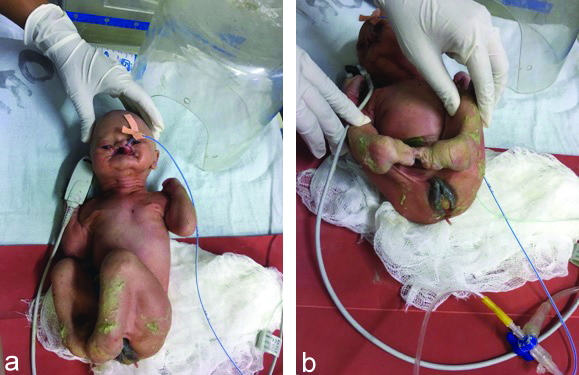
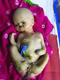
She had reported in spontaneous advanced labour and a breech vaginal delivery was conducted. The female neonate had a birth weight of 2.3 kg and was born with multiple congenital anomalies. There were severe craniofacial abnormalities including malformed nose, facial cleft with bilateral cleft lip and palate, hypoplastic maxilla and mandible, low set and cupped ears [Table/Fig-1a]. There were multiple eye anomalies in the form of bilateral micro-ophthalmia, hypotelorism, bilateral cloudy cornea (right side more than left eye) and medial canthal webbing; there was complete absence of scalp hair, eyebrows and eyelashes. There was complete syndactyly of bilateral hands (mitten hands) and lower limbs were deformed with dorsiflexion of both feet; Nails were absent in all fingers and toes [Table/Fig-1b]. Multiple webs were present at the popliteal, elbow, axillary and inguinal regions affecting majority of joints [Table/Fig-2]. Labia majora was large with absent clitoris and perianal tags were also present.
(a) Craniofacial abnormalities showing malformed nose with orofacial cleft. (b) Pterygia of hip, knee and ankle joint with dorsiflexed lower limbs, large labia majora and anal tag.
Neonate with multiple pterygia of popliteal, axillary and inguinal region.
As the neonate had all the classical features, so a clinical diagnosis of BPS was made. The baby had respiratory distress at birth with an Apgar score of 3, 4 and 6 at 1, 5 and 10 minutes and was transferred to NICU immediately. She was managed on bubble Continuous Positive Airway Pressure (CPAP) for five days and then switched over to oxygen by hood. She was also given prophylactic injectable antibiotics for 10 days (Inj. Cefotaxime 115 mg and Inj. Amikacin 6.5 mg twice daily) and was referred on 14th day in stable condition on nasogastric feeds to a tertiary hospital for further work-up.
The parents stayed in the tertiary hospital for two days and subsequently took the baby home against the advice of the treating doctors. The baby later died after 10 days. All this information was given by the mother when she came six months later to antenatal OPD (when she was three months pregnant again).
Discussion
Limb Pterygium Syndromes (LPS) are characterised by multiple ectodermal abnormalities and webbing on the neck, elbows and knees leading to limited mobility [1]. The active basal layer of the ectoderm is important for its proliferation and differentiation. Any abnormality in these proliferating cells would lead to a wide variety of epidermal development anomalies as is seen in various pterygium syndromes. BPS is an autosomal recessive form of LPS which is very rare with a frequency of 1 in 6,050,000 cases. It is the lethal form of pterygium syndrome with majority of cases dying either in-utero or in early neonatal period [2].
The inheritance of BPS is autosomal recessive but till date no specific gene has been associated with it. Molecular diagnosis in patients with BPS using whole exome sequencing have found mutations in the gene encoding Receptor Interacting Kinase 4 (RIPK4) which is required for keratinocyte differentiation [3].
The primary characteristics of BPS include multiple pterygia of the joints especially popliteal region which leads to severe ectodermal abnormalities though the visceral organs in such cases are generally unaffected and normal functioning. Abnormalities of the genitourinary system with dysplastic kidneys and absent urethra have also been found in neonates with BPS. The other ectodermal abnormalities include arthrogryposis, soft tissue syndactyly, digital hypoplasia, lack of nails, lack of scalp hair, eyelids and eyebrows, blepharitis, cleft lip and palate and hypoplastic external genitalia [4-6]. The index neonate also had these characteristic features of BPS including marked popliteal, axillary and elbow pterygia. Marked popliteal pterygium is the key finding in BPS but this case was unusual as it also showed multiple webs at the axilla and elbows. The neonate also had severe limb deformities with syndactyly of both hands (mitten hands), dorsiflexed feet and absent nails of hands and toes. BPS is also frequently associated with Fetal Growth Restriction (FGR) as was seen in this case with a low birth weight of 2.3 kg [5].
The most important differential diagnosis in BPS is the Popliteal Pterygium Syndrome (PPS), which is an autosomal dominant condition due to mutations in the IRF6 gene which controls epidermal cell proliferation. Lower lip pits and a distinctive nail anomaly comprising of a skin fold which extends from the base to the tip of the nail are part of diagnostic criteria for PPS while limb and facial abnormalities which were seen in this newborn are more common in BPS [7]. Neonates with Van Der Woude Syndrome (VWS) can also present with cleft anomalies in the form of cleft lip and/or palate with lower lip pits but they do not have the other associated anomalies seen in PPS or BPS [8]. Hay-Wells syndrome is also associated with ectodermal defects including ankylobepharonfiliform, cleft lip/palate, hypotrichosis, absent or dystrophic nails and hypodontia. However, absence of multiple pterygia distinguishes it from BPS [9]. The syndrome has a high recurrence rate of 25% and so genetic counseling and prenatal diagnosis is essential in couples with a positive family history or consanguineous marriages [10].
Conclusion(s)
Bartsocas-Papas syndrome is a rare autosomal recessive syndrome characterised by multiple pterygia of joints and this condition usually results in intrauterine fetal death or early neonatal death. All efforts should be made to diagnose the condition in early antenatal period using ultrasonography and option for termination of pregnancy should be discussed with the couple.
Author Declaration:
Financial or Other Competing Interests: None
Was informed consent obtained from the subjects involved in the study? Yes
For any images presented appropriate consent has been obtained from the subjects. Yes
Plagiarism Checking Methods: [Jain H et al.]
Plagiarism X-checker: Jul 13, 2020
Manual Googling: Nov 24, 2020
iThenticate Software: Dec 19, 2020 (11%)
[1]. Kalay E, Sezgin O, Chellappa V, Motlu M, Morsy M, Kayserili H, Mutations in RIPK4 cause the autosomal recessive form of Popliteal Pterygium syndromeThe American Journal of Human Genetics 2012 90(1):76-85.10.1016/j.ajhg.2011.11.01422197489 [Google Scholar] [CrossRef] [PubMed]
[2]. Bender RA, Tanriverdi EC, Yucel A, Erdem MG, A family from Turkey with Bartsocas- Papas SyndromeEurasian J Med 2017 49(1):74-75.10.5152/eurasianjmed.2017.1627228416941 [Google Scholar] [CrossRef] [PubMed]
[3]. Mitchell K, O’Sullivan J, Missero C, Blair Ed, Richardson R, Anderson B, Exome sequence identifies RIPK4 as the Bartsocas papas syndrome locusThe American Journal of Human Genetics 2012 90:69-75.10.1016/j.ajhg.2011.11.01322197488 [Google Scholar] [CrossRef] [PubMed]
[4]. Veenstra-Knol HE, Kleibeuker A, Timmer A, ten Kate LP, van Essen AJ, Unreported manifestations in two Dutch families with Bartsocas-Papas syndromeAm J Med Genet 2003 123A(3):243-48.10.1002/ajmg.a.2030814608644 [Google Scholar] [CrossRef] [PubMed]
[5]. Erturan G, Holton J, Wall S, Giele H, Bartsocos-papas syndrome: A case report and review of the literatureAnn Plast Surg 2016 76(4):459-62.10.1097/SAP.000000000000034825275471 [Google Scholar] [CrossRef] [PubMed]
[6]. Abdalla EM, Morsy H, Bartsocas-Papas syndrome: Unusual findings in the first reported Egyptian familyCase reports in Genetics 2011 10.1155/2011/42871423074676 [Google Scholar] [CrossRef] [PubMed]
[7]. Ratbi I, Fejjal N, Legendre M, Collot N, Amselem S, Sefiani A, Clinical and molecular findings in a Moroccan patient with popliteal pterygium syndrome: A case reportJournal of Medical Case Reports 2014 8:471-73.10.1186/1752-1947-8-47125547932 [Google Scholar] [CrossRef] [PubMed]
[8]. Leslie EJ, Mancuso JL, Schutte BC, Cooper ME, Durda KM, Heureux JL, Search for genetic modifiers of IRF6 and genotype-phenotype correlations in Van der Woude and popliteal pterygium syndromesAm J Med Genet A 2013 161A(10):2535-44.10.1002/ajmg.a.3613323949966 [Google Scholar] [CrossRef] [PubMed]
[9]. Ramya BG, Gondane S, Kumar S, Dangi S, Ankyloblepharon-ectodermal dysplasia-clefting syndromeIndian J DermatolVenereolLeprol 2015 81(6):629-30.10.4103/0378-6323.16833326515851 [Google Scholar] [CrossRef] [PubMed]
[10]. Grip KW, Ennis S, Napoli J, Exome analysis in clinical practice: Expanding the phenotype of Bartsocas-Papas syndromeAm J Med Genet A 2013 161A(5):1058-63.10.1002/ajmg.a.3591323610050 [Google Scholar] [CrossRef] [PubMed]