Low back pain is defined as pain between the costal margin and the gluteal folds. Chronic pain generally is defined as pain that has persisted beyond normal tissue healing time (about three months) [1]. Zygopophysial joints are paired synovial joints formed by adjacent articular processes of the vertebrae. The joint space is typically of about 1-2 mL of volume. Facet interventions represent the second most common type of procedure performed in pain management centers [2]. Lumbar facet arthropathy remains one of the misunderstood, misdiagnosed and improperly treated backache conditions. It involves pain originating from any structure in or around the synovial joint including the fibrous capsule, synovial membrane, hyaline cartilage surfaces, and bony articulations. Ipsilateral lumbar rotation and extension puts the joint in its closed pack position and thus, increasing the forces placed on it, causing pain in case of any pathology, this is the lumbar quadrant test which is the screening test of choice [3]. No imaging modalities have been proved to be diagnostic for facet arthropathy. The definitive diagnostic test is by anaesthetic blockade.
The treatment of lumbar zygopophysial joint pain involves a multimodal approach comprising of medications, physical therapy, modalities and in some cases, psychotherapy. If conservative treatment is ineffective, interventional treatment includes intra-articular facet joint injection using corticosteroids or medial branches block/radiofrequency ablation. Facet joint injection is most commonly performed under C-arm or fluoroscopic guidance for better accuracy and precision [4]. But, this therapeutic imaging modality has led to many side-effects in the eye, gonads and skin due to increased radiation exposure [5]. Therefore, in recent years, ultrasonography has emerged as a useful and viable alternative to C-arm which is radiation free and relatively cheaper and portable but in field of spinal pain interventions, application of USG has limited scope and scarce medical literature support [6]. Therefore, a prospective interventional study was done to compare the treatment outcomes between these two therapeutic imaging modalities (USG and C-arm) using VAS for pain intensity, ODI and time taken for intervention.
Materials and Methods
It was a prospective interventional study done at Department of Physical Medicine and Rehabilitation (PMR) at tertiary care hospital from December 2015 to May 2017. Ethical committee approval and proper inform consent was taken.
Inclusion criteria: All the patients from age 18-55 years [7] and body mass index of 18.5-29.9 kg/m2 [8] who visited PMR Outpatient Department (OPD) with chief complaints of constant/intermittent para-median low back pain, since atleast three months with no or minimal response to conservative treatment and had positive lumbar quadrant test were included in the first phase of study which involved diagnostic blockade to confirm facet arthropathy.
Exclusion criteria: Patients having non-mechanical low back pain with a history of trauma, abnormal neurological examination, previous spinal interventions or surgery, pregnancy, immuno-compromised condition, uncontrolled diabetes, known allergy to anaesthetics or radio-contrast, blood coagulation disorder and less than 50% response to diagnostic blockade were excluded from the study.
Sample size calculation: Minimum sample size was calculated on the basis of previous study by of Ackerman WE and Ahmad M, and it was 31 patients in each group (62 total patients) [9].
Study Procedure
The patients who satisfied the inclusion and exclusion criteria were required to sign a written informed consent in English, Hindi or Bengali manuscript. Then patients entered the first phase of study which involved diagnostic blockade to diagnose and determine the level and side of joint involvement. Using palpation, the level and side was determined and baseline VAS was noted at rest and movement. The higher score was recorded. Then the patients were given intra-articular facet joint injection using 0.5 mL 2% lidocaine under USG/C-arm guidance. Response was evaluated after 30 minutes using second VAS and patients whose pain reduction was less than 50% of the baseline VAS were excluded from the study and conservatively managed while patients reporting more than 50% relief were considered for second phase of the study and asked to report two weeks later. During these two weeks, patients were asked to only take paracetamol 650 mg SOS for pain relief.
The patients invited for second phase after proper informed consent of study were randomised using a sealed envelope technique into group I (Ultrasound group) and group II (Fluoroscopy group) in equal number of 31 each.
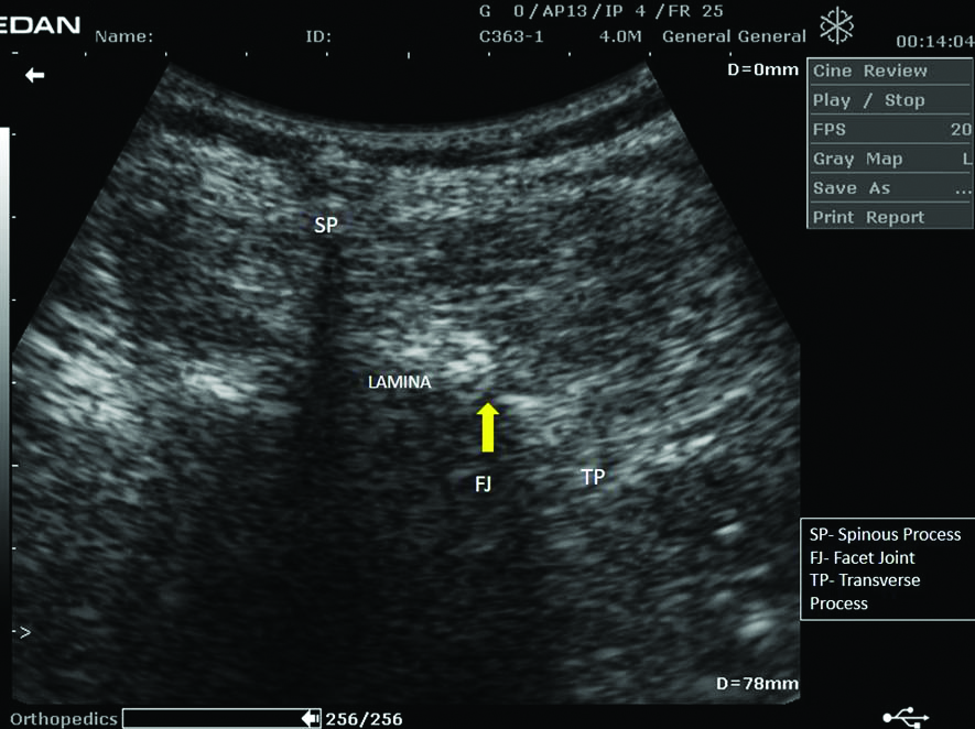
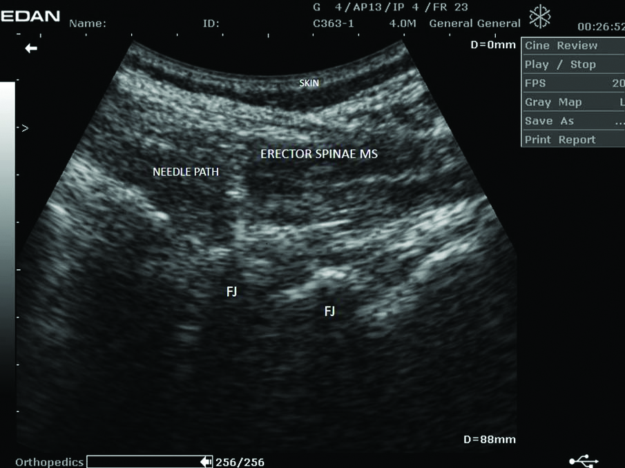
Group 1 (USG group) patients were given an ultrasound guided intra-articular facet joint injection using 0.5 mL methyl prednisolone acetate (40 mg/mL) and 0.5mL of 2% lignocaine. The patients were laid on the table in the prone position, pre-intervention VAS was noted, and level and side of facet joint involvement was reconfirmed from clinical notes. USG (EdanDus 6 100V/50 Hz) and a curvilinear probe were used. The longitudinal paraspinal sagittal image was obtained to discriminate the vertebral location. Then, transverse process was visualised (Trident Sign). After that, the spinous process was confirmed by employing an axial transverse image. Inferior displacement was then attempted, and the lamina was confirmed. The displacement was also attempted to the lateral side of the inferior border of the lamina. Thus, the desired facet joint was confirmed. After giving superficial local anaesthetic injection, a 22G spinal needle was inserted up to a level of the facet joint using the axial transverse image and in-plane approach. When the spinal needle reached the facet joint on the ultrasonography image, the drug was delivered after confirming the needle position in longitudinal as well. Thus, both transverse (short-axis) [Table/Fig-1] and longitudinal (long axis) [Table/Fig-2] views were used.
Transverse view of intra-articular facet joint injection (TP- Transverse Process, FJ- Facet Joint, SP- Spinous Process)
Longitudinal view of intra-articular facet joint injection (FJ- facet Joint, MS- Muscle)
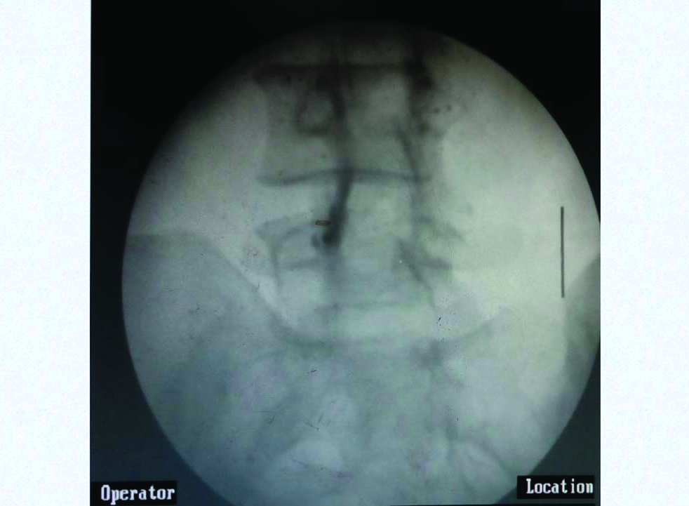
Group 2 patients were given a fluoroscopy guided intra-articular facet joint injection using 0.5 mL methyl prednisolone acetate (40 mg/mL) and 0.5 mL of 2% lignocaine. Patient was placed in prone position, pre-intervention VAS was noted and level and side of facet joint involvement was reconfirmed from clinical notes and true AP image was taken in order to check rotation and square the vertebrae. After the level was determined, the C-arm was rotated ipsilaterally to see Scottie-dog appearance, then the needle was directed towards the facet joint and after loss of resistance, 0.2 mL of radio-contrast dye was given. Then after the dye distribution confirmed needle position within the joint space, the drug was delivered [Table/Fig-3].
Dye spread in C-arm guided intra-articular facet joint injection.
Following spinal facet joint injection, the patient received approximately 30 minutes of bed rest and any abnormality was confirmed at the procedure room.
Total time taken for intervention, pre-injection VAS and ODI was noted and VAS was noted 30 minute post-intervention. Tablet paracetamol 650 mg SOS and following standard post-interventional rehabilitation program was prescribed to both the groups:
Avoidance of strenuous activities or exercises up to 48 hours post-intervention with gradual return of function, to avoid rebound effect.
Exercise programs include improving postural control by reducing any exaggerated lumbar lordosis. This is done through hip flexor stretching, pelvic tilts, and by developing the spine’s supportive musculature (including the deep abdominals, quadratuslumborum, and gluteal muscles) to stabilise the pelvis and lessen the potential shearing forces in the lumbar spine.
If land-based exercise is initially too aggravating, aquatic therapy can be the best starting place.
Following the procedure, regular follow-up was performed at the outpatient clinic on 2, 4 and 12 weeks.
Statistical Analysis
Master chart was prepared in Microsoft office excel 2007 and analysed by IBM Statistical Package for the Social Sciences (SPSS) Statistics version 20. All the variables were tested for normal distribution by Shapiro-Wilk test. Independent Sample Student t-test was applied for continuous variables while Mann-Whitney U test was applied on data which was not normally distributed. Wilcoxon Signed ranks Test was applied for intra-group improvement in pain and disability, since data was not normally distributed (as tested by Shapiro-Wilk Test). Chi-Square test was applied for discrete or categorical variables. Confidence interval (CI) was taken as 95% and p-value <0.05 was considered as statistically significant.
Results
Out of 450 patients visiting the PMR OPD and being subjected to various inclusion and exclusion criteria, 129 patients entered the phase I of the study, out of which 70 patients reported a pain relief of more than 50% after diagnostic block. Four participants each from group 1(USG group) and group 2 (C-arm group), i.e., total 8 participants were excluded from the analysis as they dropped out of the study. So, the analysis was done for total 62 participants divided equally in two groups, i.e., 31 in each group. They were randomised in two groups using sealed envelope technique.
The demographical characteristics of both the groups are given in the table below [Table/Fig-4].
Demographical characteristics and time taken for intervention of two groups. Tests used were 1) Fisher’s-exact test (two sided), 2) Mann-whitney U Test, 3) Pearson’s chi-square test, 4) Independent sample student t-test. Confidence Interval (CI) was taken as 95% and p-value <0.05 was considered as statistically significant.
| Demographics | Group 1 (USG Group) (Total-31 patients) | Group 2 (C-Arm Group) (Total-31 patients) | Comparison (p-value and test used) |
|---|
| Males | 14 | 13 | 1.0001 |
| Females | 17 | 18 |
| Average age (Years) | 37.75±8.13 (range 23-55) | 40.05±9.41 (range 20-54) | 0.6422 |
| Average BMI | 25.72±2.19 kg/m2 (range 23.1-29.7) | 24.85±3.12 kg/m2 (range 21.6-28.3) | 0.2472 |
| Level involved | 16 patients L4/L5 (9 Left and 7 Right)12 patients L5/S1 (3 Left and 9 Right)3 patients L3/L4 (3 Right) | 19 patients L4/L5 (8 Left and 11 Right)10 patients L5/S1 (6 Left and 4 Right)2 patients L3/L4 (2 Right) | 0.7273 |
| Side of facet joint involved | 19 Right12 Left | 17 Right14 Left | 0.7953 |
| Average duration of low back pain (months) | 6.25±2.51 (range 3-12) | 7.01±2.87 (range 4-13) | 0.5404 |
| Time taken for intervention | 4 minutes and 34 seconds (274±51 sec) (range, 220-338 seconds) | 6 minutes and 49 seconds (409±39 sec) (range, 342-473 seconds). | <0.0014 |
USG: Ultrasonography
The two groups did not differ significantly in terms of age, gender, BMI, level of facet joint involvement, side of facet joint involvement and duration of low back pain.
Calculation of total time taken for intervention in case of USG was time point at which paramedian longitudinal USG image was obtained using a probe to determine the level and extending to the time at which injection of drugs in the spinal needle was completed. While, that in C-arm was the time point at which first radiological image (true AP view) to determine the level was taken and extending to the time at which injection of drugs in the spinal needle was completed. USG group was quicker by about 135 seconds (2 minutes and 15 seconds).
The assessment of pain improvement between two groups is given in the table below [Table/Fig-5].
Assessment of pain between two groups. Since data was not normally distributed after testing with Shapiro-Wilk test, hence test used was Mann Whitney U-Test, where p<0.05 was considered statistically significant.
| Average VAS score | Group 1 (USG Group) | Group 2 (C-ARM Group) | Comparison (p-value) |
|---|
| Pre-injection | 7.6±0.81 points (range, 6 to 9) | 7.3±0.92 points (range, 6 to 8) | 0.850 |
| Post-injection (30 min after injection) | 3.2±0.54 points (range, 2 to 4) | 3.3±0.65 points (range, 2 to 4) | 0.656 |
| 2 weeks follow-up | 3.65±0.62 points (range, 3 to 5) | 3.25±0.56 points (range, 2 to 5) | 0.107 |
| 4 weeks follow-up | 3.05±0.65 points (range, 2 to 4) | 2.85±0.81 points (range, 2 to 4) | 0.383 |
| 12 weeks follow-up | 2.75±0.71 points (range, 1 to 5) | 2.40±0.92 points (range, 1 to 5) | 0.343 |
USG: Ultrasonography; VAS: Visual analogue score
The assessment of disability due to low back pain was evaluated using ODI [Table/Fig-6].
Assessment of Oswestry Disability Index (ODI) between two groups. Since data was not normally distributed after testing with Shapiro-Wilk test, hence test used was Mann Whitney U-Test, where p<0.05 was considered statistically significant.
| Average ODI Score | Group 1 (USG Group) | Group 2 (C-ARM Group) | Comparison (p-value) |
|---|
| Pre-injection | 28.25±3.1 (range, 21 to 33) | 27.9±2.34 (range, 23 to 32) | 0.342 |
| 2 weeks follow-up | 16.1±1.43 (range, 12 to 20) | 15.3±1.1 (range, 12 to 22) | 0.893 |
| 4 weeks follow-up | 13.85±1.68 (range, 10 to 19) | 12.3±1.94 (range, 10 to 18) | 0.408 |
| 12 weeks follow-up | 12.4±2.25 (range, 8 to 17) | 11.62±2.66 (range, 8 to 20) | 0.777 |
USG: Ultrasonography
Inter-group comparison of pain intensity and disability was first tested by Shapiro-Wilk Test for assessment of distribution pattern, the probability across all groups, at all visits was <0.05. Thus, data was not normally distributed. Hence, Mann-Whitney U test was applied which showed that there was statistically no significant difference between the two groups in terms of VAS and ODI at 2, 4 and 12 weeks.
Within group improvement was noted in both groups with the baseline and at the end of the study (p<0.001), when analysed by Wilcoxon signed ranks test. It showed significant intra-group improvement in terms of both pain intensity and ODI.
There were no major intraoperative or post-operative complications. Five patients in USG group and three patients in C-arm group experienced vasovagal reactions which were managed conservatively. Two patients in each group faced rebound pain (pain returned after intervention) because of non-compliance to lifestyle modifications or rehabilitation program. They were put on supervised rehabilitation program other than home-based exercises.
Discussion
All the patient included in the study were from the age of 18-55 years because new onset low back pain in children less than 18 years and in those more than 55 years is considered a red flag sign [7]. Underweight (BMI <18.5) and Obese (BMI > 29.9) patients were excluded from the study, since obesity can be a confounding factor and an independent cause of back pain while underweight patients can have malnutrition or fatigue as an independent cause [8]. Since, this was an interventional study, in order to reduce complications, obese patients were excluded. The mean age of participants of this study was 38.9 years while Eubanks JD et al. concluded that prevalence of lumbar facet arthropathy increases with age with more than 82% in 30-39-year-old and 93% in 40-49-year-old [10]. Study by Kalichman L et al., found that facet arthropathy is more common in women as compared to men which is same as this study (35 females and 27 males) [11]. Most of the studies including ones by Eubanks JD et al., and Kalichman L et al., show highest prevalence at L4/L5 level [10,11]. Thirty five out of 62 patients in this study had pain at similar level. The prevalence of facet arthropathy, in study by Abbas J et al., was greater for the right side [12]. This study also had a similar finding (36 on right side and 26 on left side).
The study showed that ultrasonography guided intra-articular lumbar facet joint injection is significantly quicker as compared to fluoroscopic guidance while previous study by Ha DH et al., reported no significant difference between two groups [13]. This is significant since longer procedure time increases the occurrence of vasovagal complication(s) [14]. Intra-articular facet joint injections are widely used and very effective in treatment of low back pain due to lumbar facet arthropathy [15]. In this study, there was significant intra-group improvement in both pain and disability at the end of three months which proves the effectiveness of intra-articular steroid injections in facet joint whether given using fluoroscopy or ultrasound guidance. As far as comparison of both imaging modalities (ultrasound and C-arm) are concerned, there was no significant difference in between the two groups at the end of 2, 4 or 12 weeks. These results are similar to the conclusions of previous studies by Ha DH et al., and Wu T et al., [13,16].
Interventional pain management of lumbar facet arthropathy involves intra-articular steroid injection or dual medial branch block/ablation. As the facet joints are deeply located and cannot be accessed using anatomic landmarks, radiologic assistance under fluoroscopy has been regularly required for all procedures to maximise medication placement success and to avoid neurovascular complications [17]. Despite so many advantages, major disadvantage of C-arm is exposure to radiation, large-sized equipment and high cost [13]. Pack GT and Davis J, reported skin cancer due to radiations [18] while another study by Lee EW et al., reported hand lesions due to irradiation [19]. To minimise these side effects, intermittent fluoroscopy, collimation, grid removal, dose level setting, trained operators and safety equipment are used. Due to these adverse effects of C-arm, USG has recently emerged as a very useful alternative imaging modality for diagnostic and therapeutic purposes. Smaller sized equipment, dynamicity in assessing, avoidance of radiation exposure and safety for pregnant patients are some of the important advantages of ultrasound. Besides, there is no requirement of any additional safety equipment against radiation and it can be moved easily and thus, can be used in outpatient setting. But all this comes with various limitations like low accuracy due to improper imaging of spinal structures, lack of proper and standardised training, low supporting evidence and difficult intervention in obese patients. However, with proper training and knowledge of anatomy, more use and standardisation of ultrasonography in spinal interventions, use of doppler to ascertain synovitis in facet joint and use of sonography in ablation of medial branch blocks, these issues can be avoided.
Limitation(s)
Sample size was very low and a longer follow-up is required. To ascertain better accuracy, no confirmation was performed using radiological images. Also, in obese patients, there was a great gap between the skin and the facet joint. High-quality ultrasonographic image cannot always be obtained while in thin patients, the probe cannot be closely contacted to the skin. This posed a difficulty in obtaining a high quality image. No control group was taken. Psychological and biomechanical factors were not considered in this study. Time taken for intervention could vary from interventionist to interventionist. It is purely based on skill and proper knowledge of Osseo-anatomy and fluoro-anatomy. There could’ve been a sample and interviewer bias.
Conclusion(s)
Both the groups reported significant reduction in pain intensity and disability after 2, 4 and 12 weeks of follow-up, however there is no significant difference between facet joint injections given using Ultrasonography and fluoroscopy guidance. But since ultrasonography is quicker, radiation free, portable, feasible and less expensive, intra-articular facet joint injection using transverse followed by longitudinal view of ultrasonography can be preferred over fluoroscopic guidance.
USG: Ultrasonography
USG: Ultrasonography; VAS: Visual analogue score
USG: Ultrasonography