Teeth selection, being a crucial step reflects aesthetic quality of complete dentures and the prime responsibility of the dentist to ensure their selection with maximum precision [1]. Teeth selection in dentulous patients is rendered simpler due to the existing dentition, and comparison can be made between the natural teeth and the prosthesis. However, in an edentulous patient, the tooth mould has to be carefully selected in order to compliment the personality of the patient. Ahila SC et al., conducted a study and stated there was a significant correlation between the maxillary and mandibular anterior teeth with the index and little finger length [1]. However, only the total mesiodistal width was considered. Kern BE stated they cannot designate the skull length measurement as a reliable means for determining the length of the maxillary central incisor crowns, or the bizygomatic measurement as, a means of determining the width of the maxillary anterior teeth for edentulous patients [2]. The ratios between the mesiodistal width of the maxillary central incisor and the interpupillary distance were statistically similar [3]. However, this study represented only the central incisors and location of the interpupillary line may not be always accurate [3]. The evaluation of the collated results demonstrated by Mavroskoufis F and Ritchie GM showed that 86-90% of the subjects examined did not have identical dimensions or form of the left and right maxillary central incisors [4]. Latta GH et al., showed that there was a lack of strong correlation between anatomic measurements of the width of the mouth, interalar width, bizygomatic width, and interpupillary distance and indicated that the use of any one measurement by itself in the selection of anterior denture teeth for edentulous patients is contraindicated. On the other hand, when all four measurements were used, a good probability exists that the selection of a prosthetic tooth mould will coincide for two or more of the methods [5]. Hence, this study aimed to provide an additional parameter that can be used for selection of anterior teeth by comparing them with the dimensions of finger nail beds.
Materials and Methods
The study was approved by the Ethical Committee of National Council of Medical Research (KIMSDU/ICMR/STS/2015). This was an observational cross-sectional type of study carried out for a time period of three months (April-June 2015). A total number of 402 (201 females and 201 males) subjects ranging between 20-25 years of age were included in the study. According to the institute criteria, the authors were provided a three month study period within which the study had to be completed. A convenience sampling method was used, and to eliminate bias, the maximum number of samples were collected within the stipulated period which amounted to a sample size of 402. Subjects were selected depending on time and as per convenience. The subjects were the undergraduate students and patients visiting School of Dental Sciences, Krishna Institute of Medical Sciences Deemed to be University, Karad, Maharashtra, India. Informed consent was obtained from the subjects.
A pilot study was done on 20 subjects wherein length and width of all finger nail beds of right hand, and length and width of central and lateral incisors of 1st quadrant were measured. The mean obtained of each finger nail bed and both incisors were calculated respectively and were compared to find closest correlation between dimensions of finger nail beds and maxillary incisors. The results of the pilot study showed that the ring finger, index finger, and little finger nail beds showed maximum correlation with the maxillary incisors, and hence they were chosen for the final study.
Inclusion criteria: Dentate individuals with permanent dentition, periodontally healthy teeth.
Exclusion criteria: Restoration/prosthesis involving central and lateral incisors, anomalies of the teeth (size, shape, number), clubbing of fingers and Koilonychia.
Study Procedure
The null hypothesis stated that no correlation exists between dimensions of maxillary central and lateral incisors with dimensions of index, ring and little finger nail beds. The patients were seated in an upright position and the chair position was adjusted to the operator’s level. The blades of the Vernier calliper were disinfected using a disinfecting solution (Aquaclore; Aarsha chemicals Pvt., Ltd., Mumbai; India) and the dimensions of central and lateral incisors were measured using the same [Table/Fig-1,2].
Measurement of the width of right lateral incisor (in mm) using Vernier calipers.
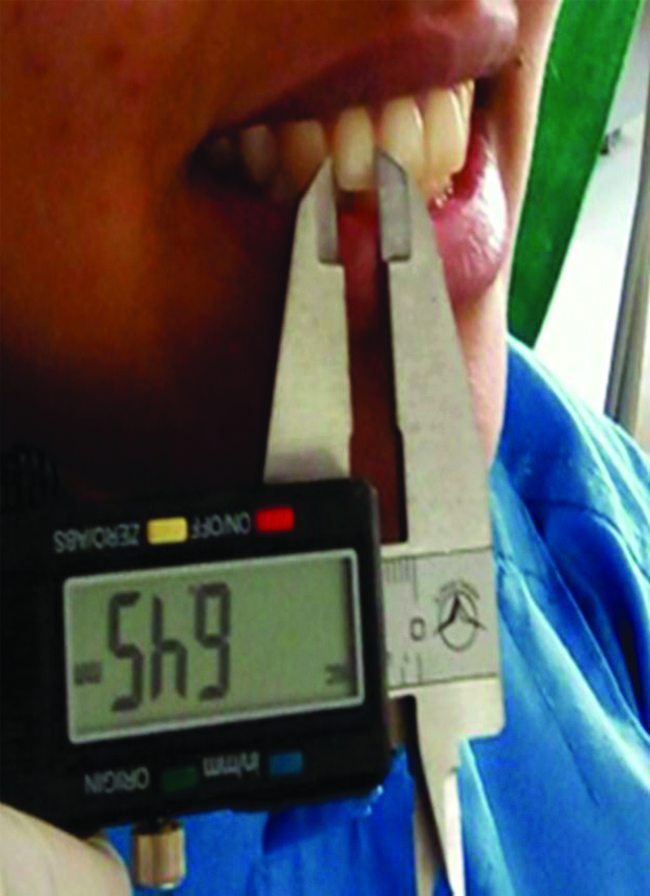
Measurement of the length of right central incisor (in mm) using Vernier calipers.
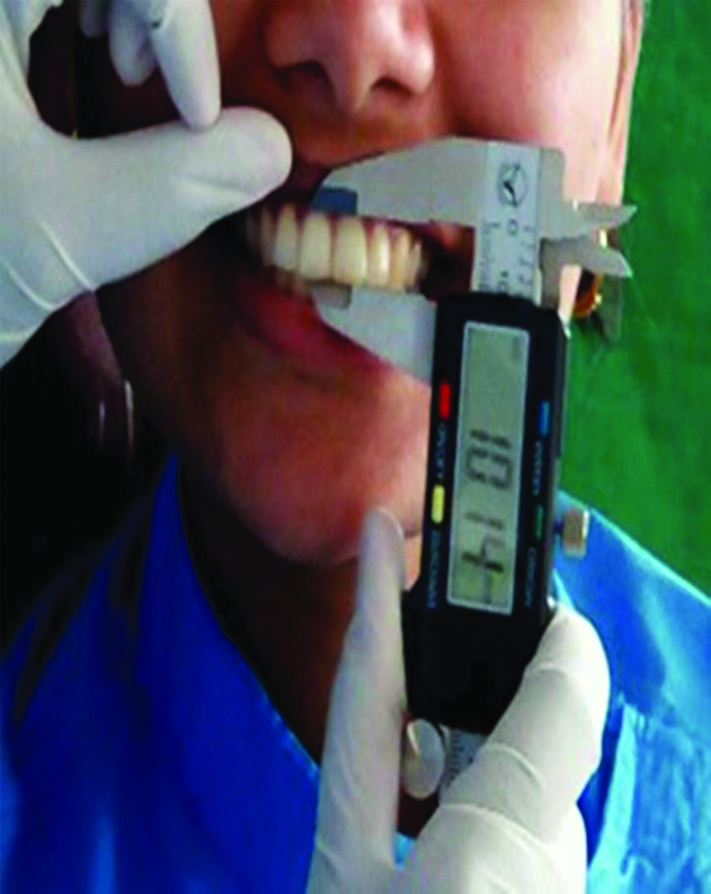
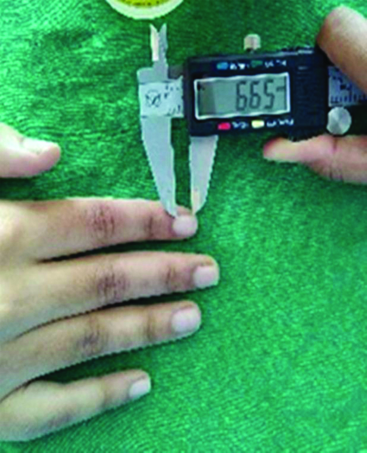
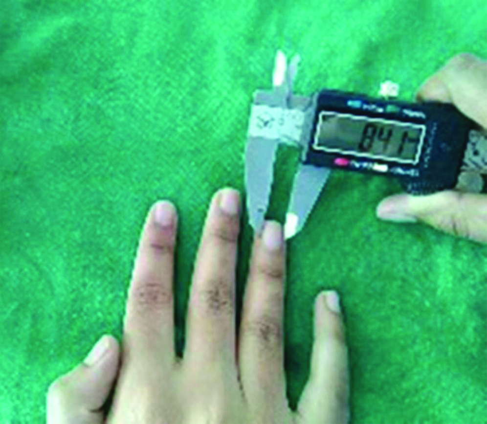
The superior point selected was the gingival zenith of the teeth and the inferior point was the mid-point of the incisal edge. A line connecting these points determined the teeth length. Similarly, width was determined by marking the midpoint of the proximal surfaces and a line connecting them. The measurements recorded were tabulated as the control group. The right hand of the subject was rested on a stable surface and it was ensured that the subjects presented with trimmed nails. The points considered for recording the dimensions of the index, ring and little finger nail bed were the mid-points of the mesial and lateral surfaces of the nail bed and mid-points of the superior and inferior edges of the nail bed, respectively [Table/Fig-3,4].
Measurement of the length of right index finger nail bed (in mm) using Vernier Calipers.
Measurement of the width of right ring finger nail bed (in mm) using Vernier callipers.
These measurements acted as variables. The length and width of maxillary right central incisor was correlated with the length and width of the right nail beds of the index, ring and little fingers. Similar procedure was followed for the left central incisor; left and right lateral incisors; respectively.
Statistical Analysis
The recorded data were statistically analysed using Statistical Package for the Social Sciences (SPSS) version 20 to find out Pearson’s correlation coefficient (r) between dimensions of maxillary incisors and finger nail beds. Further regression equation was derived to determine dimensions of maxillary incisors (dependent variable) from dimensions of nail beds (independent variables) for variables with significant correlations. The p-value less than or equal to 0.05 was taken significant.
Results
[Table/Fig-5] shows significant correlation was found in male subjects between central incisor and lateral incisor dimensions with index, ring, and little finger nail bed dimensions. In males, the central incisor length significantly correlated with little finger nail bed width (p=0.01*). The lateral incisor length had significant correlation with index finger nail bed width (p=0.03*), and little finger nail bed width (p=0.02*). The lateral incisor width had significant correlation with index finger nail bed width (p=0.01*).
Correlation of length and width in male incisors and finger nail beds.
| Length and width | Male IF length | Male IF width | Male RF length | Male RF width | Male LF length | Male LF width |
|---|
| Male CI length | Pearson correlation | 0.059 | 0.151 | 0.036 | 0.267 | 0.075 | 0.358** |
| p-value | 0.681 | 0.290 | 0.801 | 0.058 | 0.599 | 0.01 |
| Male CI width | Pearson correlation | 0.054 | 0.119 | 0.024 | 0.058 | -0.014 | 0.166 |
| p-value | 0.706 | 0.405 | 0.866 | 0.688 | 0.920 | 0.245 |
| Male LI length | Pearson correlation | 0.196 | 0.305* | 0.197 | 0.178 | 0.229 | 0.306* |
| p-value | 0.169 | 0.03 (S) | 0.165 | 0.212 | 0.105 | 0.02 |
| Male LI width | Pearson correlation | 0.115 | 0.339* | 0.199 | 0.217 | 0.204 | 0.213 |
| p-value | 0.423 | 0.01 (S) | 0.162 | 0.126 | 0.152 | 0.134 |
CI: Central incisor; LI: Lateral incisor; IF: Index finger; RF: Ring finger; LF: Little finger; S: Significant; *p-value ≤0.05 significant, ** p-value ≤0.01 highly significant
[Table/Fig-6] shows significant correlation found in female subjects between central incisor and lateral incisor dimensions with index, ring, and little finger nail bed dimensions. In females, the lateral incisor width has significant correlation with index finger nail bed length (p=0.01*) and ring finger nail bed length (p=0.002*).
Correlation of length and width in female incisors and finger nail beds.
| Length and width | Female IF length | Female IF width | Female RF length | Female RF width | Female LF length | Female LF width |
|---|
| Female CI length | Pearson correlation | 0.083 | 0.188 | 0.156 | 0.116 | -0.003 | 0.016 |
| p-value | 0.564 | 0.187 | 0.275 | 0.417 | 0.981 | 0.911 |
| Female CI width | Pearson correlation | 0.105 | -0.064 | 0.207 | 0.087 | 0.119 | -0.066 |
| p-value | 0.463 | 0.658 | 0.145 | 0.545 | 0.404 | 0.647 |
| Female LI length | Pearson correlation | 0.218 | 0.155 | 0.226 | 0.144 | 0.159 | 0.136 |
| p-value | 0.124 | 0.277 | 0.111 | 0.313 | 0.266 | 0.341 |
| Female LI width | Pearson correlation | 0.358** | 0.270 | 0.421** | 0.203 | 0.227 | -0.063 |
| p-value | 0.01 (S) | 0.055 | 0.002 (S) | 0.153 | 0.110 | 0.662 |
CI: central incisor; LI: Lateral incisor; IF: index finger; RF: Ring finger; LF: Little finger; S: Significant; **p-value ≤0.01 highly significant
Further, regression formula was developed respectively for males and females for acquiring dimensions of the incisors with the help of dimensions of finger nail beds. Regression formula was developed based on the main study involving 402 subjects.
[Table/Fig-7] shows the skeleton for the regression formula for determining lateral incisor width in females. The regression equation was as follows
Regression skeleton for lateral incisor width (Female).
| Model | Unstandardised coefficients | Standardised coefficients | T | Sig. |
|---|
| B | Std. error | Beta |
|---|
| 1 | (Constant) | 4.946 | 0.813 | | 6.084 | 0.000 |
| Female IF length | -0.003 | 0.092 | -0.007 | -0.032 | 0.975 |
| Female IF width | 0.091 | 0.108 | 0.161 | 0.842 | 0.404 |
| Female RF length | 0.125 | 0.098 | 0.329 | 1.271 | 0.210 |
| Female RF width | 0.158 | 0.136 | 0.222 | 1.158 | 0.253 |
| Female LF length | 0.031 | 0.101 | 0.067 | 0.308 | 0.760 |
| Female LF width | -0.316 | 0.157 | -0.392 | -2.017 | 0.050 |
IF: Index finger; RF: Ring finger; LF: Little finger; p-value ≤0.01 highly significant
Female lateral incisor width: 4.946 - 0.003 (IF* Length) + 0.091 (IF* width) + 0.125 (RF* Length) + 0.158 (RF* width) + 0.031 (LF* Length) - 0.316(LF* width)
Where, (*IF-Index finger; RF-Ring finger; LF-Little finger)
Similarly, regression equation was formed in males. [Table/Fig-8] shows the skeleton for the regression formula for determining central incisor length in males. In males, the regression equation was as follows
Regression skeleton for central incisor length (Male).
| Model | Unstandardised coefficients | Standardised coefficients | T | Sig. |
|---|
| B | Std. error | Beta |
|---|
| 1 | (Constant) | 6.545 | 1.612 | | 4.061 | 0.000 |
| Male IF length | 0.095 | 0.184 | 0.144 | 0.514 | 0.610 |
| Male IF width | -0.204 | 0.257 | -0.197 | -0.795 | 0.431 |
| Male RF length | -0.180 | 0.212 | -0.309 | -0.849 | 0.400 |
| Male RF width | 0.245 | 0.201 | 0.277 | 1.220 | 0.229 |
| Male LF length | -0.023 | 0.206 | -0.043 | -0.114 | 0.910 |
| Male LF width | -0.579 | 0.268 | 0.434 | 2.163 | 0.036 |
IF: Index finger; RF: Ring finger; LF: Little finger; p-value ≤0.01 highly significant
Male Central incisor length: 6.545 + 0.095 (IF* Length) - 0.204 (IF* width) - 0.18 (RF* Length) + 0.245 (RF* width) - 0.023 (LF* Length) - 0.579 (LF* width)
Where, (*IF-Index finger; RF-Ring finger; LF-Little finger)
[Table/Fig-9] shows the skeleton for the regression formula for determining lateral incisor length in males. In males, the regression equation was as follows
Regression skeleton for lateral incisor length (Male).
| Model | Unstandardised coefficients | Standardised coefficients | T | Sig. |
|---|
| B | Std. error | Beta |
|---|
| 1 | (Constant) | 4.189 | 1.667 | | 2.512 | 0.016 |
| Male IF length | -0.049 | 0.191 | -0.075 | -0.257 | 0.798 |
| Male IF width | 0.364 | 0.265 | 0.352 | 1.370 | 0.177 |
| Male RF length | -0.055 | 0.219 | -0.094 | -0.248 | 0.805 |
| Male RF width | -0.182 | 0.208 | -0.206 | -0.875 | 0.386 |
| Male LF length | 0.073 | 0.213 | 0.133 | 0.343 | 0.733 |
| Male LF width | 0.293 | 0.277 | 0.220 | 1.059 | 0.295 |
IF: Index finger; RF: Ring finger; LF: Little finger; p-value ≤0.01 highly significant
Male lateral incisor length: 4.189 - 0.049 (IF* Length) + 0.364 (IF* width) - 0.055 (RF* Length) -0.182 (RF* width) + 0.073 (LF* Length) + 0.293 (LF* width)
Where, (*IF-Index finger; RF-Ring finger; LF-Little finger)
[Table/Fig-10] shows the skeleton for the regression formula for determining lateral incisor width in males. In males, the regression equation was as follows
Regression skeleton for lateral incisor width (Male).
| Model | Unstandardised coefficients | Standardised coefficients | T | Sig. |
|---|
| B | Std. error | Beta |
|---|
| 1 | (Constant) | 4.699 | 1.047 | | 4.487 | 0.000 |
| Male IF length | -0.178 | 0.120 | -0.424 | -1.487 | 0.144 |
| Male IF width | 0.356 | 0.167 | 0.539 | 2.135 | 0.038 |
| Male RF length | 0.014 | 0.138 | 0.038 | 0.102 | 0.919 |
| Male RF width | -0.083 | 0.131 | -0.147 | -0.637 | 0.527 |
| Male LF length | 0.100 | 0.134 | 0.285 | 0.748 | 0.459 |
| Male LF width | -0.019 | 0.174 | -0.022 | -0.108 | 0.915 |
IF: Index finger; RF: Ring finger; LF: Little finger; p-value ≤0.01 highly significant
Male lateral incisor width: 4.699 - 0.178 (IF* Length) + 0.356 (IF* width) + 0.014 (RF* Length) -0.083 (RF* width) + 0.1 (LF* Length) -0.019 (LF* width)
Where, (*IF-Index finger; RF-Ring finger; LF-Little finger)
Discussion
The study results obtained showed a significant correlation between some teeth dimensions (lateral incisor width in females, and central incisor length in males, lateral incisor length and width in males) and the finger nail beds. Objective analysis of teeth selection can have interoperator variability depending upon the experience of the operator. This method of teeth selection eliminates the above factors. Several studies have been done in the past to determine methods of teeth selection. Ahila SC et al., concluded that significant correlation exists between the total width of the maxillary and mandibular teeth with the index and little finger lengths and a formula for the same was calculated [1]. Cesario VA and Latta GH conducted a study that proved that a ratio of 6.6 existed between the interpupillary distance and the central incisor width both in white men and women, and also in black women [3].
Latta GH et al., concluded that relationships among interalar width of the mouth, bizygomatic width and interpupillary distance might be used as references if applied in combination, however, when anatomic measurements were evaluated individually, racial and gender differences were noted [5]. Sears VH created an Anthropometric-Cephalic Index method and stated that if individual tooth breadth is disregarded and only the average breadths of the incisors and cuspids was considered, a greater consistency in their relation to bizygomatic breadth and cranial circumference was found [6]. Thus, more than one variable was needed to predict the width of the maxillary anterior teeth and central incisors since no best single predictors were accurate enough for clinical application [7].
Sellen PN and Harrison A determined if a correlation exists between tooth dimensions, face, arch forms and palatal contour [8]. This method comprised of incorporation of a computer program to analyse above mentioned aesthetic factors. The correlation was insignificant between face, tooth dimensions and arch forms [8]. There were attempts made before to quantify anterior teeth selection for edentulous patients, but a consensus wasn’t achieved. Previous investigators focussed attention on ratios between the measured distance of several anthropometric parameters and width of the maxillary central incisors [9]. These anthropometric parametric measurements haven’t been evaluated with geometric progression to find out proportionality between them and maxillary central incisor width [9].
Proportion is basically a harmony of structures in space [10-12]. Given that mathematical progressions are evident in nature, it is worthwhile to discuss their simple role in relation to dentistry [9]. There are different types of progressions, including arithmetic, geometric and harmonic among others [13]. Among them, geometric progressions have a special place in dentistry [14]. The vital aspects of aesthetic dentistry are forming a mathematical proportion to relate dimensions of anterior teeth [15]. Lombardi RE first suggested the application of golden proportion in dentistry. He said that the mould assigned should have a pleasing proportion with corresponding anatomy of face, and therefore harmonise with factors necessary to unify it with realism [12]. According to Murthy BV et al., golden proportion of 1.618:1 was not found to be correlated with the relationship between the maxillary central incisor and the mandibular lateral incisor [16].
Hence, the current study conducted not only measured, correlated significantly but also derived a regression model to determine the individual dimensions of the central and lateral incisors from the dimensions of the finger nail beds of index, ring and little fingers for both the genders.
Limitation(s)
Further studies with greater sample sizes and with inclusivity of other fingers/regions are required to determine a more precise regression model. Nevertheless, the current regression model also stands true as a potential method of teeth selection.
Conclusion(s)
The results of the study can still be applied in clinical practice to determine maxillary incisor dimension for edentulous patients. It is less time consuming and is a chair side procedure which can be carried out during routine patient appointment. It also gives a sense of customisation of teeth selection which heightens patient satisfaction. This alternate/additional procedure of teeth selection can be used as a procedure for teeth selection and adds to the scarce literature available comparing teeth dimensions with parameters outside of the orofacial region.
The regression model can be used as an alternate extra-oral method to determine the dimensions of lateral incisor width in females; and central incisor length, lateral incisor length and width in males for edentulous patients from the dimensions of index, ring and little fingers.
CI: Central incisor; LI: Lateral incisor; IF: Index finger; RF: Ring finger; LF: Little finger; S: Significant; *p-value ≤0.05 significant, ** p-value ≤0.01 highly significant
CI: central incisor; LI: Lateral incisor; IF: index finger; RF: Ring finger; LF: Little finger; S: Significant; **p-value ≤0.01 highly significant
IF: Index finger; RF: Ring finger; LF: Little finger; p-value ≤0.01 highly significant
IF: Index finger; RF: Ring finger; LF: Little finger; p-value ≤0.01 highly significant
IF: Index finger; RF: Ring finger; LF: Little finger; p-value ≤0.01 highly significant
IF: Index finger; RF: Ring finger; LF: Little finger; p-value ≤0.01 highly significant