Microleakage is a clinical condition wherein the bacteria, fluids, molecules or ions pass undetectably between a prepared wall of the cavity and the restorative materials which are applied to the cavity [8]. Various restorative materials show varying amount of marginal microleakage either due to some kind of dimensional change during setting or due to lack of good adaptability to the prepared cavity wall of the tooth [9]. The major drawbacks of leakage are postoperative sensitivity, recurrent caries, staining of the restoration and finally failure of the tooth/restoration interface [10]. Plaque on the surface of the restoration contains bacteria which releases nutrients or hydrogen ions thus causing leaking into interfacial spaces [11].
Since Biodentine has many outstanding properties like shorter setting time, ease of manipulation and good biocompatibility to the tooth surface [15], so a need of in-vitro evaluation of this material is required to examine its clinical use as a restorative material. Therefore, the aim of this study is to determine the sealing ability of BiodentineTM as bulk filling material and a base material in Class II restorations. Null hypothesis tested in this study was that the microleakage does not affect the sealing ability of Biodentine when used as a restorative material.
Materials and Methods
Study Design
This in-vitro study was carried out in the Department of Conservative Dentistry and Endodontics, Institute of Dental Sciences, Sehora, Jammu, for about four months from March 2019 to June 2019. Ethical clearance for the study was obtained from the Institute’s head.
Tooth Selection and Preparation
Ten freshly extracted, periodontally compromised, intact, non-carious maxillary human molars were used for this study. These ten samples were later divided into two equal halves making the sample size of 20 (each group 10) was considered significant; the power of the study was 90% at 5% significance. This was done in order to obtain a statistically significant result. The samples were thoroughly cleaned by scaler and pumice slurry. To ensure that the teeth did not present with any cracks or caries, a light microscopic inspection was done. The extracted teeth were kept in saline solution at room temperature for not more than one month. This was done in order to maintain hydration of the samples being used for mechanical testing.
Cavity Preparation and Restorative Procedure
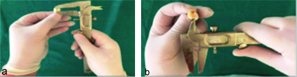
Before cavity preparation, the samples were cleaned to remove any kind of hard and soft deposit over the tooth surfaces. Twenty Class II box-only cavities were prepared on mesial and distal surfaces of each tooth which were randomly assigned to one of two groups (n=10 per group) by using random numbers table for assigning teeth [Table/Fig-1]. Standardised Class II (box only) cavities were made using 106-125 μm diamond bur SF-41 (Straight Flat End, DIA-BURS MANI, INC.) under a water-cooled, high speed airotor handpiece. The dimensions of the cavity were 3 mm in the buccolingual dimension at the occlusal level and gingival floor, 2 mm mesiodistally and 5 mm depth of proximal box. Cavity dimensions were measured using a Vernier Caliper (SSEA Stainless Iron Nickel Vernier Caliper, India) in millimeters [Table/Fig-1]. All the specimens were prepared by a single expert operator in order to ensure consistent depth and size of cavity preparation.
a) Prepared cavities measured by Vernier Calliper in millimetres in proximal box; b) Prepared cavity in Buccolingual direction.
After the cavity preparation, the samples were randomly divided into two groups (G1 and G2):
G1 (Biodentine group): Biodentine (Biodentine”, Septodont®, France) was manipulated according to manufacturer’s instructions. The powder and liquid were manipulated in a capsule using an amalgamator for about 30 seconds. Cavities were filled with an amalgam plugger and cement spatula without any enamel or dentine surface treatment before placement. The restoration was finished and polished using stone burs under copious water spray [Table/Fig-2-a,b].
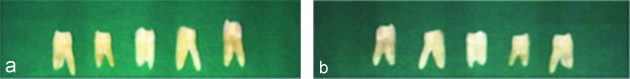
Restored Cavities with Biodentine as Bulk filling material and as a dentine substitute. a) Prepared samples restored with Biodentine (Mesial aspect); b) Prepared samples restored with Biodentine (Distal aspect).
G2 (Biodentine + Tetric N-Ceram Group): Open sandwich technique was used to restore Class II cavities in this group. Biodentine (Biodentine™, Septodont®, France) was manipulated as described for G1 and used as base, leaving 1 mm (measured by Willam’s graduated periodontal probe) (Hu-Friedy, Chicago, USA) of the cavity unfilled from the occlusal surface. Acid etching of the surface was done using 37% phosphoric acid (3M ESPE) gel for 30 seconds, before thoroughly rinsing (10 s) and drying (3s). Prime and Bond NT (Dentsply De Trey, Konstanz, Germany) was applied on all the surfaces with a microbrush and light cured for 20 seconds. The resin composite Tetric N-Ceram (Dentsply De Trey, Konstanz, Germany) was applied for final restoration with Teflon coated composite instrument. A Tofflemire matrix band retainer was adapted to the preparation in order to prevent gingival overhang of the restoration. This was light cured (Celalux 2 High-Power LED curing-light, Voco, GmbH, Germany) for 40 seconds, followed by finishing the restorations using extra fine/ 20- 30 μm diamond burs TR- 13EF (Taper Round End, DIA- BURS MANI, INC.) and EX-21EF (Special Extra Shape, DIA- BURS MANI, INC.) with a water spray. For final finishing, the samples were finished with composite polishing kit (SHOFU INC.) used with a slow speed micromotor [Table/Fig-2c,d]. All the specimens were prepared by a single expert operator to ensure consistent depth and size of the cavity preparation.
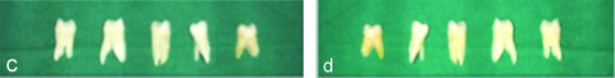
Restored Cavities with Biodentine as Bulk filling material and as a dentine substitute. c) Prepared samples restored with Biodentine + Tetric N-Ceram (Mesial aspect); d) Prepared samples restored with Biodentine + Tetric N-Ceram (Distal aspect).
Thermocycling Test and Dye Penetration
Thermocycling was performed first followed by dye penetration procedure. Following restoration, the samples were stored in saline solution at 37°C for 24 hours and then subjected to 500 thermocycles in a thermocycling machine (Thermomixer comfort by Eppendorf) at temperature range of 5°C±2°C and 55°C±2°C with a dwell time of 30 secs. This was done in order to stimulate temperature fluctuations found in oral cavity [17].
To prepare the samples for dye penetration test, the apices of the specimens were sealed with epoxy resin (Araldite, Brascola Ltda, Sao Bernardo do Campo, Brazil) and coated with fingernail polish except for an area around the periphery of the restoration. 1% methylene blue solution was used to immerse the coated teeth (fisher scientific by Thermo Fisher Scientific) for 24 hours at normal room temperature [18].
Microleakage Evaluation
The samples were removed out of the dye and thoroughly washed for five minutes to remove extra dye on the external surface of the teeth. Under copious amount of water spray, the teeth were sectioned in a buccolingual direction through the centre of the tooth to separate the two Approximal (mesial and distal) Class II restorations with the help of a diamond disk attached to slow speed micromotor. In order to evaluate, the dye penetration at tooth/restoration interface, the samples were also sectioned in a mesiodistal direction [19].
The photographs of sectioned specimens were observed under a stereomicroscope (Nikon SMZ 1500 Zoom Stereomicroscope) at 30x magnification to measure the dye penetration depth at tooth-restoration interface at the cervical levels in both the groups G1 and G2. The scores were given using the ISO (International Organisation for Standardisation) microleakage scoring system (ISO/TS 11405:2003) [Table/Fig-3].
The microleakage scoring criterion [20].
| Score | Tooth-restoration interface | Score criteria (in proportions) |
|---|
| 0 | No dye penetration | 0.00 |
| 1 | Dye penetration into 1/3 of the cervical wall | 0.25 |
| 2 | Dye penetration into 2/3 of the cervical wall | 0.50 |
| 3 | Dye penetration into all of the cervical | 0.75 |
| 4 | Dye penetration into the cervical region and pulpal wall | 1.00 |
There was 94% inter-rater agreement (Cohen’s Kappa=0.94, p<0.01) After a week, the scores of the two examiners were compared, and both of the examiners together re-evaluated restorations with inconsistent microleakage scores.
Statistical Analysis
Data was analysed using Statistical Package for the Social Sciences (SPSS) version 24 (SPSS Inc., Chicago, IL, USA). Descriptive statistics was used to obtain microleakage associated with material when used as dentin substitutes in class II open sandwich technique and also when this material was used as bulk fill material. Comparison mean microleakage was done using Analysis of Variance. In this, test p-value less than 0.05 were taken to be statistically significant.
Results
The results of the null hypothesis determined that microleakage occurred in the specimens of both the groups with variability within the groups [Table/Fig-4]. One group (Group1- Biodentine) exhibited higher microleakage i.e., less sealing ability where as other group (Group2- Biodentine + Tetric N Cearm) showed limited microleakage i.e., higher sealing ability of the material with the tooth surface at the cervical levels. The microleakage in both groups for each sample was measured by ISO/TS 11405:2003 microleakage scoring system at the cervical levels [Table/Fig-5] [20].
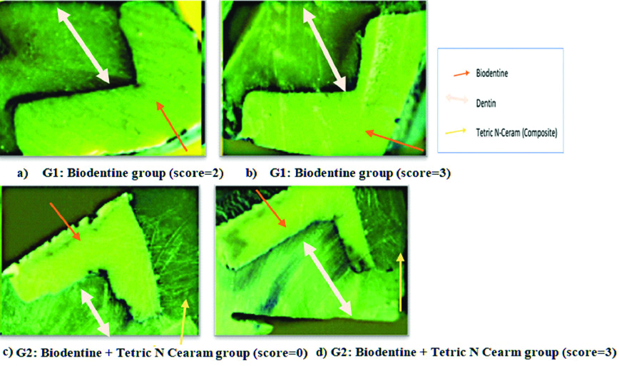
Microleakage evaluation as observed under stereomicroscope at 30X magnification.
Various distribution of microleakage scores along the cervical margin.
| Dye penetration scores | G1 (n=10) | G2 (n=10) |
|---|
| Score 0 | 1 | 2 |
| Score 1 | 1 | 5 |
| Score 2 | 3 | 0 |
| Score 3 | 3 | 3 |
| Score 4 | 2 | 0 |
On taking the statistical analysis (One-way ANOVA) of mean the microleakage score in both the groups, it was interpreted that group G1 and G2 showed sufficient amount of microleakage. Also, group G2 (0.35) indicated comparatively less microleakage as compared to group G1 (0.6) [Table/Fig-6].
Statistical analysis of mean microleakage of both the groups.
| Group | N | Sum of mean microleakage | Mean microleakage | Variance | Standard deviation |
|---|
| G1 | 10 | 6 | 0.6 | 4.5 | 0.3162 |
| G2 | 10 | 3.5 | 0.35 | 2 | 0.2934 |
| Total | 20 | 9.5 | 0.475 | 6.5 | 0.3234 |
One-way ANOVA test; N, number of samples
On comparison of both the groups with help of Post-hoc Test showed that there is no significant difference between group G1 and G2. Thus, both the groups shows that microleakage occurred with less difference when used as dentin substitute or bulk filling restorative materials [Table/Fig-7].
Comparisons between Biodentine G1 and Biodentine + Tetric N-Ceram G2 group (Post hoc-test).
| Source | SS | df | MS | f-ratio |
|---|
| Between groups | 0.3125 | 1 | 0.3106 | 3.35821 |
| Within groups | 1.675 | 18 | 0.0931 | |
| Total | 1.9875 | 19 | | |
SS: Sum of squares; df: Degree of freedom; MS: Mean square. p>0.05; The f-ratio is 3.35821. The p-value is.083467.
Discussion
This study determined the in-vitro sealing ability of calcium silicate-based material when used as a base material in an open sandwich restoration and also when used as a bulk filling material. It was concluded from the study that no statistically difference exists between the two techniques of restoration. The new calcium silicate-based material performed well as the base material than as a bulk fill material. A brief summary of in-vitro/in-vivo human studies presenting microleakage in Biodentine when compared with other restorative materials was tabulated [Table/Fig-8] [16,19,26-28,33-38].
Studies on comparative evaluation of microleakage in Biodentine when used as restorative material [16,19,26-28,33-38].
| Author | Place of study | Year/Type of study | Number of samples (n) | Method of evaluation | Outcome of the study |
|---|
| Raskin A et al., [26] | Marseille, France | 2012/In-vitro study | n=60 | Silver nitrate dye penetration | Biodentine performed well as dentin substitute or restorative material in approximal cavities |
| Koubi S et al., [16] | Marseille, France | 2012/In-vitro study | n=30 | Glucose diffusion from glucose solution | Biodentine performed well in open-sandwich restoration. |
| Koubi G et al., [28] | Marseille, France | 2013/In-vivo study | n=397 | 3-year prospective study | Biodentine is well tolerated dentine substitute under composite |
| Solomon RV et al., [19] | Hyderabad, India | 2014/In-vitro study | n=50 | 2% methylene blue dye penetration | Biodentine scored better than resin modified-GIC in Class II open-sandwich restoration |
| Aggarwal V et al., [33] | Gurgaon, India | 2015/In-vitro study | n=60 | Aging in phosphate buffered saline | The study reports a better adaptation obtained with Biodentine than MTA plus |
| Niranjan B et al., [27] | Bhopal, India | 2016-In-vitro-study | n=60 | 2% methylene blue dye | Biodentine exhibits superior marginal sealing ability under composite as compared to MTA and GIC |
| Darsan J et al., [34] | Bengaluru, India | 2018/In-vitro study | n=40 | 0.5% aqueous solution of rhodamine B dye | Theracal LC and Biodentine performed better than RMGIC as liner in deep Class II closed sandwich restorations |
| Choudhary D and Verma P [35] | Jammu, India | 2019/In-vitro study | n=30 | 1% methylene blue dye penetration | Sealing ability of SDR was best at occlusal and cervical levels followed by Biodentine and Fuji II LC |
| Bhullar KK et al., [36] | Amritsar, India | 2019/In-vitro study | n=50 | Rhodamine-B | Biodentine proved to be the best coronal sealing material followed by GIC, Cention N. |
| Kusumvalli S et al., [37] | Bengaluru, India | 2019/In-vivo study | n=20 | One year clinical and radiographic evaluation | Biodentine proved at maintaining pulp vitality. |
| Soliman AF et al., [38] | Tanta, Egypt | 2019/In-vitro study | n=30 | 1% methylene blue dye | Biodentine revealed higher sealing ability as compared to Dycal in deep carious lesion. |
| Choudhary D [present study] | Jammu, India | 2019/In-vitro study | n=20 | 1% methylene blue dye | Biodentine showed no significant difference when used as dentine substitute or as a bulk filling material. |
GIC: Glass ionomer cement; MTA: Mineral trioxide aggregate; SDR: Smart dentin replacement; RMGIC: Resin-modified glass ionomer cements; LC: Light cured
There is continuous search for the restorative material which provides good sealing with the tooth in order to reduce microleakage and also have excellent physical properties. Biodentine (Septodont, Saint Maur des Fosses, France) is composed of modified composition of MTA by addition of setting accelerators and softners. Being a biocompatible material, Biodentine has said to revolutionise the restorative dentistry [15]. It causes mineralisation after placement in the cavity, during the setting it forms osteodentine [21]. It has an inhibitory effect on bacteria due to its alkaline nature. In addition to many appreciable properties of biodentine like tissue regeneration, early mineralisation, short setting time, antibacterial, high push-out bond strength etc., the major properties of Biodentine which has been a boon to the adhesive dentistry are the exceptional good biocompatibility to the tooth and a good marginal sealing ability of the material [21,22]. The elastic modulus of this material is 22.0 Gpa which quite near to dentine at 18.5 Gpa, compressive strength is 220Mpa which is same as the average for dentine of 290 Mpa and microhardness of Biodentine is equal to natural dentin (60 HVN). All these mechanical properties makes this material a best suitable dentin substitute for Class II restorations [23]. A similar kind of study was performed and Biodentine was compared to two other restorative materials i.e., (Smart Dentin Replacement) and Fuji II LC. The results of the study were different from the present study and they concluded that Biodentine showed maximum microleakage when used as dentin substitute [35]. Biodentine forms crystals with hydroxyapatite at the surface of the tooth [24]. These crystals promote the sealing efficiency. As the cement ages, it has a capacity to produce hydroxyapatite crystal which subsequently closes the gap between the material with tooth or other restoration [19]. It micromechanically bonds to the tooth without surface treatment of the tooth surface done prior. It adheres to the surface of the tooth because of its highly alkaline nature which leads to erosion of dentin and its penetration into the dentinal tubules. On mixing, there is an initial contraction of cement during hydration but later there will be secondary expansion of the cement. This mechanism explains the sealing ability [25].
The results of this study are quite similar to an in-vitro study, which concluded that Biodentine performed well when used as dentin substitute in cervical lining restorations or as a restorative material in Class II cavities where they extend cervical under the Cemento-Enamel Junction (CEJ) without any conditioning treatment [26]. Another study documented that Biodentine has a superior marginal sealing ability than MTA and Glass ionomers and thus is the best suitable dentin substituent under composite resin [27]. Biodentine was introduced in the market with a view to overcome the drawbacks of MTA like long setting time, discoloration, difficult handling and composition containing toxic elements but Biodentine also had limitations in particular to its lower wash out resistance and poor radioopacity [39]. A study by Koubi G et al., concluded that Biodentine was a superior material for posterior restorations as it revealed good marginal adaptability even after six months of its placement under a layer of composite [28]. Another study evaluating the sealing ability of Biodentine in cervical lining or as a restorative material in proximal cavities reported that it performed well without any prior conditioning over the tooth surface [16].
The experiment protocol should be quite similar to clinical conditions, in order to reproduce a valid outcome of the study. The teeth were subjected to themocycling which is a method of subjecting the restoration on the tooth to temperature extremes in-vitro, compatible with the oral cavity. The coefficient of thermal expansion between the tooth and restorative material is suggested by introducing hot and cold temperatures [29].
Over the years there are various methods to access microleakage to evaluate the performance of the material at the tooth-restoration interface [30]. Dyes and radioisotopes are the most common methods to evaluate the sealing ability among the others like air pressure, neutron activation analysis, scanning electron microscopy and pH changes [31]. In this study, 1% methylene blue was used. Dye penetration test is one of the oldest and reliable method to study microleakage. Methylene blue has a small molecular weight thus has a high penetration rate [32].
The observation of the dye penetration at the tooth/restoration interface was observed under stereomicroscope which is a well-established method to provide clear images. Stereomicroscopic images revealed that significant amount of microleakage occurred in Biodentine as a dentin substitute and also as bulk material.
Biodentine is an interesting and rising material in the field of adhesive dentistry, which has the potential to maintain pulp vitality and thus has increased the life of a restored tooth.
Limitation(s)
The present research work was carried out on a small sample size. Dye penetration is used for evaluating microleakage; using advanced method can be helpful for better result for restorative materials routinely used. Also, since no control group was advocated in this study the results of groups were difficult to compare. Biodentine was also not compared with other restorative materials, which can be one of the drawbacks to the present study.
Conclusion(s)
The present study concluded that BiodentineTM (Septodont, Saint Maur des Fossés, France) showed some kind of microleakage when used as dentin substitute or as a restorative material in Class II restorations. It exhibited slightly superior sealing ability as a dentin substitute when placed under a rigid composite resin, thus confirms as one of the good biocompatible material. Biodentine is a miracle material in all its physical properties thus further research is required to consider it as good restorative option for Class II cavities. Further, future in-vivo studies are recommended to provide more valid results.
One-way ANOVA test; N, number of samples
SS: Sum of squares; df: Degree of freedom; MS: Mean square. p>0.05; The f-ratio is 3.35821. The p-value is.083467.
GIC: Glass ionomer cement; MTA: Mineral trioxide aggregate; SDR: Smart dentin replacement; RMGIC: Resin-modified glass ionomer cements; LC: Light cured