Autopsy is a Greek word that means to see with ones own eyes. Medico-legal Autopsy is a special type of scientific examination of dead body carried out mainly for the identification, prosecution of the guilty and cause of death [1]. At around 3000 BC, the autopsy evolved. During the process of mummification, the ancient Egyptian remove and examine the internal organs of dead bodies. But they related changes to magic rather than pathologic anatomy. It is also called as Necropsy/Post-mortem, but the widely used terminology is autopsy [2]. There are two types of Autopsies. 1) Forensic/Medico-legal/ Coroner’s Autopsy, is ordered by law, when death occurs under suspicious circumstances, sudden death or accidental death. 2) Medical/Clinical/Pathological Autopsy, is performed to find the cause of death and for research purposes. The second most common cause of unexpected and sudden death is due to pathological condition in the respiratory system affecting thousands of people in the world [3]. Respiratory disease progress rapidly, and is particularly common in extremes of ages such as neonate, old ages and immunocompromised individuals. There is rapid increase in respiratory morbidity in todays population due to increase in air pollution, smoking, chemical substances that leads to preventable chronic respiratory diseases affecting millions of people all over the world [4,5]. Lungs are secondarily involved in almost all form of Terminal Diseases [6]. The lungs are pyramid shaped paired organs that are connected to the trachea by the right and left bronchi, progressive branching of the bronchi forms bronchioles that leads to terminal bronchioles. The parts of the lungs distal to the terminal bronchioles is called the acinus and are located in the thoracic cavity. The right lung has three lobes and left lung has two lobes. On the inferior surface the lungs are bordered by the diaphragm. The main function of lungs is carry out exchange of gases between inspired air and blood. Histopathological examination of lung autopsy is very useful to diagnose respiratory cause of death [6].
The study on autopsy findings of lungs has not been conducted in rural area of Bastar region, hence this study was conducted, to find out the disease/changes in lungs. The aim of the present study was to study the spectrum of histopathological finding in lung autopsy and the incidental findings identified during histopathological examination gain a prime importance in academic and research purpose. The objective of the study was to study the histopathological patterns of lung lesions in autopsy.
Materials and Methods
This retrospective study was conducted in Department of Pathology in Lt. BRKM Govt Medical College, Jagdalpur, Bastar, Chhattisgarh, India. In this study, 513 Lung specimens were received over period of 10 years i.e., from January 2010 to December 2019 irrespective of age and sex, 39 specimens were autolysed, so 474 specimens were included in the study. Department of Pathology receives post-mortem specimens from District hospitals, Primary health centres and Community health centres of district Jagdalpur, Kanker, Kondagav, Narayanpur, Sukma, Bijapur, Bhairamgarh, Dantewada, Kirandul, Bhailadila for histopathological examination. As lung specimens were medico-legal, ethical clearance was not taken. In medico-legal autopsy, consent from the relatives of deceased is not required.
Inclusion criteria: Autopsy usually from medico-legal cases, custodial deaths, road side accidents, poisoning and drowning irrespective of cause of death.
Exclusion criteria: Autolysed specimens. All the data were taken from autopsy records bought by police constables. Most of the specimens were received in pieces, fixed in 10% formalin. Gross examination included recording of weight, assessment of size, colour change and texture. Routine tissue processing technique was done and paraffin blocks were prepared. Tissue sections of 3-5 microns thickness were cut. These sections were stained with routine Hemotoxylin and Eosin stains. The stained sections were mounted with cover slip using DPX. The slides were prepared and studied under light microscope.
Statistical Analysis
Descriptive statistics were used was data analysis and data were enter into microsoft excel sheet.
Results
A total of 474 specimens were included in the study. Histopathological examination was carried out. Relevant tables were made. Age wise distributions of these autopsy cases is shown in [Table/Fig-1]. Majority of cases 141 (29.70%) were in the 30-39 years of age followed by 20-29 years 106 cases (22.45%) and 40-49 years 87 cases (18.36%). Among the lung lesion detected in 474 lung autopsies, there were 345 (73%) males and 129 (27%) females. Among these 474 lung lesions in 33 cases (7%) histopathology were unremarkable, significant microscopic findings were found in 441 cases (93%). [Table/Fig-2] shows most common findings were Congestion with Pulmonary oedema seen in 352 (74.25%) followed by Congestion with Pulmonary oedema and Intra-alveolar Haemorrhage 41 (8.64%), Bronchopneumonia 14 (2.94%), Lobar Pneumonia 11 (2.32%), Tuberculosis 7 (1.48%), and Chronic Bronchitis 1 (0.21%).
Age group distribution in lung lesions.
| Age (Years) | Cases | % |
|---|
| 0-09 | 14 | 2.95 |
| 10-19 | 60 | 12.70 |
| 20-29 | 106 | 22.45 |
| 30-39 | 141 | 29.70 |
| 40-49 | 87 | 18.36 |
| 50-59 | 42 | 8.85 |
| >60 | 24 | 4.99 |
| Total | 474 | 100 |
Histopathological finding in lung tissue.
| Lung lesion | N | % |
|---|
| Congestion+Pulmonary oedema | 352 | 74.25 |
| Congestion+Pulmonary oedema+Intra-alveolar haemorrhage | 41 | 8.64 |
| Bronchopneumonia | 14 | 2.94 |
| Lobar pneumonia | 11 | 2.32 |
| Tuberculosis | 7 | 1.48 |
| Interstitial pneumonia | 7 | 1.48 |
| Vegetative material | 3 | 0.63 |
| Emphysema | 3 | 0.63 |
| Aspiration pneumonitis | 2 | 0.42 |
| Chronic bronchitis | 1 | 0.21 |
| Normal lung | 33 | 7 |
| Total | 474 | 100 |
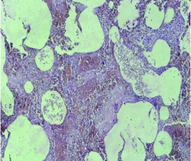
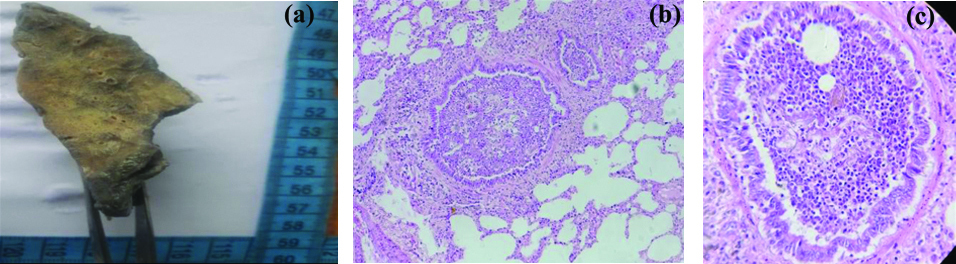
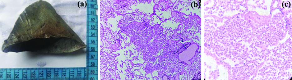
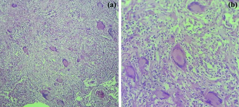
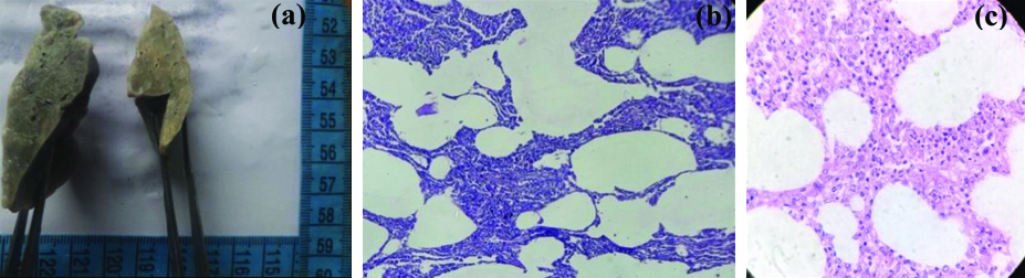
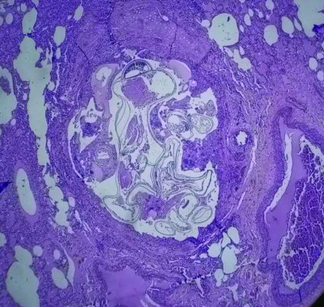
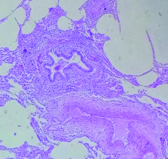
We found varieties of microscopic findings seen in lungs which included oedema and congestion, inflammation (acute and granulomatous), changes in interstitium, emphysema, vegetative material aspiration. In current study, most common pathological finding was Pulmonary Oedema associated with Congestion [Table/Fig-3], [Table/Fig-4] microscopic picture shows congested blood vessels with pinkish fluid (oedema) present intra-alveolar spaces and septae. Next pathology was intra-alveolar haemorrhage [Table/Fig-5] microscopic picture shows area of haemorrhage present in alveolar spaces and septa. The next commonest pathology was bronchopneumonia, which was characterised by gross picture showing patchy discoloration [Table/Fig-6a] and microscopic picture shows bronchioles and peribronchiolar alveoli are filled with inflammatory exudates [Table/Fig-6b,c]. Lobar pneumonia was found in 11 cases, in which gross picture shows patchy discoloration [Table/Fig-7a] and microscopic pictures show intra-alveolar exudates of polymorphonuclear cells, red cells [Table/Fig-7b,c]. In this study seven cases of tuberculosis was found, microscopic pictures shows granuloma formation with Langhans giant cell [Table/Fig-8a,b] and seven cases of Interstitial Pneumonia was also found in this study, gross picture shows patchy discolouration [Table/Fig-9a] and microscopic pictures show interstitial inflammatory cells [Table/Fig-9b,c]. Vegetative material was found in three cases, which is characterised by microscopic picture shows plugging of bronchioles with vegetative material [Table/Fig-10] and Aspiration pneumonitis was found in two cases which is characterised by microscopic picture shows vegetative material with inflammatory exudates [Table/Fig-11]. Emphysematous changes were found in three cases, in which microscopic picture shows abnormally large alveoli with focal destruction of alveoli separated by thin septa [Table/Fig-12]. One case of chronic bronchitis was found in this study, microscopic picture shows secretion within bronchioles and chronic inflammatory infiltrates around bronchioles [Table/Fig-13].
Shows congested blood vessels. H&E (4X).
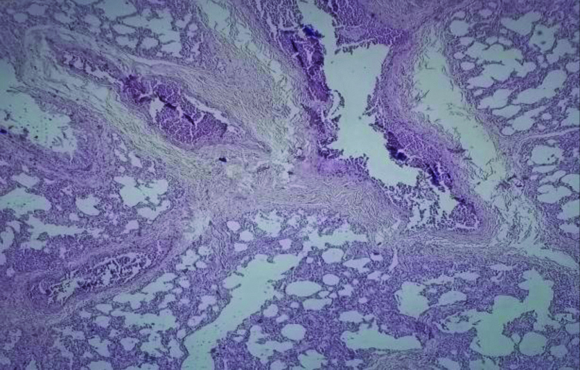
Shows congested blood vessels with pinkish fluid (Oedema) present intra-alveolar spaces and septae. H&E (10X). Congestion and pulmonary oedema.
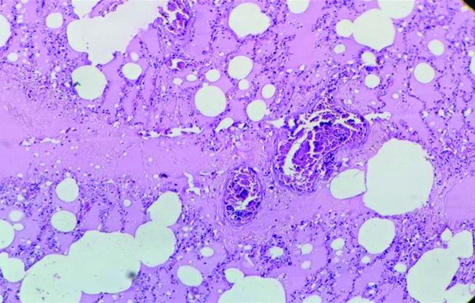
Intra-alveolar hemorrhage image showing hemorrhage present in alveolar spaces and septa. H&E (10X).
Bronchopneumonia Gross- a) Shows patchy discoloration; b,c) shows bronchioles and peribronchiolar alveoli filled with inflammatory exudates. H&E b) (10X), c) (40X).
Lobar pneumonia Gross- a) Shows patchy discoloration; b,c) Shows intra-alveolar exudates of polymorphonuclear cells, red cells. H&E b) (4X), c) (10X).
Shows granuloma formation with langhans giant cell. H&E a) (4X), b) (10X).
Interstitial pneumonia Gross a)-Shows patchy discolouration; b,c) Shows interstitial inflammatory cells. H&E b) (4X), c) (10X).
Microscopic picture shows plugging of bronchioles with Vegetative material. H&E (4X).
Aspiration pneumonitis image Showing vegetative material with inflammatory exudates. H&E (40X).

Emphysema image showing abnormally large alveoli with focal destruction of alveoli separated by thin septa. H&E (4X).

Chronic bronchitis image showing secretion within bronchioles and chronic inflammatory infiltrates around bronchioles. H&E (4X).
Discussion
Medico-legal autopsy is the detailed examination of the dead body mainly done to find out the cause of death. Many a times cause of death having been established antemortem, the histopathological examination is done to study the hidden disease process, which are missed during gross examination thus enriching medical knowledge. Gross examination of the organs combine with histopathology gives better diagnosis. The present study was compared with the other similar studies. In present study, most affected age group was 30-39 years, i.e., 141 cases were obtained in Histopathology, followed by 20-29 years and 40-49 years. Study by Patel CB et al., [7] also found the maximum number of cases of 30-39 years. Kurawar RR and Vasaikar MS [4], Sumaya et al., [8], Chauhan G et al., [9] and Amin NS et al., [10] found the third, third, seven and six decade respectively the most common age group [Table/Fig-14]. In the present study, out of 474 cases, lung lesion was present in 441 cases i.e., 93% and absent in 7% cases, which was also comparable with study done by Shweta et al., [1], Kour B et al., [6], Amin NS et al., [10], Momin YA et al., [11], Hanmante RD et al., [12], Khare P et al., [13], Gunja RS et al., [14] [Table/Fig-15]. Male predominance was noted 345 (73%), which was also comparable with study done by Kurawar RR and Vasaikar MS [4], Patel CB et al., [7], Sumaya et al., [8], Amin NS et al., [10], Gunja RS et al., [14], Bal MS et al., [15], Udayashankar SK et al., [16], Garg P et al., [17] and Selvambigai G et al., [18] [Table/Fig-16]. In the current study, the most common histopathological lesion was Pulmonary Oedema associated with Congestion and Intra-alveolar Haemorrhage. These are the terminal events and the most common findings. These changes could be due to pollution, smoking. Present study findings were comparable to studies done by Sumaya et al., [8], Chauhan G et al., [9], Amin NS et al., [10], Momin YA et al., [11], Khare P et al., [13], Gunja RS et al., [14], Garg P et al., [17], Jhavery S and Dudhatra S [19], Kaur B et al., [20], [Table/Fig-17]. Among all cases, Pneumonia was the next most common cause of death after terminal events, could be due to lack of knowledge, self ignorance or treatment by “jadi-buties” as our area is bastar region and most of the people live in forest. Present study is comparable with Selvambigai G et al., [18], Nazish S et al., [21] found Pneumonia to be the first most common finding and Chauhan G et al., [9], Amin NS et al., [10], Gunja RS et al., [14], Jhavery S and Dudhatra S [19], Kaur B et al., [20] found Pneumonia to be second most common finding [Table/Fig-17]. Though tuberculosis is very common in our area, we get daily patients of Tuberculosis, but in Autopsy only seven cases of Tuberculosis (1.4%) were found. Kurawar RR and Vasaikar MS [4], Hanmante RD et al., [12], Kaur B et al., [20], Momin YA et al., [11], Sumaya et al., [8] also found the low percentage of Tuberculosis in their study [Table/Fig-18]. Three cases of Aspiration of vegetative material and two cases of Aspiration pneumonitis were found which were comparable to study by Momin YA et al., [11] which had aspiration of vegetative material in two cases and aspiration pnemonitis in two cases. Three cases of Emphysema and it was comparable to study by Sumaya et al., [8], Khare P et al., [13], which had five, five cases of Emphysema respectively. One case of Chronic bronchitis was found. Thirty three cases showed morphology of a Normal Lung. No malignant lesion was found in the study.
Comparison of age wise distribution of lung lesion in autopsy cases reported by other authors [4,7-10].
| Age (Years) | Present study | Kurawar RR and Vasaikar MS [4] | Chauhan G et al., [9] | Amin NS et al., [10] | Patel CB et al., [7] | Sumaya et al., [8] |
|---|
| 0-9 | 14 | 98 | 12 | 17 | 20 | 0 |
| 10-19 | 60 | 132 | 24 | 41 | 16 | 1 |
| 20-29 | 106 | 273 | 33 | 49 | 43 | 31 |
| 30-39 | 141 | 244 | 59 | 54 | 76 | 20 |
| 40-49 | 87 | 194 | 61 | 73 | 74 | 22 |
| 50-59 | 42 | 162 | 66 | 96 | 57 | 19 |
| >60 | 24 | 160 | 80 | 80 | 62 | 22 |
| Total | 474 | 1263 | 335 | 410 | 348 | 115 |
Comparison of distribution of lung lesions in autopsy cases reported by other authors [1,6,10-14].
| Study group | Year | Total cases | Lung (Pathological lesions) present | Lung (Pathological lesions) absent |
|---|
| Hanmante RD et al., [12] | 2014 | 120 | 110 (91.7%) | 10 (8.3%) |
| Shweta et al., [1] | 2015 | 150 | 138 (92%) | 12 (8%) |
| Amin NS et al., [10] | 2017 | 410 | 353 (86.10%) | 57 (13.90%) |
| Khare P et al., [13] | 2017 | 86 | 60 (69.77%) | 26 (30.23%) |
| Momin YA et al., [11] | 2018 | 300 | 270 (90%) | 30 (10%) |
| Kour B et al., [6] | 2019 | 200 | 150 (75%) | 50 (25%) |
| Gunja RS et al., [14] | 2019 | 35 | 29 (82.8%) | 6 (17.2%) |
| Present study | 2020 | 474 | 441 (93%) | 33 (7%) |
Comparison of sex wise distribution of lung lesions reported by other authors [4,7,8,10,14-18].
| Study group | Year | Sex | Total case |
|---|
| Male | Female |
|---|
| Bal MS et al., [15] | 2008 | 120 (80%) | 30(20%) | 150 |
| Udayashankar SK et al., [16] | 2015 | 17 (77.27%) | 5 (22.73%) | 22 |
| Selvambigai G et al., [18] | 2016 | 62 (62%) | 38 (38%) | 100 |
| Amin NS et al., [10] | 2017 | 326 (79.51%) | 84 (20.49%) | 410 |
| Kurawar RR and Vasaikar MS [4] | 2017 | 709 (56.13%) | 554 (43.86%) | 1263 |
| Garg P et al., [17] | 2017 | 84 (84%) | 16(16%) | 100 |
| Patel CB et al., [7] | 2018 | 285 (81.9%) | 68 (18.1%) | 348 |
| Gunja RS et al., [14] | 2019 | 20 (58.6%) | 15 (41.3%) | 35 |
| Sumaya et al., [8] | 2020 | 89 (77.39%) | 26 (22.6%) | 115 |
| Present study | 2020 | 345 (73%) | 129 (23%) | 474 |
Comparison of histopathological findings of lung reported by other authors [8-11,13,14,17,19,20].
| Study group | Years | Histopathological findings |
|---|
| Most common | 2nd Common | 3rd Common |
|---|
| Chauhan G et al., [9] | 2015 | Congestion, Pulmonary oedema and haemorrhage | Pneumonia | Emphysema |
| Kaur B et al., [20] | 2017 | Congestion, Pulmonary oedema and haemorrhage | Pneumonia | Tuberculosis |
| Khare P et al., [13] | 2017 | Congestion and Pulmonary oedema | Changes in interstitium | Inflammation |
| Jhavery S and Dudhatra S [19] | 2017 | Congestion, Pulmonary oedema and haemorrhage | Pneumonia | Tuberculosis |
| Amin NS et al., [10] | 2017 | Congestion, Pulmonary oedema and haemorrhage (Terminal events) | Pneumonia | Tuberculosis |
| Garg P et al., [17] | 2017 | Congestion, Pulmonary oedema and haemorrhage (Terminal events) | Emphysema | Granulomatous lesion (tuberculosis) |
| Momin Y A et al., [11] | 2018 | Pulmonary oedema | Alveolar haemorrhage | Pneumonia |
| Gunja RS et al., [14] | 2019 | Congestion and Pulmonary oedema | Inflammatory (Pneumonia) | Emphysema/ARDS |
| Sumaya et al., [8] | 2020 | Combined lesion | Pulmonary oedema | Pulmonary congestion |
| Present study | 2020 | Congestion and Pulmonary oedema | Congestion, Pulmonary oedema and haemorrhage | Bronchopneumonia |
ARDS-Acute respiratory distress syndrome
Comparative study of tuberculosis lesions reported by other authors [4,8,11,12,20].
| Study | Year | No. of cases (%) |
|---|
| Hanmante RD et al., [12] | 2014 | 02/120 (1.7%) |
| Kurawar RR and Vasaikar MS [4] | 2017 | 32/1263 (2.53%) |
| Kaur B et al., [20] | 2017 | 07/250 (2.8%) |
| Momin YA et al., [11] | 2018 | 09/300 (3%) |
| Sumaya et al., [8] | 2020 | 07/115 (6.08%) |
| Present study | 2020 | 7/474 (1.48%) |
Limitation(s)
The limitation of the study was that only a part of the organ was received. Histopathological reporting would be more precise if we got the whole organ. Also autolysed specimen and lack of detailed clinical history hamper the diagnosis and cause of death.
Conclusion(s)
Even in the era of advanced technology in medicine, the autopsy remains an important tool for quality assessment of clinical diagnosis. Histopathological examination of the autopsied specimen helps to highlight many incidental findings. Many pathological processes are clinically asymptomatic. The present study showed the terminal events and Pneumonia were the most common findings. Lung lesions were more prevalent in males as compared to females. This study highlights the various lesions in the lungs which were either incidental or direct cause of death.
ARDS-Acute respiratory distress syndrome