Successful root canal therapy requires thorough knowledge of the root canal anatomy and an understanding of its variations from the normal. The root canal morphology of teeth is extremely complex and highly variable [1]. Multiple root canals or tortuous path can lead to inappropriate cleaning and obturation resulting in treatment failure. A canal is often left untreated because of failure to recognise its presence. There may be branching, division and rejoining of canals. The presence of multiple foramina, additional canals, fins, deltas, intercanal connections, loops, C-shaped canals and accessory canals have been shown by various studies. Every practitioner must be aware of the complex anatomy of the root canal system [2].
Various techniques have been used till date for the visualisation of root canals like radiographs, sectioning, Cone Beam Computed Tomography (CBCT), Micro Computed Tomography (Micro CT), Scanning Electron Microscope (SEM) and clearing. Each of the technique has advantages and limitations. Sectioning of teeth does not provide a continuous view of the canal system as a whole whereas radiographs provide only a two-dimensional view of the root canals. These methods rarely showed the presence of lateral canals or apical ramifications [3].
Conventional radiography is being used in various stages of root canal treatment. It may demonstrate the main features but it is unlikely to show the complexities of root canal anatomy like multiple canals, lateral canals and cannot distinguish centrally placed apical foramina from those that are eccentrically placed [4]. Clearing technique is also known as diaphanization. It is a simple and inexpensive technique which gives a three-dimensional view of the root canal with the exterior. Original form and relationship of the canals is maintained as minimal instrumentation is done in this technique which has an edge over other techniques. The technique includes decalcification, dehydration and clearing to render the teeth transparent [5]. Root canal anatomy of the tooth is viewed by injecting dye into the root canal spaces, first done by Okumara T, who also classified the root canals in relation to their anatomical distribution [6].
Transparent tooth model can be used in the following ways; as an aid in teaching dental anatomy, preclinical endodontic teaching aid, root canal treatment model to patients, insight into postendodontic treatment failure cases [7], canal instrumentation techniques, sealer placement techniques in curved canals, effect of post design and its influence on root fracture, penetration of human saliva through dentinal tubules, microleakage studies [5], analysis of broken instruments and perforations.
Shrinkage of organic tooth tissue has been reported during the demineralisation process and it is anticipated that it can be avoided if a weak concentration of acid is used [8]. This study was aimed to access the efficacy of two decalcifying agents (5% nitric acid and 10% trichloroacetic acid) and two clearing agents (methyl salicylate and eugenol) to prepare a transparent tooth model for the study of root canal morphology.
Materials and Methods
The in-vitro research study was carried out in the Department of Conservative Dentistry and Endodontics, Vyas Dental College and Hospital, Jodhpur, Rajasthan, India from August 2, 2019 to August 27, 2019 with the approval of the Institutional Ethical Committee (VDCH/IEC/30/2019). After collecting 40 freshly extracted teeth (18 maxillary and 22 mandibular), extracted due to periodontal and orthodontic reasons from the Oral and Maxillofacial Surgery Department of the centre during June 2019 to July 2019.
Inclusion criteria: Maxillary teeth included 4 central incisors, 3 canines, 9 premolars and 2 first molars while mandibular teeth included 3 central incisors, 2 lateral incisors, 10 premolars, 4 first molars and 3 second molars. Teeth having at least two-third of the entire crown with intact root canal morphology were included in the study.
Exclusion criteria: Teeth with calcified canals, resorbed roots or with any periapical pathology were excluded from the study.
Teeth were cleaned of tissue and debris and randomly divided into two groups (n=20). Sample size was decided based on the similar studies published earlier [3,9]. Access cavity was prepared using round diamond bur (Dentsply Maillefer, Ballaigues, Switzerland) at high speed, under cooling with distilled water. Orifices of the canals were evaluated using endodontic explorer avoiding any alteration of the pulpal floor and the root canal anatomy. Patency of the root canal was checked using No.8 K-file (Dentsply Maillefer, Ballaigues, Switzerland). Then group A was decalcified in 5% Nitric Acid (NA) (Shri Sai Company, Jodhpur, Rajasthan, India) for 3-4 days and group B was decalcified using 10% Trichloroacetic Acid (TCA) (Organo Biotech Laboratories (P) Ltd, New Delhi, India) for 5-6 days. The nitric acid and trichloroacetic acid were changed daily and specimens in the beaker were agitated by hand several times a day. The end point of decalcification was checked by careful piercing with the needle. Bulky teeth took longer time for decalcification than the slender ones. After complete decalcification was achieved, the samples were rinsed in running water for 4 hours [10].
Dehydration was carried out using ascending grades of isopropyl alcohol (JK Chemicals, New Delhi, India) starting with 70% alcohol overnight, 80% for 4 hours, 90% for 2 hours and 100% alcohol for 1 hour. Half of the dehydrated samples were placed in Methyl Salicylate (MS) {Bharat Heavy Chemicals, New Delhi, India} (10 NA+10 TCA) for 60-90 minutes and the other half (10 NA+10 TCA) were cleared in Eugenol (E) (Natural Aroma Products Private Limited, New Delhi, India) for 48 hours [10]. Insulin syringes were used to inject eosin (Shubhmets, Mumbai, India) and methylene blue (Samarth Industries, Sonepat, Haryana, India) dye into the pulp chambers drop by drop until whole of the pulp cavity got filled with the dye solution. Any excess dye at the apical end of the tooth was cleaned with gauze soaked in methyl salicylate. The teeth were left in their respective clearing agents (methyl salicylate or eugenol) indefinitely until they were examined [11].
The study included the following subgroups:
NA/MS- decalcification by nitric acid and clearing by methyl salicylate
NA/E- decalcification by nitric acid and clearing by eugenol
TCA/MS- decalcification by trichloroacetic acid and clearing by methyl salicylate
TCA/E- decalcification by trichloroacetic acid and clearing by eugenol
The results were examined based on the criteria mentioned in [Table/Fig-1] [10].
Grading Criteria for transparency and haziness [10].
| Transparency criteria | Grade |
| Whole root canal visible | 2 |
| Partial root canal visible | 1 |
| Root canal not visible | 0 |
| Haziness criteria | |
| Haziness present | 1 |
| Haziness absent | 0 |
Statistical Analysis
Statistical analysis was done using SPSS version 20 software. The transparency level and haziness level was compared among various subgroups using Chi-square test, keeping value of significance at p<0.05.
Results
The results of the current study revealed that time taken by nitric acid for decalcification was less (3-4 days) as compared to trichloroacetic acid (6-7 days). As a clearing agent, methyl salicylate took 90-120 minutes while E took approximately 48 hours for complete clearing.
When transparency criteria was compared between the subgroups, it was observed that methyl salicylate showed better transparency in combination with nitric acid as shown in [Table/Fig-2].
Comparison of transparency level of the teeth for four different groups.
| Groups | Transparency | χ2 | p-value |
|---|
| Partially visible (%) | Visible (%) |
|---|
| NA/MS | 10.0 | 90.0 | 5.812 | 0.121 |
| NA/E | 30.0 | 70.0 |
| TCA/MS | 30.0 | 70.0 |
| TCA/E | 60.0 | 40.0 |
Chi-square test, p<0.05 Significant; NA: Nitric acid, MS: Methyl salicylate, E: Eugenol, TCA: Trichloroacetic acid
Ninety percent of the samples in the NA/MS subgroup showed transparency as compared to 70% of the samples in TCA/MS subgroup; however the difference was not significant, the p-value being 0.121.
When haziness criteria were compared, most of the samples decalcified with TCA showed haziness irrespective of the clearing agent used [Table/Fig-3]. Haziness was absent in 80% of the samples in the subgroup NA+MS. A 100% of the samples showed haziness when eugenol was used with TCA. There is a significant association between different groups of decalcification and the level of haziness developed in the teeth (p-value 0.002).
Comparison of haziness level of the teeth for four different groups.
| Groups | Haziness | χ2 | p-value |
|---|
| Absent | Present |
|---|
| NA/MS | 80.0% | 20.0% | 15.038 | 0.002 |
| NA/E | 60.0% | 40.0% |
| TCA/MS | 30.0% | 70.0% |
| TCA/E | 0.0% | 100.0% |
Chi-square test, p<0.05 Significant; NA: Nitric acid, MS: Methyl salicylate, E: Eugenol, TCA: Trichloroacetic acid
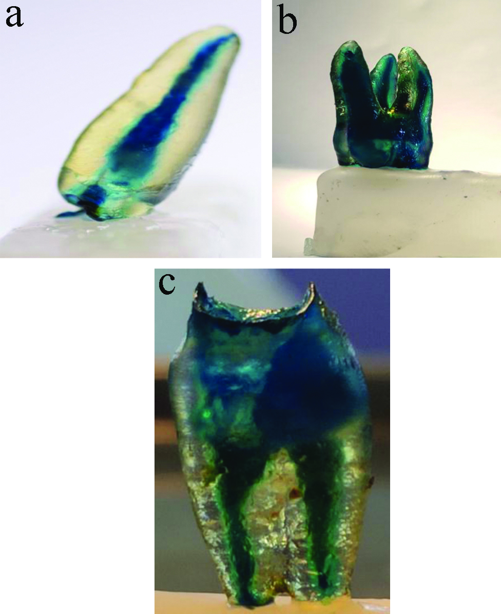
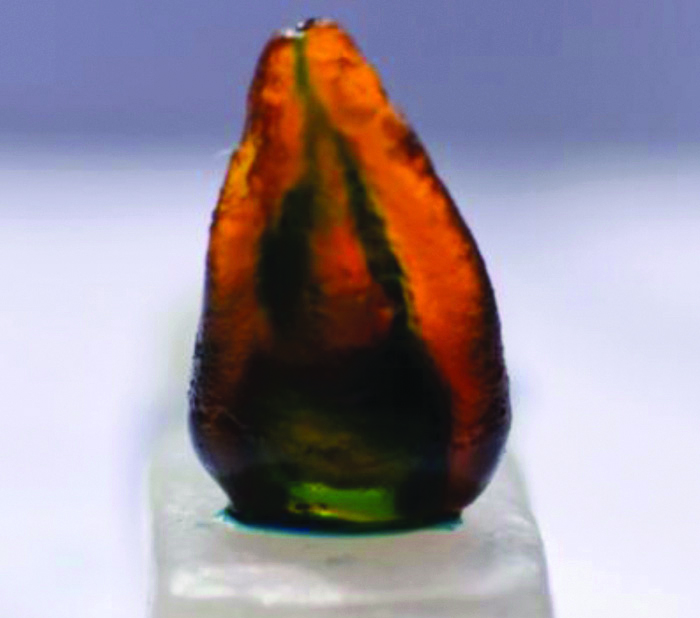
Though nitric acid caused some discoloration of teeth [Table/Fig-4] as compared to trichloroacetic acid, samples decalcified with trichloroacetic acid (group B) showed haziness in most of the samples [Table/Fig-5]. Few samples showed post decalcification clogging of root canals when eugenol was used as clearing agent irrespective of the decalcifying agent used [Table/Fig-6]. Eugenol gave some orange or yellowish tinge to the samples as shown in [Table/Fig-7].
NA/MS combination used i.e., decalcification by nitric acid and clearing by methyl salicylate in a) single rooted tooth; b) and c) multirooted teeth.
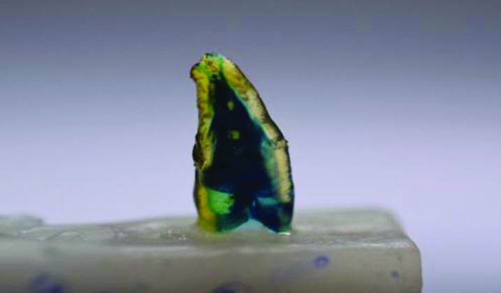
TCA/MS combination used i.e., decalcification by trichloroacetic acid and clearing by methyl salicylate.
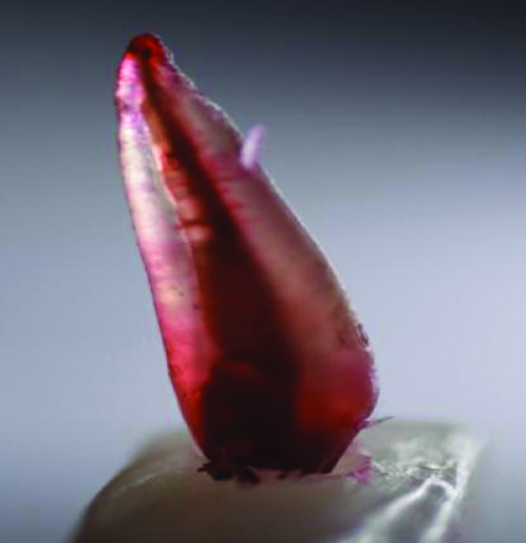
NA/E combination used i.e., decalcification by nitric acid and clearing by eugenol.
TCA/E combination used i.e., decalcification by trichloroacetic acid and clearing by eugenol.
Discussion
The pulp space has been studied using various techniques till date like macroscopic sections, radiographs of extracted teeth, polyester resin casts, transparencies of previously stained samples; including transparent tooth model for its complexity and to have a three-dimensional view of the canals. Vulcanite replicas of root canals of the tooth were done. These helped in better understanding of the teeth structures, however could not provide any information about the pulp chambers [9]. The transparent tooth model is an effective addition to the endodontic evaluation armamentarium [12]. Clearing technique is a reliable method for viewing the root canal system as it has revealed lateral canals, transverse anastomoses, apical deltas and other canal complexities [13]. Many physical and chemical changes are involved in the process of making the teeth transparent. Decalcification by acids causes dissolution of the inorganic constituents of the teeth and dehydration removes the remaining water, air and lipid components. The teeth become transparent after immersion in clearing agents as the tooth becomes permeable to the clearing agent and takes up its refractive index [14]. As the clearing agents are volatile in nature, once the teeth are taken out of the clearing agents they again become opaque. The solvent evaporates and the teeth are invaded by air and moisture rendering them opaque [5].
The acids used for decalcification in various studies are nitric acid, formic acid, Ethylenediamine Tetraacetic Acid (EDTA), citric acid, carbonic acid and trichloroacetic acid. Nitric acid has been proved to be an efficient decalcifying agent as it balances both tissue integrity and time factor. It preserves the tissue integrity with a reasonable speed of decalcification [15]. Xylene, benzene, methyl salicylate and eugenol have been used as clearing agents in routine histological techniques and decalcification studies [5]. In the present study, we used nitric acid and trichloroacetic acid for decalcification and methyl salicylate and eugenol for clearing. Methyl salicylate is non-toxic, economical, non-inflammable and causes minimum distortion of the tissue. It also possesses high tolerance for water which may not be removed in tissue processing [16]. This might correspond with the results of the present study that methyl salicylate gave the good transparency and haziness results when used in combination with nitric acid as decalcifying agent.
The results of this study revealed that the combination of nitric acid and methyl salicylate produced the best transparent tooth model which is in accordance with the study conducted by Gupta B et al., in 2014. They compared the efficacy of nitric acid and formic acid as decalcifying agents along with methyl salicylate and eugenol as clearing agents. They concluded that methyl salicylate showed better transparency, less haziness and better canal morphology when compared to eugenol [10].
Although the new technologies like micro CT, CBCT, visualisation endogram, fibreoptic endoscope are being used to view the pulpal spaces [1]; still the advantages of this technique provide it an edge over the other techniques which includes the speed (5-7 days), simplicity, inexpensive nature, and use of relatively less toxic chemicals as compared to numerous sophisticated and expensive methodologies [3]. A precise assessment of the root canal morphology is of extreme importance, which presents itself as a myriad of shapes, configurations and patterns. Understanding of the variations from the normal root canal morphology prepares the students for the unexpected presentations [7].
Limitation(s)
Firstly, the technique has little clinical implications, can be used only as a teaching aid. Secondly, samples become opaque after they are taken out of the clearing agent. Thirdly, incomplete dehydration can lead to opaque areas. Fourthly, some damage to the tooth structure occurs because of decalcification by acid.
Conclusion(s)
The results of the study revealed that nitric acid took lesser time to decalcify the teeth and also showed better transparency without haziness when used with methyl salicylate as clearing agent. The transparent tooth model hence produced provides an increased appreciation of the anatomy of the root canals in a three-dimensional view with the exterior of the tooth. Studies on clearing techniques can be further advanced by comparison of refractive indices of teeth using various clearing agents, and elementary analysis of the cleared teeth. A method having the accuracy of the canal staining and clearing technique, which is clinically feasible is essential in endodontic practice.
Chi-square test, p<0.05 Significant; NA: Nitric acid, MS: Methyl salicylate, E: Eugenol, TCA: Trichloroacetic acid
Chi-square test, p<0.05 Significant; NA: Nitric acid, MS: Methyl salicylate, E: Eugenol, TCA: Trichloroacetic acid