Cutaneous Horn of Prepucial Skin on Pre-existing Lichen Sclerosus in a Young Male: A Rare Presentation
Amit Kumar Singh1, Nitin Kumar Agarwal2, Dev Kumar Sao3
1 Assistant Professor, Department of Surgery, Datta Meghe Institute of Medical Sciences, Wardha, Maharashtra, India.
2 Consultant Pathologist, Department of Pathology, Nishkarsh Pathology, Bilaspur, Chhattisgarh, India.
3 Consultant Surgeon, Department of Surgery, Jan Swasthya Sahyog, Bilaspur, Chhattisgarh, India.
NAME, ADDRESS, E-MAIL ID OF THE CORRESPONDING AUTHOR: Amit Kumar Singh, Sawangi (Meghe), Wardha, Maharashtra, India.
E-mail: endosurgamit@gmail.com
Cutaneous horn of penis is rare. Cutaneous horns are generally found over sun exposed areas of body after fifth decade of life. Penile horns are hyperkeratotic lesions which are secondary to chronic irritation. Hereby, the author reports a case of a young male with cutaneous horn over phimotic prepucial skin with lichen sclerosus which was successfully treated by circumcision.
Circumcision, Penile, Premalignant
Case Report
A 24-year-old unmarried male, presented with pruritis, thickening and whitish discolouration of penile foreskin for two years and a lesion over it for one year. The lesion had gradually tripled in size after first being noticed one year back. Patient also gave history of non retractability of prepucial skin since childhood. There was no history of surgery or trauma and patient was sexually not active. He had taken treatment in form of steroid cream for local application and antihistaminic without much relief.
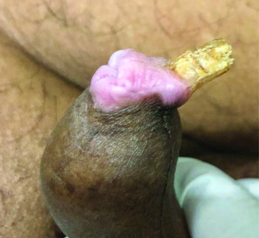
Physical examination revealed a 2×1 cm conical horn like light coloured hard lesion over ventral aspect of prepucial skin [Table/Fig-1]. There was thickening and white discolouration of inner aspect of prepucial skin and mucosa, suggestive of lichen sclerosus. The prepuce was minimally retractable. There was no induration around the lesion and the lesion was localised over the prepucial skin, glans was free of any lesion. There was active tinea cruris infection in both groins. There was no palpable inguinal lymphadenopathy. Systemic examination was within normal limits.
Clinical Photograph showing penile cutaneous horn with lichen sclerosus over prepuce.
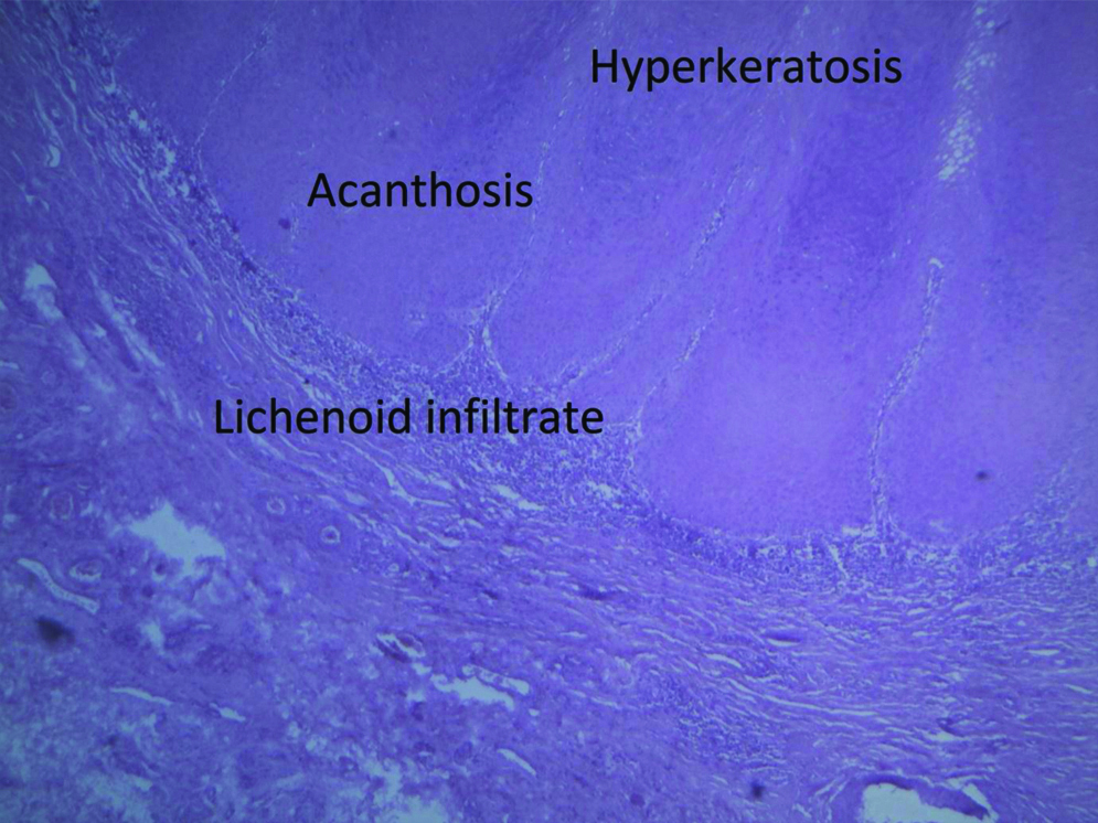
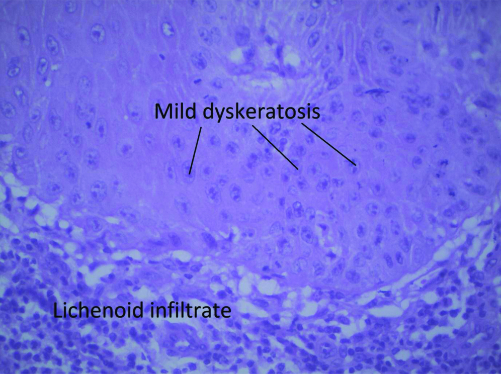
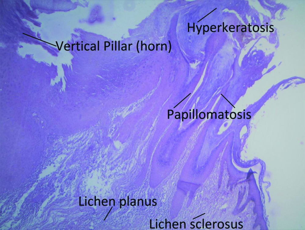
Based on clinical diagnosis of cutaneous horn of penis with lichen sclerosus, circumcision was done with adequate skin margin. Postoperative recovery was uneventful. Histopathology report revealed pigmented epidermis showing acanthosis, hyperkeratosis [Table/Fig-2] and dyskeratosis [Table/Fig-3] with a single vertical pillar of a cellular thick keratinous layer with lichen planus and lichen sclerosus [Table/Fig-4]. There was no evidence of malignancy. Patient was followed for a year and has been asymptomatic.
Section showing acanthosis, hyperkeratosis and lichenoid infiltrate. (Haematoxylin and Eosin stain, x4).
Section showing dyskeratosis (Haematoxylin and Eosin stain, x40).
Section showing horn, lichen planus, lichen sclerosus and hyperkeratosis. (Haematoxylin and Eosin stain, x4).
Discussion
Clinically, cutaneous horn is a term used to describe conical hyperkeratotic protrusion above skin surface which mimics horn of an animal [1]. Cutaneous horns are not uncommon in light exposed areas of body however they are uncommon over covered areas and are unusual and rare over penis [1,2]. The earliest case of cutaneous horn documented was of an elderly woman from Wales in 1588 and was conceived as natural body anomaly while the first penile cutaneous horn was reported in 1854 [2]. Penile cutaneous horn is rarely reported in medical literature since first case reported more than one and half century back [1]. Long standing Lichen sclerosus has been associated with development of penile cutaneous horn [3]. It is a worrisome condition for patient for organ disfigurement and the clinician as it may be a window to serious underlying pathology. As nearly one third of the penile cutaneous horns are associated with malignancy, prompt surgical intervention is warranted [4,5].
Fahmy MAB has described preputial cutaneous horn as a rare prepucial anomaly [6]. Cutaneous horn is not a pathological diagnosis but a morphological designation of dense cohesive keratinised material forming a cone like structure without underlying bone [4]. Aetiology of cutaneous horn is not clear however various factors like viral infection, chronic irritation, surgical trauma, long standing phimosis, malignancy or radiation therapy have been implicated [1,4]. Penile cutaneous horns have been shown to be associated with pre-existing lichen sclerosus [3]. Penile lichen sclerosus is most common in age group 30-49 years while the penile cutaneous horns are common in males aged above 50 years, however it has been reported in young males as well, though rarely as in present case [2,7]. Prabhakar G and Bhat A reported a case of prepucial cutaneous horns in 22-month-old child and was successfully treated with circumcision [8].
Lowe FC and McCullough AR in their study reported that penile cutaneous horns may be benign in 42-56% cases, premalignant in 22-37% cases or malignant in 20-22% cases [5] while a study by Yu RC et al., on review of histopathology of 643 cases of keratinous horns reported 61.1% to be benign, 23.2% to be premalignant and 15.7% were malignant [9]. The European Association of Urology (EAU) 2009 guidelines have classified penile lichen sclerosus and penile cutaneous horn as a premalignant lesion [10]. As penile cutaneous horns are premalignant lesions, definitive management involves surgical excision with prudent histopathological examination of specimen and long term follow-up, as postcircumcision recurrences and malignant transformations on long standing lichen planus have been reported. If histopathology report of specimen is suggestive of malignancy then penectomy (partial or total) is recommended [1,4]. Although electrocauterisation, laser and cryosurgery have been described as effective therapies, they alter histopathology of lesion hence, not preferred [1]. The index patient was managed with circumcision and postoperative recovery and follow-up has been uneventful.
Conclusion(s)
This is a rare case of prepucial cutaneous horn in a young male, developing on long standing phimosis with lichen sclerosus, as a probable aetiology. The patient was treated with circumcision and histopathology report was confirmatory of keratinous horn with lichen sclerosus. After a year of follow-up, the patient has been asymptomatic. Cutaneous horn is a premalignant lesion, definitive management and follow-up is necessary.
Author Declaration:
Financial or Other Competing Interests: None
Was informed consent obtained from the subjects involved in the study? Yes
For any images presented appropriate consent has been obtained from the subjects. Yes
Plagiarism Checking Methods: [Jain H et al.]
Plagiarism X-checker: May 25, 2020
Manual Googling: Jun 05, 2020
iThenticate Software: Aug 10, 2020 (11%)
[1]. Wang Y, Tu MQ, Li XJ, Fu Q, Shi GW, Penile cutaneous horn: A rare case report and review of the literatureAsian J Androl 2018 20:407-08.10.4103/aja.aja_48_1729086760 [Google Scholar] [CrossRef] [PubMed]
[2]. Karthikeyan K, Penile cutaneous horn: An enigma-newer insights and perspectivesIndian J Sex Transm Dis AIDS 2015 36:26-29.10.4103/0253-7184.15669226392650 [Google Scholar] [CrossRef] [PubMed]
[3]. Singhal RR, Patel TM, Pariath KA, Vora RV, Premalignant male genital dermatosesIndian J Sex Transm Dis AIDS 2019 40:9710.4103/ijstd.IJSTD_106_1731922098 [Google Scholar] [CrossRef] [PubMed]
[4]. Aggarwal A, Pandey S, Agarwal S, Garg G, Penile cutaneous horn: Still an enigmaCase Rep 2018 2018:225930.bcr10.1136/bcr-2018-22593030206068 [Google Scholar] [CrossRef] [PubMed]
[5]. Lowe FC, McCullough AR, Cutaneous horns of the penis: An approach to management. Case report and review of the literatureJ Am Acad Dermatol 1985 13:369-73.10.1016/S0190-9622(85)70177-6 [Google Scholar] [CrossRef]
[6]. Fahmy MAB, Rare Preputial Anomalies. In: Fahmy MAB, edNormal and Abnormal Prepuce 2020 ChamSpringer International Publishing:141-49.10.1007/978-3-030-37621-5_16 [Google Scholar] [CrossRef]
[7]. Singh JP, Priyadarshi V, Goel HK, Vijay MK, Pal DK, Chakraborty S, Penile lichen sclerosus: An urologist’s nightmare! A single center experienceUrol Ann 2015 7:303-0.10.4103/0974-7796.15049026229314 [Google Scholar] [CrossRef] [PubMed]
[8]. Prabhakar G, Bhat A, Penile cutaneous horn in a 22-month-old child: A rare case reportJ Indian Assoc Pediatr Surg 2018 23:16710.4103/jiaps.JIAPS_176_1730050270 [Google Scholar] [CrossRef] [PubMed]
[9]. Yu RC, Pryce DW, Macfarlane AW, Stewart TW, A histopathological study of 643 cutaneous hornsBr J Dermatol 1991 124:449-52.10.1111/j.1365-2133.1991.tb00624.x2039721 [Google Scholar] [CrossRef] [PubMed]
[10]. Pizzocaro G, Algaba F, Horenblas S, Solsona E, Tana S, Poel HVD, EAU penile cancer guidelines 2009Eur Urol 2010 57:1002-12.10.1016/j.eururo.2010.01.03920163910 [Google Scholar] [CrossRef] [PubMed]