JCDR - Register at Journal of Clinical and Diagnostic Research
Journal of Clinical and Diagnostic Research, ISSN - 0973 - 709X
Microbiology Section DOI : 10.7860/JCDR/2020/44130.13947
Year : 2020 | Month : Aug | Volume : 14 | Issue : 08 Full Version Page : DC10 - DC12

Phenotypic Pattern of Vibrio cholerae Isolates from a Tertiary Care Hospital in Vadodara, Gujarat, India

Sonia Sandeep Barve1, Srujana Prabhala2, Tanuja Bakul Javadekar3, Sandeep Om Nanda4

1 Associate Professor, Department of Microbiology, Medical College Baroda, Vadodara, Gujarat, India.
2 Resident, Department of Microbiology, Medical College Baroda, Vadodara, Gujarat, India.
3 Professor, Department of Microbiology, Medical College Baroda, Vadodara, Gujarat, India.
4 Assistant Professor, Department of Microbiology, Medical College Baroda, Vadodara, Gujarat, India.


NAME, ADDRESS, E-MAIL ID OF THE CORRESPONDING AUTHOR: Dr. Sandeep Om Nanda, A/303, Lilleria Avenue, Sama Savli Road, Vadodara, Gujarat, India.
E-mail: nandasand@gmail.com
Abstract

Introduction

Cholera is an acute diarrhoeal disease caused by Vibrio cholerae (V.cholerae). Based on antigenic differences of O antigen, O1 serogroup can be divided into three serotypes. In addition, by performing various biochemical reactions, O1 Serogroup can be differentiated into two biotypes. Outbreaks of Cholera occur seasonally. It is associated with monsoon season, warm temperature, heavy rainfall and increased plankton population.

Aim

The aim was to determine the trends in resistance pattern and phenotypic Pattern of Vibrio cholerae.

Materials and Methods

A retrospective study was conducted during the period from June 2019-December 2019. Culture of Stool specimens were done on different agar media. Biotyping was done by conventional methods. Serotyping and phage typing was also done along with the Antibiotic susceptibility testing. Descriptive analysis was used and presented in terms of percentage.

Results

V.cholerae was isolated in 72 patients and they belonged to serogroup O1 and biotype El Tor. The most common serotype was Ogawa. The predominant phage type were T2 by old scheme and T27 by new scheme of phage typing. The maximum number of V. cholerae isolates was seen in the month of November, 2019 followed by October, 2019.

Conclusion

The phenotypic pattern and fluctuating seasonal trend of V. cholerae and antimicrobial resistance encourage the continued epidemiological and microbiological surveillance of the disease.

Keywords

Introduction

Cholera is a diarrhoeal illness caused by Vibrio cholerae, transmitted by ingestion of contaminated food or water [1]. The causative agent, V. cholerae, has two biotypes, classical and El Tor. Classical biotype was responsible for the first six pandemics of cholera worldwide. El Tor biotype replaced the classical biotype by 1961 and caused the seventh pandemic of cholera [2]. Each biotype has three serotypes: Ogawa, Inaba and Hikojima [3]. Prevalent biotypes and their antibiotic resistance changes over time [4,5]. Presently, O1 serogroup which is belonged to El Tor biotype is most common in India [6]. The use of phage typing as a method of classifying V. cholerae has contributed greatly to the understanding of the epidemiology of cholera. Phage typing at the National Institute of Cholera and Enteric Diseases (NICED), Kolkata, India includes the Basu S and Mukherjee S typing and new phage typing schemes. These two phage-typing schemes are specific for V. cholerae O1 and O139 and are being routinely used for the classification of strains at this institute [7,8]. Cholera can occur throughout the year, but maximum transmission is associated with high temperature, heavy rainfall, and flooding [9]. A change in seasonal pattern was observed in the present study, since the maximum number of cases in the study was seen during the month of November. Hence, this study was done with an objective to determine any change in the trend of the resistance pattern and phenotypic pattern of V. cholerae isolates.

Materials and Methods

A retrospective study was conducted on the V. cholerae isolates from the stool samples during the period from June 2019 to December 2019 at the Department of Microbiology, Tertiary Care Hospital, Vadodara, Gujarat, India. Permission was taken from Ethics Committee (No. IECBHR-50/2020 dated 05/06/2020). During this study, all clinically suspected cases of cholera were included. As a routine procedure, informed consent from patients was obtained when sample was collected. Stool samples received in the laboratory were analysed and V. cholerae was isolated in 72 patients. Repeat isolates from the same patients were not included in this study.

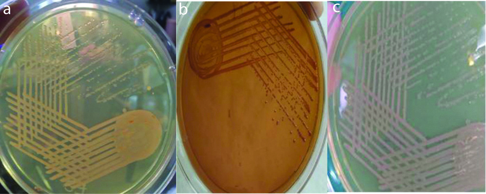
Stool specimens were collected on admission in a clean, wide mouthed, leak proof container preferably before starting antibiotics and transported immediately to the laboratory for processing. Specimens were cultured directly on Nutrient agar, Blood agar, MacConkey’s agar, Thiosulfate-citrate-bile salts (TCBS) sucrose agar [Table/Fig-1]. Nutrient agar was used to perform biochemical tests on the isolates and Blood agar was used for biotyping. Further, the specimens were sub cultured on MacConkey’s agar, TCBS agar after enrichment in Alkaline peptone water and were examined after overnight incubation at 37°C [10].

a) Nutrient agar, b) Mac Conkey agar, c) Nutrient agar showing isolated colonies of Vibrio cholerae.

Colonies suggestive of V. cholerae were identified by standard biochemical tests. Serotyping of isolates was done for agglutination using V. cholerae polyvalent, V. cholerae Ogawa and V. cholerae Inaba antisera (Denka Seiken Co., Ltd., Japan). Biotyping was done by conventional Methods such as Polymyxin-B sensitivity and Sheep RBC haemolysis [10].

Phage typing was done by Basu S and Mukherjee S method and New Phage Typing method and susceptibility to phage IV and V for biotyping was done at NICED, Kolkata, West Bengal, India [7,8].

Antibiotic susceptibility testing was performed by Kirby Bauer’s Method as per Clinical and Laboratory Standards Institute (CLSI) guidelines [11]. The control used for antimicrobial sensitivity testing was ATCC strain Escherichia coli 25922. The following antibiotics were used-Ceftriaxone (30 μg), Doxycycline (30 μg), Levofloxacin (10 μg), Ampicillin (30 μg) and Cotrimoxazole (25 μg). The results were seen after 16-18 hours of incubation at 37°C. The zone of inhibition for each antibiotic was interpreted as per CLSI guidelines [11].

Statistical Analysis

Descriptive analysis was used and presented in terms of percentage.

Results

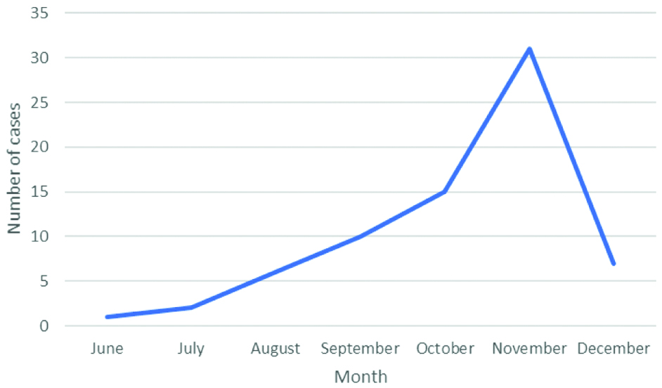
V. cholerae was isolated from 72 patients. Of the 72 patients, 53 (73.6%) were adults and 19 (26.4%) were children. Male to female ratio was 1.4:1. Maximum number of cases was in the month of November which were 32 isolates followed by 15 isolates in October [Table/Fig-2].

Seasonal pattern of occurrence of cases.

All the 72 isolates of V. cholerae belonged to serogroup O1, biotype El Tor and serotype Ogawa. None of the isolates belonged to serotype Inaba. No discordance was found for serotyping and biotyping done in the laboratory and results provided by NICED, Kolkata, West Bengal, India.

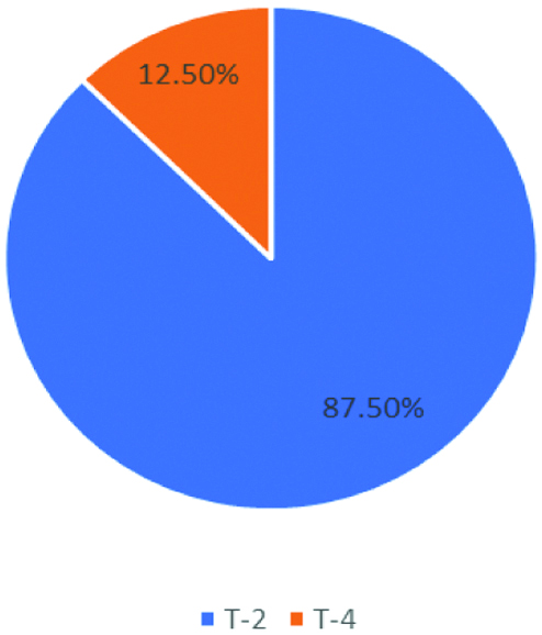
Phage typing was done at Vibrio Phage Reference Lab, NICED, Kolkata by Basu S and Mukherjee S method and New Phage Typing method. According to Basu S and Mukherjee S method, T2 was the most prevalent phage type. Among the isolates, 63 isolates belonged to T-2 phage and 9 isolates were from T-4 phage. None of the strains were untypable [Table/Fig-3] [7].

Distribution of isolates according to Basu S and Mukherjee S classification.

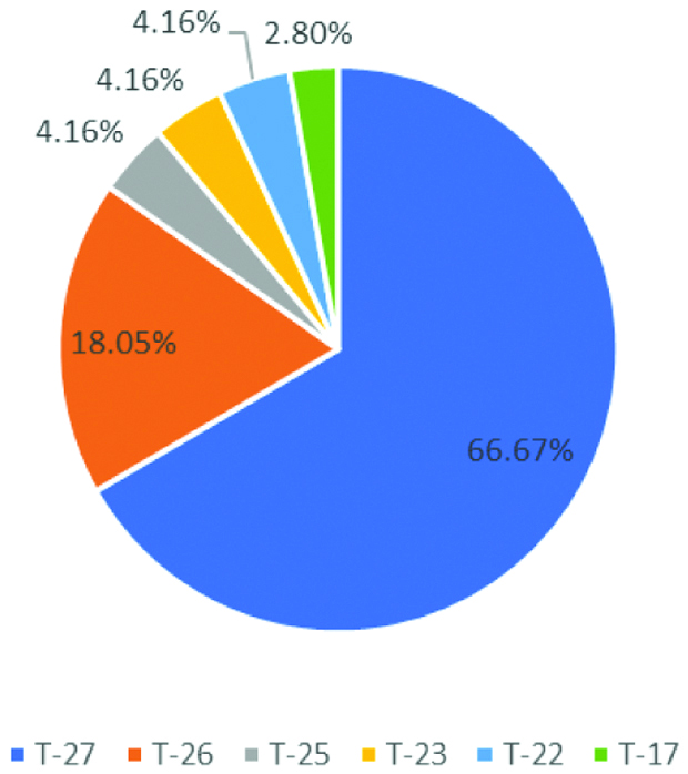
According to New Typing Scheme, 48 isolates belong to T-27 phage. It was the most predominant phage type followed by 13 isolates belonging to T-26, 3 isolates to T-25, T-23, T-22 each and 2 isolates to T-17 [Table/Fig-4].

Distribution of isolates according to the new scheme classification.

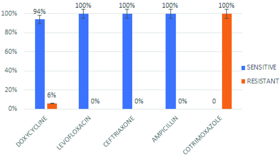
Antibiotic susceptibility was performed by Kirby Bauer disc diffusion method. Resistance to Doxycycline was shown by 6% of isolates. All the isolates were sensitive to Ceftriaxone, Ampicillin and Levofloxacin. Whereas, resistance to Co-trimoxazole was observed in all isolates [Table/Fig-5].

Antibiogram of the Vibrio cholerae O1 isolates.

Discussion

In this study, all the 72 V. cholerae isolates belonged to Serogroup O1, Biotype El Tor and Serotype Ogawa which is in correlation with other studies [12-14]. Kanungo S et al found that V. cholerae O1 belonging to the El Tor was the most common biotype in India and frequency of O139 biotype has been reduced [15]. Phage typing done by old and New method is widely used for epidemiological characterisation of V. cholerae isolates. According to Basu S and Mukherjee S method, the study showed T2 as the predominant Phage type which coincides with studies by Torane V et al., and Turbadkar SD et al., [7,12,16]. However, Sarkar BL et al., reported T4 as predominant phage type [17].

According to New Typing Method, 100% strains are typable. Similar to the study by Turbadkar SD et al., and Sarkar BL et al., with the New Phage Typing method, the study showed T27 as the predominant phage type [16,17]. However, T27 and T25 were reported as predominant strains by Bhowmick TS et al., [18]. Since the study also reported that variation in phage type is associated with resistance to tetracycline, change in antibiotic resistance pattern in V. cholerae isolates can be studied by phage typing [18].

Usually, a seasonal trend in the isolation of V. cholerae is seen. Cases peak in July, taper after September and are negligible after October which closely mimics the seasonal trends in monsoon [12]. However, in present study though the onset of cases coincided with monsoon, they peaked in August and continued till December.

Doxycycline or tetracycline is preferred for the treatment of cholera. This is consistent with the susceptibility pattern over the years [19]. However, the irrational use of these antibiotics could have led to the emergence and spread of the resistant isolates. All the strains showed uniform sensitivity to ampicillin, ceftriaxone and levofloxacin with high level resistance to co-trimoxazole which correlated with other studies [12,14,19]. The phenotypic pattern of the V. cholerae isolates was consistent with other studies. However, a change in the trend of antibiotic susceptibility and seasonal pattern of cases has been observed.

Limitation(s)

The major limitation was that it was conducted only for period of six months. To monitor change in trend of the V. cholerae isolates, continuous monitoring of cases should be carried out which will help to compare the phenotypic trend. Molecular studies are important and should be looked upon for the virulence-associated genes to establish a correlation between biotype and virulence factors.

Conclusion(s)

V. cholerae isolates in this study continue to demonstrate the predominant phenotypic pattern. However, the change in the disease pattern encourage the continued epidemiological and microbiological surveillance. Periodic survey should be carried out to study the trend of phenotypic pattern in the community. The isolates should also be sent to a reference laboratory for routine phage typing.

References

[1]Cholera- Fact sheet No.107 published by W.H.O accessed from www.who.int/mediacentre/factsheets/fs107/en  [Google Scholar]

[2]Gardner AD, Venkatraman KV, The antigens of the cholera group of Vibrio J Hyg (London) 1935 35(2):262-82.10.1017/S002217240003226520475277  [Google Scholar]  [CrossRef]  [PubMed]

[3]Nobechi K, Contributions to the knowledge of vibrio cholerae i. fermentation of carbohydrates and polyatomic alcohols by Vibrio cholerae J Bacteriol 1925 10(3):197-215.10.1128/JB.10.3.197-215.19251655914  [Google Scholar]  [CrossRef]  [PubMed]

[4]Faruque ASG, Alam K, Malek MA, Khan MGY, Ahmed S, Saha D, Emergence of multidrug-resistant strain of vibrio cholerae O1 in Bangladesh and reversal of their susceptibility to tetracycline after two years J Health Popul Nutr 2007 25(2):241-43.  [Google Scholar]

[5]Safa A, Nair GB, Kong RY, Evolution of new variants of Vibrio cholerae O1 Trends Microbiol 2010 18(1):46-54.10.1016/j.tim.2009.10.00319942436  [Google Scholar]  [CrossRef]  [PubMed]

[6]Narang P, Mendiratta DK, Deotale VS, Narang R, Changing patterns of Vibrio cholerae in sevagram between 1990 and 2005 Indian J Med Microbiol 2008 26(1):40-44.10.1016/S0255-0857(21)01990-3  [Google Scholar]  [CrossRef]

[7]Basu S, Mukherjee S, Bacteriophage typing of Vibrio El Tor Experientia 1968 24:299-300.10.1007/BF021528325661438  [Google Scholar]  [CrossRef]  [PubMed]

[8]Chattopadhyay DJ, Sarkar BL, Ansari MQ, Chakrabarti BK, Roy MK, Ghosh AN, New phage typing scheme for Vibrio cholerae O1 biotype El Tor strains J Clin Microbiol 1993 31(6):1579-85.10.1128/JCM.31.6.1579-1585.19938315000  [Google Scholar]  [CrossRef]  [PubMed]

[9]Sastry AS, Bhat S, Essentials of Medical Microbiology. Vibrio and Aeromonas 2019 2nd editionNew DelhiJaypee Brothers Medical Publishers:341-51.  [Google Scholar]

[10] Mackie & McCartney. Practical Medical Microbiology. Vibrio, Aeromonas, Pseudomonas, Campylobacter, Wolinella 2016 14th editionNew DelhiRelx India Pvt Ltd:425-48.  [Google Scholar]

[11]Clinical Laboratory Standard Institute (CLSI) Performance standards for Antimicrobial Susceptibility Testing. Twenty-fourth Informational Supplement. CLSI document M100-S24. Wayne, PA: Clinical and Laboratory Standards Institute; 2014  [Google Scholar]

[12]Torane V, Kuyare S, Nataraj G, Mehta P, Dutta S, Sarkar B, Phenotypic and antibiogram pattern of V. cholerae isolates from a tertiary care hospital in Mumbai during 2004-2013 a retrospective cross-sectional study BMJ Open 2016 6(11):e01263810.1136/bmjopen-2016-01263827888174  [Google Scholar]  [CrossRef]  [PubMed]

[13]Shah AJ, Revdiwala SB, Vaghela GM, Mulla SA, Retrospective analysis to study trend of biotype, serotype and phage type of Vibrio cholerae isolates in South Gujarat over last 17 years Int J Med Public Health 2014 4(2):155-58.10.4103/2230-8598.133116  [Google Scholar]  [CrossRef]

[14]Madhavi Gayatri A, Bio typing, serotyping and phenotyping characterisation of Vibrio cholerae O1 isolated in Hyderabad World Journal of Pharmaceutical Research 2015 4(7):670-78.  [Google Scholar]

[15]Kanungo S, Sah BK, Lopez AL, Sung JS, Paisley AM, Sur D, Cholera in India an analysis of reports 1997-2006 Bull World Health Organ 2010 88(3):185-91./10.2471/BLT.09.07346020428385  [Google Scholar]  [CrossRef]  [PubMed]

[16]Turbadkar SD, Ghadge DP, Patil S, Chowdhary AS, Bharadwaj R, Circulating phage type of Vibrio cholerae in Mumbai Indian J Med Microbiol 2007 25(2):177-78.10.1016/S0255-0857(21)02190-3  [Google Scholar]  [CrossRef]

[17]Sarkar BL, Bhowmick TS, Das M, Rajendran K, Nair GB, Phage types of Vibrio cholerae O1 and O139 in the past decade in India Jpn J Infect Dis 2011 64(4):312-15.  [Google Scholar]

[18]Bhowmick TS, Das M, Ruppitsch W, Stoeger A, Pietzka AT, Allerberger F, Detection of virulence-associated and regulatory protein genes in association with phage typing of human from several geographical regions of the world J Med Microbiol 2009 58(9):1160-67.10.1099/jmm.0.008466-019528176  [Google Scholar]  [CrossRef]  [PubMed]

[19]Kumar P, Jain M, Goel AK, Kamboj DV, Kumar O, Tetracycline resistant V. cholera O1 biotype El Tor serotype Ogawa with classical ctx B from a recent cholera outbreak in Orissa, Eastern India J Infect Public Health 2012 5(2):217-19.10.1016/j.jiph.2011.09.00722541274  [Google Scholar]  [CrossRef]  [PubMed]