Comparative Evaluation of Root Dentin Microhardness after using Different Standard Irrigating Solutions and Stevia Leaf Extract- An In vitro Study
Avani Paresh Shah1, Rushikesh Mahaparale2, TM Mangala3, Adish Anand Saraf4, Sneha Mali5, Sagar Pawar6, Urmila Chauhan7, Vincia Dsouza8
1 Postgraduate Student, Department of Conservative Dentistry and Endodontics, School of Dental Sciences, Karad, Maharashtra, India.
2 Reader, Department of Conservative Dentistry and Endodontics, School of Dental Sciences, Karad, Maharashtra, India.
3 Professor and Head, Department of Conservative Dentistry and Endodontics, School of Dental Sciences, Karad, Maharashtra, India.
4 Senior Lecturer, Department of Conservative Dentistry and Endodontics, School of Dental Sciences, Karad, Maharashtra, India.
5 Senior Lecturer, Department of Conservative Dentistry and Endodontics, School of Dental Sciences, Karad, Maharashtra, India.
6 Senior Lecturer, Department of Conservative Dentistry and Endodontics, School of Dental Sciences, Karad, Maharashtra, India.
7 Postgraduate Student, Department of Conservative Dentistry and Endodontics, School of Dental Sciences, Karad, Maharashtra, India.
8 Postgraduate Student, Department of Conservative Dentistry and Endodontics, School of Dental Sciences, Karad, Maharashtra, India.
NAME, ADDRESS, E-MAIL ID OF THE CORRESPONDING AUTHOR: Avani Paresh Shah, School of Dental Sciences, Karad, Maharashtra, India.
E-mail: shah.aps21@gmail.com
Introduction
Success in endodontic therapy largely depends on mechanical and chemical debridement of the root canals by using instruments and effective irrigating solutions which are not only important for cleaning and disinfecting the root canals, but also, are capable of altering the chemical and structural properties of dentin. As the microhardness test is sensitive to surface changes of tooth structure, it is useful in making a correlation between irrigating solutions and root dentin microhardness.
Aim
To evaluate the effect of different standard irrigating solutions at standard concentrations and Stevia leaf extract (2.5%) on the microhardness of root canal dentin.
Materials and Methods
This is an in-vitro comparative study where forty intact single rooted teeth were selected and decoronated to get an apico-coronal length of 10 mm and were randomly divided into four groups as per the irrigant used; Group 1 (control): Irrigation with Normal saline, Group 2: Irrigation with 2.5% Sodium Hypochloride (NaOCl) followed by 17% EDTA, Group 3: Irrigation with 2.5% Stevia extract solution, Group 4: Irrigation with SmearClear solution. They were prepared using ProTaper Universal Rotary Files with intermittent irrigation with the respective irrigating solution. The teeth were then embedded in acrylic resin and subjected to Vicker’s Hardness test and the data obtained were analysed using one way ANOVA test. p<0.05 was taken to be statistically significant.
Results
At 500 microns, Vickers Hardness Number (VHN) value was less than at 1000 micron, but was not statistically significant, (p>0.05). Between the groups, the control group showed the highest microhardness at 500 and 1000 microns, namely, 51.27±4.36 VHN and 53.60±5.12 VHN, respectively. Group 3 and 4 showed a comparable reduction in microhardness with Group 3 showing slightly better results (47.98±4.34 VHN and 48.89±5.26 VHN, respectively) as compared to Group 4 (47.36±5.50 VHN and 48.62±5.84 VHN, respectively). Group 2 showed the least value (36.60±5.71 VHN and 37.11±5.82 VHN, respectively).
Conclusion
Within the limitations of this study, teeth irrigated with normal saline showed least reduction in microhardness followed by irrigation with Stevia leaf extract solution, SmearClear and Hypochlorite followed by EDTA.
Dentin, ProTaper universal, SmearClear, Sodium hypochlorite, Vicker’s hardness testing
Introduction
Cleaning is the removal of all contents of the root canal system before and during shaping in endodontic treatment [1]. Successful cleaning requires the use of instruments to physically remove substances. It also requires various irrigating systems to flush loosened materials away and chemicals which dissolve contents from regions which are not conventionally accessible. Currently, the best method for the removal of tissue remnants from the canals and dentin debris during instrumentation is the procedure of irrigation. Irrigation is an imperative step in endodontic therapy as it helps to get rid of any loose and necrotic tissue and contaminated materials before they are pushed deeper into the canal and the periapical tissues [2]. Irrigation solutions are also efficient in providing gross debridement of the canals, lubrication for the instruments against the canal walls with destruction of microbes and dissolution of organic and inorganic tissues [3].
An irrigating solution should have many physiochemical properties, however no such irrigant with ideal properties exists. Hence other auxiliary solutions should be combined to get the desired effect [4]. There are high chances of a successful endodontic therapy when more debris and smear layer are eliminated. It is believed that removing the smear layer enables the detachment of microbiota and their associated toxins from root canals, helps in the improvement of the seal of root fillings and reduces the chances of survival of any potential bacteria and their subsequent reproduction [5].
One of the most widely used chemical solutions in the biomechanical preparation of the root canal system is sodium hypochlorite (NaOCl) and it has been systematically used in endodontics in a broad range of concentrations ranging from 0.5%-5.25%. Although, it has excellent antimicrobial action and an inherent capacity to dissolve organic materials, sodium hypochlorite alone is ineffective against the smear layer and thus it needs to be used in association with chelating agents that are expedient against organic matter [6]. It also has an unpleasant odour and bitter taste, which makes it unacceptable in the patient’s oral cavity.
The demineralising effect of chelators acts on the smear layer and the root dentin, resulting in exposure of collagen fibers and a decrease in dentin microhardness [7]. It is desirable to greatly reduce the microhardness of the superficial canal dentine layer of the root canal lumen. Chelating agents which are used during biomechanical preparation of root canals effectively remove the smear layer and increase the access of the irrigant into the dentin tubules. This not only allows adequate canal disinfection but also reduces dentin microhardness, which facilitates the access and action of endodontic instruments in narrow, calcified root canals [8].
Stevia rebaudiana is a perennial shrub which has been used worldwide as a medicine and a natural sweetener to lower blood sugar [9]. Stevioside, its white crystalline compound, is 100-300 times sweeter than table sugar. Various studies regarding the anti-cariogenicity of Stevia are being carried out which evaluated its antimicrobial potential against many pathogens [10]. Ethanolic and methanolic extracts of stevia leaves have been found to be effective against the Gram-negative and Gram-positive organisms [11]. It has added advantages, that it is odourless, colourless and sweet to taste, which counteract the disadvantages of hypochlorite. The fact that it has never been used as an irrigant, warrants further studies which employ the use of Stevia in this particular role in endodontic treatment.
SmearClear consists of 17% EDTA, Cetrimide and a surfactant. The combination of surfactant agents with antiseptics or chelating agents (MTAD, Tetraclean, SmearClear, Cetrexidin) is recommended to reduce the surface tension of irrigants and facilitate their penetration into places of difficult access [12]. It is of interest to investigate to what extent the dentine of the root canal is affected by the use of several chemical and herbal irrigants. Therefore, the aim of the present study was to evaluate the effect of different standard endodontic irrigating solutions like normal saline, 2.5% NaOCl (Prime Dental Products Pvt., Ltd., India) followed by 17% EDTA (Prime Dental Products Pvt., Ltd., India), SmearClear (Kerr) and a novel irrigant 2.5% Stevia extract solution (Herboveda, India) on the microhardness of root canal dentin.
Materials and Methods
Teeth Selection
The in-vitro study was carried out in 2020 in the Department of Conservative Dentistry and Endodontics, School of Dental Sciences, Karad within 1 week. Ethical clearance for the study was obtained from the Institutional Ethical Committee (269/2019-2020). Forty noncarious, intact, single rooted premolar teeth extracted for periodontic or orthodontic reasons were selected for the study. Selection of teeth was made on the basis of relative dimensions, similarity in morphology and absence of any cracks or caries defects especially within the root portions, which was verified radiographically. The teeth were stored in normal saline until use. The crowns were sectioned by using a high-speed bur under water cooling, such that the apico-coronal length of the root was 10 mm for the sake of standardisation. The mesio-distal and bucco-lingual diameters of the coronal planes were measured with a digital Vernier caliper and the mean mesio-distal and bucco-lingual dimensions were obtained. Thereafter, roots presenting a difference of 20% from the mean were discarded, leaving a total of 40 root samples.
Specimen Preparation
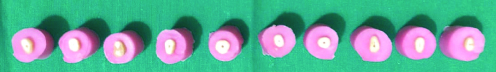
All the specimens were prepared and testing was done by a single operator. Length of the canal was determined by visual inspection using a #10 K file till it was just visible at the apex. The working length was then established by deducting 0.5 mm from that length. 2.5% of Stevia solution was prepared by diluting 2.5 ml of Stevia extract in 100 ml of deionised water and was then used as irrigant. At this stage the teeth were randomly divided into 4 groups (n=10) as per the irrigants used; Group 1 (control): Irrigation with Normal saline, Group 2: Irrigation with 2.5% NaOCl (Prime Dental Products Pvt., Ltd., India) followed by 17% EDTA (Prime Dental Products Pvt., Ltd., India), Group 3: Irrigation with 2.5% Stevia extract solution (Herboveda, India), Group 4: Irrigation with SmearClear (Kerr) solution [Table/Fig-1]. Each of the root canals were prepared using rotary ProTaper Universal Files (Dentsply, Maillefer) upto size F2 with intermittent irrigation between consecutive files, with 2 mL of the respective irrigant. Each of the irrigants used were delivered through a 27 gauge needle reaching about 2/3rds the working length. Finally, each of the specimens was rinsed with normal saline to wash off any remnants before testing.
Specimen distribution as per irrigating solutions used (n=40).
| Group | Irrigating solution |
|---|
| 1 | Normal saline (control) |
| 2 | 2.5% NaOCl followed by 17% EDTA |
| 3 | 2.5% Stevia extract solution |
| 4 | SmearClear |
Microhardness Testing
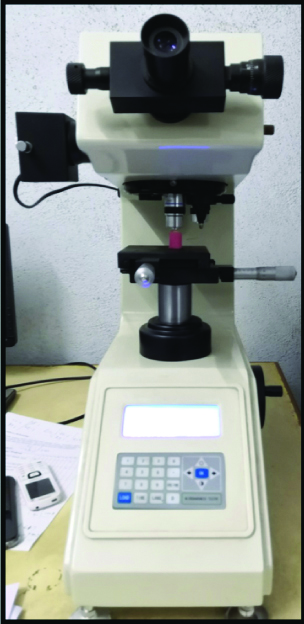
Using self cure acrylic resin (DPI Pvt., Ltd., India), the teeth were embedded along their long axes upto 8 mm, exposing 2 mm of their remaining length [Table/Fig-2]. The exposed flat surfaces were then polished using pumice slurry. All the specimens were then subjected to Vicker’s Hardness testing [Table/Fig-3] at 500 and 1000 microns from the pulpo-dentinal junction on the polished surfaces (Mitutoyo America Corporation, Load= 100 gram force, Dwell time=15 sec). Three readings (in VHN) were taken at 500 and 1000 microns respectively and an average was taken as the final reading at both distances.
Microhardness tester used for VHN with sample loaded on it.
Statistical Analysis
Data was collected and statistical analysis was done using one-way ANOVA at p <0.05 which was taken to be statistically significant. The data was entered into Microsoft Excel 2010. All analyses were performed using SPSS (Statistical Package for Social Sciences) software version 20.
Results
At 500 microns, Vickers Hardness Number value was less than at 1000 micron, but was not statistically significant, (p>0.05) [Table/Fig-4].
Microhardness values in VHN (p<0.05 is significant).
| Groups of study | 500 microns | 1000 microns | p-value | Significance |
|---|
| Group 1: Normal saline | 51.27±4.36 | 53.60±5.12 | p=0.8 | Non-significant |
| Group 2: 2.5% NaOCl + 17% EDTA | 36.60±5.71 | 37.11±5.82 |
| Group 3: Stevia leaf extract | 47.98±4.34 | 48.89±5.26 |
| Group 4: SmearClear | 47.36±5.50 | 48.62±5.84 |
Between the groups, the control group showed the highest microhardness at 500 and 1000 microns, namely, 51.27±4.36 VHN and 53.60±5.12 VHN, respectively. Group 3 and 4 showed a comparable reduction in microhardness with Group 3 showing slightly better results (47.98±4.34 VHN and 48.89±5.26 VHN, respectively) as compared to Group 4 (47.36±5.50 VHN and 48.62±5.84 VHN respectively). Group 2 showed the least microhardness (36.60±5.71 VHN and 37.11±5.82 VHN respectively). Tukey’s Honestly Significant Difference (HSD) test was carried out for inter-group comparison [Table/Fig-5]. When Group 1 was compared with Group 2, Group 2 with Group 3 and Group 2 with Group 4, the values showed a statistically significant difference (p<0.05). When Group 1 was compared with Group 3, Group 1 with Group 4 and Group 3 with Group 4, the values did not show any statistically significant difference (p>0.05).
Tukey’s HSD values for intergroup comparison (p<0.05 is significant).
| Pairs | Tukey’s HSD p-value | Tukey’s HSD inference |
|---|
| Normal saline- 2.5% NaOCl + 17% EDTA (Group 1&2) | 0.0010053 | p<0.01 |
| Normal saline-Stevia leaf extract (Group 1&3) | 0.4698817 | Non-significant |
| Normal saline- SmearClear (Group 1&4) | 0.3184225 | Non-significant |
| 2.5% NaOCl + 17% EDTA-Stevia leaf extract (Group 2&3) | 0.0010053 | p<0.01 |
| 2.5% NaOCl + 17% EDTA-SmearClear (Group 2&4) | 0.0010053 | p<0.01 |
| Stevia leaf extract- SmearClear (Group 3&4) | 0.8999947 | Non-significant |
Discussion
The results of this study depict that dentin microhardness was reduced with root canal irrigation with either solution except normal saline. At 500 microns lower values of vickers microhardness was obtained from the pulp space. This in coordination with previous findings wherein the dentin microhardness is related to the location and its value decreases as indentations get nearer to the pulp. This could be due to the widely opened dentinal tubules near the pulp which are free of peritubular dentin offer less resistance to the indenter. As per Pashley D et al., there is an inverse correlation between the microhardness of dentin and tubular density. To determine the intrinsic hardness profile of dentin structure, the degree of mineralisation and amount of hydroxyapatite in the intertubular substance are to be considered [13].
The evidence of mineral loss or gain in dental hard tissue can be determined by microhardness. In the present study, Vickers microhardness test was used as practicality for evaluating surface changes of dental tissues treated with chemical agents of it has been shown by previous studies [14]. The significant alteration in the microhardness of dentin, following the irrigation treatment depicts that they directly affect the dentin structure and its components. Reduced microhardness lowers modulus of elasticity and flexural strength of dentin. Hence, the determination of microhardness provides an arbitrary assessment of the change in any mineral content of dental hard tissues [13]. According to a study by Doğan H and Çalt S, 2.5% NaOCl used as an irrigant for 15 minutes altered the mineral content of root dentin to a significant extent [17]. It has been concluded that NaOCl treatment caused mineral accumulation in human root dentin [15,16,17], increased the amount of carbonate and reduced the amount of phosphate [16,17]. In a study by Slutzky-Goldberg I et al., indicated that change in the biomechanical properties of the dentin was possible with irrigation with 2.5% NaOCI. As 2.5% NaOCI is the most common concentraion used as an endodontic irrigant it was used in the current study [18]. Sayin et al., reported that the use of EDTA alone or with NaOCl resulted in the maximum decrease in dentin microhardness [19]. Since the combination of 2.5% NaOCl and 17% EDTA are most commonly used in everyday endodontic practice and are the standard irrigants, they were chosen for this study.
Ulusoy ÖI et al., used SmearClear in their study to evaluate its effect on root dentin microhardness, smear layer removal and erosion among other endodontic irrigants and found that it was effective in removing the smear layer in the middle and cervical thirds and also did not cause a significant reduction in the root dentin microhardness [20]. Aranda-Garcia AJ et al., reported a decrease in the microhardness after using SmearClear. SmearClear contains 17% EDTA, cetrimide and surfactant. This substance is able to remove the smear layer of the root canals however its chelating ability is lower than the 17% EDTA [21]. This probably explains a lesser reduction in the microhardness as compared to the 2.5% NaOCl + 17% EDTA group. Studies suggest that Stevia has been used since ancient times as a sweetener and a medicine. It is a small shrub that has been used as a bio-sweetener and for other medicinal uses such as to decrease blood sugar levels [9]. It is colourless, odourless and is sweet, which beat the disadvantages of NaOCl, which has a bitter taste and an unpleasant odour. Also, its anti-cariogenic property is an added advantage, which is unavailable in any other endodontic irrigant presently. In a study by Usha C et al., which assessed the anticariogenicity of microwave-assisted 0.5% extract of Stevia rebaudiana leaves in high caries risk patients, found that in 0.5% concentration, it can be used as a mouthwash for moderate to high caries risk patients [22].
Escobar E et al., conducted a study to evaluate the growth and viability of Streptococcus mutans in sucrose with different concentrations of Stevia rebaudiana Bertoni and found that S. mutans total growth and biofilm formation showed a marked decrease with reduced concentrations of Stevia along with reduced biofilm and acid production [23]. Kishta-Derani MA et al., reported a loss of microhardness of bovine dentin in their study where they used 0.5% and 5% stevia extract and Phosphate Buffered Saline (PBS) as control, 0.5% stevia extract had a greater loss of microhardness than the other groups but the reason behind the demineralisation is unknown [24]. The above studies prove the anti cariogenic potential of Stevia when used as a mouth rinse [22] but there have been no studies regarding its use as an endodontic irrigant. An irrigant like Stevia which has anti-cariogenic potential, showing a reduction in the biofilm formation and acid production [23] along with a palatable taste that does not significantly reduce the root dentin microhardness [24] could revolutionise the field of endodontics.
Limitation(s)
The microhardness determination can only give an indirect picture of mineral loss. Also, in-vitro conditions may or may not accurately predict the performance of the irrigant in-vivo.
Conclusion(s)
Irrigation with various endodontic irrigants changes the structure of root dentin as seen according to the obtained values. It could be concluded that combination of NaOCl and EDTA significantly reduced the root dentin microhardnes at 500 and 1000 microns from the pulpo-dentinal junction. SmearClear and Stevia extract had a similar effect on root dentin microhardness as seen by the obtained values, meaning, that these irrigants reduced the microhardness of root dentin to a lesser extent as compared to NaOCl and EDTA combination, which proves that with future studies and further improvements in the concentration of Stevia, it could be used as an effective endodontic irrigant with added anti-cariogenic properties.
Author Declaration:
Financial or Other Competing Interests: None
Was Ethics Committee Approval obtained for this study? Yes
Was informed consent obtained from the subjects involved in the study? NA
For any images presented appropriate consent has been obtained from the subjects. NA
Plagiarism Checking Methods: [Jain H et al.]
Plagiarism X-checker: Feb 13, 2020
Manual Googling: May 11, 2020
iThenticate Software: Jul 24, 2020 (22%)
[1]. Hargreaves KM, Cohen’s Pathways of the Pulp 2016 10thSt. Louis, Mo, USAMosby [Google Scholar]
[2]. Haapasalo M, Shen Y, Qian W, Gao Y, Irrigation in endodonticsDent Clin North Am 2010 54(2):291-312.10.1016/j.cden.2009.12.00120433979 [Google Scholar] [CrossRef] [PubMed]
[3]. Handa A, Handa RS, Influence of root canal irrigants on dental tissues: A review of literatureIndian J Dent Sci 2013 5(3):73-76. [Google Scholar]
[4]. Saleh AA, Ettman WM, Effect of endodontic irrigation solutions on microhardness of root canal dentineJ Dent 1999 27(1):43-46.10.1016/S0300-5712(98)00018-9 [Google Scholar] [CrossRef]
[5]. Kandaswamy D, Venkateshbabu N, Root canal irrigantsJ Conserv Dent: JCD 2010 13(4):256-64.10.4103/0972-0707.7337821217955 [Google Scholar] [CrossRef] [PubMed]
[6]. da Silva LA, Sanguino AC, Rocha CT, Leonardo MR, Silva RA, Scanning electron microscopic preliminary study of the efficacy of SmearClear and EDTA for smear layer removal after root canal instrumentation in permanent teethJ Endod 2008 34(12):1541-44.10.1016/j.joen.2008.08.00719026891 [Google Scholar] [CrossRef] [PubMed]
[7]. De-Deus G, Paciornik S, Mauricio MH, Evaluation of the effect of EDTA, EDTAC and citric acid on the microhardness of root dentineInt Endod J 2006 39(5):401-07.10.1111/j.1365-2591.2006.01094.x16640640 [Google Scholar] [CrossRef] [PubMed]
[8]. Cruz-Filho AM, Sousa-Neto MD, Savioli RN, Silva RG, Vansan LP, Pécora JD, Effect of chelating solutions on the microhardness of root canal lumen dentinJ Endod 2011 37(3):358-62.10.1016/j.joen.2010.12.00121329821 [Google Scholar] [CrossRef] [PubMed]
[9]. Goyal SK, Samsher GR, Goyal RK, Stevia (Stevia rebaudiana) a bio-sweetener: A reviewInt J Food Sci Nutr 2010 61(1):01-10.10.3109/0963748090319304919961353 [Google Scholar] [CrossRef] [PubMed]
[10]. Mohammadi-Sichani M, Karbasizadeh V, Aghai F, Mofid MR, Effect of different extracts of Stevia rebaudiana leaves on Streptococcus mutans growthJ Med Plants Res 2012 6(22):4731-34.10.5897/JMPR11.1622 [Google Scholar] [CrossRef]
[11]. Ajagannanavar SL, Shamarao S, Battur H, Tikare S, Al-Kheraif AA, Al Sayed MS, Effect of aqueous and alcoholic Stevia (Stevia rebaudiana) extracts against Streptococcus mutans and Lactobacillus acidophilus in comparison to chlorhexidine: An invitro studyJ Int Soc Prevent Communit Dent 2014 4(Suppl 2):S116-21.10.4103/2231-0762.14621525558451 [Google Scholar] [CrossRef] [PubMed]
[12]. Ferrer-Luque CM, Perez-Heredia M, Baca P, Arias-Moliz MT, González-Rodríguez MP, Decalcifying effects of antimicrobial irrigating solutions on root canal dentinMedicina Oral, Patologia Oral Y Cirugiabucal 2013 18(1):e158-161.10.4317/medoral.1820722926482 [Google Scholar] [CrossRef] [PubMed]
[13]. Pashley D, Okabe A, Parham P, The relationship between dentin microhardness and tubule densityDent Traumatol 1985 1(5):176-79.10.1111/j.1600-9657.1985.tb00653.x3865764 [Google Scholar] [CrossRef] [PubMed]
[14]. Clark-Holke D, Drake D, Walton R, Rivera E, Guthmiller JM, Bacterial penetration through canals of endodontically treated teeth in the presence or absence of the smear layerJ Dent 2003 31(4):275-81.10.1016/S0300-5712(03)00032-0 [Google Scholar] [CrossRef]
[15]. Inaba D, Ruben J, Takagi O, Arends J, Effect of sodium hypochlorite treatment on remineralisation of human root dentine invitroCaries Res 1996 30(3):218-24.10.1159/0002621638860033 [Google Scholar] [CrossRef] [PubMed]
[16]. Tsuda H, Ruben J, Arends J, Raman spectra of human dentin mineralEuropean Journal of Oral Sciences 1996 104(2):123-31.10.1111/j.1600-0722.1996.tb00056.x8804900 [Google Scholar] [CrossRef] [PubMed]
[17]. Doğan H, Çalt S, Effects of chelating agents and sodium hypochlorite on mineral content of root dentinJ Endod 2001 27(9):578-80.10.1097/00004770-200109000-0000611556562 [Google Scholar] [CrossRef] [PubMed]
[18]. Slutzky-Goldberg I, Liberman R, Heling I, The effect of instrumentation with two different file types, each with 2.5% NaOCl irrigation on the microhardness of root dentinJ Endod 2002 28(4):311-12.10.1097/00004770-200204000-0001212043870 [Google Scholar] [CrossRef] [PubMed]
[19]. Taner Cem Sayin, Ahmet Serper, Zafer C Cehreli, Harika G, The effect of EDTA, EGTA, EDTAC, and tetracycline-HCl withand without subsequent NaOCl treatment on themicrohardness of root canal dentinOtlu Oral Surg Oral Med Oral Pathol Oral Radiol Endod 2007 104(3):418-24.10.1016/j.tripleo.2007.03.02117709073 [Google Scholar] [CrossRef] [PubMed]
[20]. Ulusoy ÖI, Görgül G, Effects of different irrigation solutions on root dentine microhardness, smear layer removal and erosionAust Endod J 2013 39(2):66-72.10.1111/j.1747-4477.2010.00291.x23890262 [Google Scholar] [CrossRef] [PubMed]
[21]. Aranda-Garcia AJ, Kuga MC, Chavéz-Andrade GM, Kalatzis-Sousa NG, Hungaro Duarte MA, Faria G, Effect of final irrigation protocols on microhardness and erosion of root canal dentinMicrosc Res Tech 2013 76(10):1079-83.10.1002/jemt.2226823897860 [Google Scholar] [CrossRef] [PubMed]
[22]. Usha C, Ramarao S, John BM, Babu ME, Anticariogenicity of stevia rebaudiana extract when used as a mouthwash in high caries risk patients: Randomized controlled clinical trialWorld J Dent 2017 8(5):364-69.10.5005/jp-journals-10015-1466 [Google Scholar] [CrossRef]
[23]. Escobar E, Piedrahita M, Gregory RL, Growth and viability of Streptococcus mutans in sucrose with different concentrations of Stevia rebaudiana BertoniClin Oral Investig 2020 :1-6.10.1007/s00784-020-03197-532189073 [Google Scholar] [CrossRef] [PubMed]
[24]. Kishta-Derani MA, Neiva GF, Boynton JR, Kim YE, Fontana M, The antimicrobial potential of stevia in an invitro microbial caries modelAm J Dent 2012 29(2):87-92. [Google Scholar]