Williams syndrome is a rare genetic disorder that occurs due to deletion on chromosome 7q11.23. The prevalence is said to be 1:7500-1:20,000. The typical presentation of the syndrome includes dysmorphic facial features, cardiovascular malformations and intellectual disability. Craniofacial features of these children include short anterior cranial base, protrusive maxilla, increased Mandibular Plane- Occlusal angle, steep mandibular plane and reduced ratio of posterior to anterior facial height. The unique characteristics of these children are over friendliness, out-going personality, hyper-acusis and tendency to get easily distracted. This is a unique case of Williams syndrome in an eight-year-old female patient.
Case Report
An eight-year-old female patient reported to the Department of Paediatric and Preventive dentistry with the chief complaint of mobile teeth in relation to lower right back tooth region for the past three months. On eliciting the medical history, it was revealed that the patient had been diagnosed of Williams syndrome right after birth. Mutational analysis had been carried out which revealed the presence of heterozygous deletion of exons 20 and 33 in the Elastin (ELN) gene. The patient has cardiac anomalies namely Osteum Secundum, Atrial Septal Defect (ASD), Ventricular Septal Defect (VSD) and Moderate Pulmonary Arterial Hypertension (PAH) which were diagnosed by Echocardiogram 2 days after birth. The patient also had mild intellectual disability with delayed developmental milestones. It was reported by the parent that the patient had difficulty in comprehending speech and was undergoing speech therapy. There was no history of consanguinity. The patient exhibited a definitely positive behaviour (Frankl’s behaviour rating- 4)
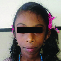
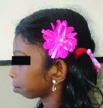
On extraoral examination, it was observed that the patient had the following facial features namely, Brachycephaly, Leptoprosopic face, Broad forehead, Periorbital fullness, Short stubby nose, Short palpebral fissures, Long philtrum, Broad, thick lips that were potentially competent, Convex profile, Severe prognathism of both the jaws [Table/Fig-1,2].
Facial features of the patient.
Convex profile of the patient.
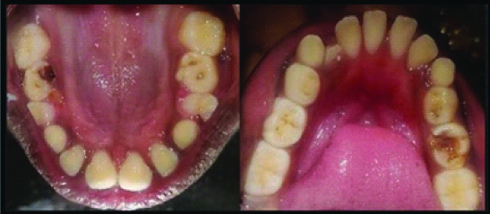
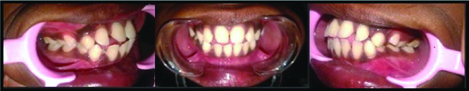
Intraoral examination revealed the presence of mixed dentition with dental caries in relation to 55, 65 with no soft tissue anomalies, retained root stumps in relation to 54 and physiologic mobility with respect to 73, 84, 85 [Table/Fig-3] with Bilateral Class I permanent first molar relation, Bilateral Mesial step terminal plane relation, proclined and spaced upper and lower anteriors, anterior open bite and tongue thrusting habit [Table/Fig-4].
Intraoral mirror images of maxillary and mandibular arches.
Intraoral occlusal relationships showing angle’s class I molar relation and anterior open bite.
Lateral cephalogram revealed the presence of short anterior cranial base, protrusive maxilla, increased Mandibular plane–Occlusal (MP- Occ) angle, steep mandibular plane, reduced ratio of posterior to anterior facial height and proclination of upper and lower incisors [Table/Fig-5,6].
Lateral cephalogram showing the presence of short anterior cranial base, protrusive maxilla, increased Mandibular Plane – Occlusal (MP- OCC) angle, steep mandibular plane, reduced ratio of posterior to anterior facial height and proclination of upper and lower incisors.
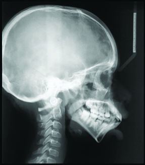
Lateral cephalogram values (N-S-Ar- Nasion- Sella- Articulare); S-Ar-Go- Sella- Articulare- Gonion; Ar-Go-Me- Articulare- Gonion- Menton; S-N-A- Sella- Nasion- Point A; S-N-B- Sella- Nasion- Point B; A- N- B – Point A-Nasion- Point B; Pal-Mp- Palatal plane- Mandibular plane angle; Mp-Occ- Mandibular plane- Occlusal plane angle; S-N-Mp- Sella- Nasion- Mandibular plane angle; N-S-Gn- Nasion- Sella- Gnathion;Ar- Go- N- Articulare- Gonion- Nasion; N- Go- Me- Nasion- Gonion- Menton; S-Go- Sella- Gonion; N-Me- Nasion- Menton).
| S. No. | Measure | Values in degrees |
|---|
| 1 | N-S-Ar | 119 |
| 2 | S-Ar-Go | 146 |
| 3 | Ar-Go-Me | 144 |
| 4 | Bjork’s Sum | 409 |
| 5 | S-N-A | 90 |
| 6 | S-N-B | 77 |
| 7 | A-N-B | 13 |
| 8 | Pal-Mp | 39 |
| 9 | Mp-Occ | 24 |
| 10 | S-N-Mp | 48 |
| 11 | N-S-Gn (Y axis) | 70 |
| 12 | Ar- Go- N (Upper gonial angle) | 61 |
| 13 | N- Go- Me (Lower gonial angle) | 82 |
| 14 | S- Go (Anterior facial height) | 46 |
| 15 | N- Me (Posterior facial height) | 87 |
| 16 | Jaraback’s ratio | 52.87 |
Since, tooth number 54 was a retained root stump and 73, 84, 85 exhibited severe physiological mobility and could not be salvaged, extraction of these teeth were planned. However, since 55 and 65 did not exhibit mobility, restoration of 55 and 65 was planned. Since patients with William’s syndrome are prone to cardiac abnormalities and as the patient had given a history of a similar condition at birth, in order to rule out any underlying cardiac abnormality and also to evaluate the need for antibiotic prophylaxis, Paediatric consultation was sought prior to the treatment. Echocardiogram revealed that there was no cardiac abnormality. Paediatrician opinion was sought and after the medical consent was given, the treatment was carried out under local anaesthesia. Tell-Show-Do behaviour management technique was employed. Although the patient had a positive behaviour, the patient was extremely sensitive to the sound of the airotor which posed a slight difficulty during the restorative treatment. The patient was asked to report every 3 months for recall visits.
Discussion
Williams syndrome (Mendelian inheritance number: 194050) named after William JC (1961) is a rare genetic disorder characterised by Autosomal dominant/Sporadic deletion on chromosome 7q11.23 which encodes for protein ELN [1]. The diagnosis is made by Fluorescent In-Situ Hybridisation (FISH). The global prevalence is said to be 1:7500–1: 20,000 with no gender predilection. It is otherwise known by the terms Elfin facies syndrome, Idiopathic hypercalcaemia of infancy, William-Bueren syndrome, Supravalvular aortic stenosis syndrome [1,2]. It is typically characterised by dysmorphic facial features, intellectual disability and cardiovascular abnormalities. The overall clinical features of the syndrome include: Dysmorphic facial features- Periorbital fullness, wide mouth, full lips, micrognathia- called as ‘Elfin facies’, Cardiovascular anomalies (Mainly Supravalvular aortic stenosis (55%), Peripheral pulmonary stenosis (60%), VSD (8-21%), MVP (15%), Aortic insufficiency (10%), Intellectual disability, Hypercalcaemia, Outgoing personality, Hoarse voice, Hyperacusis, Easy distractibility and Renal malformations [3-5]. Although the genetic and psychological aspects of the syndrome has been well explained by several studies however, there seem to be paucity of literature that discuss the oral manifestations of Williams Syndrome and also the behaviour of these children in a dental setting [Table/Fig-7] [6-10]. Due to the unique behavioural traits and underlying cardiac and renal abnormalities, it is imperative for the Paediatric dentist to have an adequate understanding regarding the syndrome so as to evaluate the need for antibiotic prophylaxis, drug dose alteration, choice of treatment (local/general anaesthesia) and risk stratification for treatment under general anaesthesia [6,11-14]. The general management of these patients include: Regular medical evaluation for cardiovascular and renal abnormalities, speech therapy, behavioural counselling, dietary modification in case of hypercalcaemia and special education programs for varying degrees of developmental disabilities. These patients are said to have a reduced life expectancy due to the complications that arise due to cardiovascular abnormalities [15].
Review of literature [6-10].
| Author and Year | Reference | Presentation |
|---|
| Torres CP et al., 2015 | [6] | A seven-year-old male with Williams Syndrome was treated with restorative and endodontic procedures under antibiotic cover for underlying heart defect. |
| Wong D et al., 2015 | [7] | A 17-year-old female patient with comorbid conditions of epilepsy and transient hypertension provided with restorative and preventive treatment in dental setting. |
| Andrade NS et al., 2015 | [8] | A report of four cases of Williams Syndrome that emphasise the fact that underlying heart condition and behaviour are important challenges in treating patients with Williams Syndrome. |
| Ribeiro MG et al., 2018 | [9] | A case report of a 20-year-old male that discusses the orthodontic and orthognathic considerations in treating a patient with Williams Syndrome. Heart defects, behaviour and periodontal status dictate the treatment strategy to be followed. |
| Ferreira SB et al., 2018 | [10] | An evaluation of the orofacial characteristics of seventeen patients with Williams syndrome that concludes that dental midline deviation and high arched palate to be the most common findings. |
The General Transcription Factor II- I Repeat Domain- containing protein I (GTF21RD1) and General Transcription Factor II-I (GTF21) genes are responsible for the dental and craniofacial abnormalities. The dental anomalies include: Broad upper arch, Abnormal tooth morphology, Enamel defects, Excessive spacing, Tongue thrusting, Open bite, Increased gingival and periodontal problems [16,17]. Whereas the craniofacial findings are: Short anterior cranial base, Pointed chin, Small but protrusive maxilla, Increased MP-Occ angle, Steep mandibular plane, Reduced ratio of posterior to anterior facial height, Proclination of upper and lower incisors and Dysmorphic sella [9,18,19].
The presence of cardiac abnormalities pose a great challenge for treatment in these sorts of cases so the dentist should carefully assess the cardiac condition present in the patient and seek medical consultation prior to initiating any dental treatment, so as to prevent the risk of infective endocarditis. In the present case, the cardiac condition was found to be normal and thereby did not require any antibiotic prophylaxis prior to treatment. This is of utmost importance even if the child has to be treated under General anaesthesia since reports of sudden cardiac arrest has been reported in patients with William’s syndrome during anaesthetic administration. So, a risk stratification has to be done based on the age of the patient and severity of underlying cardiac condition prior to general anaesthesia [3,4]. In this case since the dental treatment needs of the patient was minimal comprising of extraction of mobile teeth and restorations, an in-office treatment under local anaesthesia was preferred over general anaesthesia.
A typical feature of patients with Williams syndrome is over friendly behaviour/hypersociability. This is due to polymorphism in GTF2I gene leading to oxytocin dysregulation and decreased threat related amygdala reactivity [20]. This is a distinguishing feature when compared to those with intellectual disability. Also, these patients have hyper acusis- i.e., they are extremely sensitive to sounds and are easily distractible. The dental operatory can be threatening to the child with Williams syndrome which was encountered in the present case also as the child was extremely sensitive to the sound of the airotor during restorative procedure. In such situations, techniques like music distraction can be employed to mask the noise of the airotor and thereby reduce the anxiety during dental treatment.
Since, it is a genetic disorder, definitive treatment of the abnormal maxillo-mandibular relationships requires orthognathic surgical correction at a later stage after growth completion. However, prior to subjecting the child for surgery under general anaesthesia, the renal condition should also be assessed. Renal impairment, if present manifests as hypercalcaemia, elevated blood urea and creatinine levels. This is extremely important since drug dosage alteration is required in case of renal impairment.
Conclusion(s)
The paediatric dentist is the first person to identify a child with Williams Syndrome which has a typical facial presentation. So an understanding about this syndrome and its features would help the dentist in seeking medical opinion when needed and employ suitable behaviour management techniques to manage these children.
[1]. Kelly JR, Barr ES, The elfin facies syndromeOral Surg Oral Med Oral Pathol 1975 40(2):205-17.10.1016/0030-4220(75)90153-X [Google Scholar] [CrossRef]
[2]. Sharma P, Gupta N, Chowdhury MR, Phadke SR, Sapra S, Hadler A, Williams-Beuren syndrome: Experience of 43 patients and a report of an atypical case from a tertiary care center in IndiaCytogenet Genome Res 2015 146(3):187-94.10.1159/00043920526352091 [Google Scholar] [CrossRef] [PubMed]
[3]. Brown ML, Nasr VG, Toohey R, DiNardo JA, Williams syndrome and anaesthesia for non-cardiac surgery: High risk can be mitigated with appropriate planningPediatr Cardiol 2018 39(6):1123-28.10.1007/s00246-018-1864-129572733 [Google Scholar] [CrossRef] [PubMed]
[4]. Twite MD, Stenquist S, Ing RJ, Williams syndrome: Educational reviewPediatr Anaesth 2019 29(5):483-90.10.1111/pan.1362030811742 [Google Scholar] [CrossRef] [PubMed]
[5]. Nowak AJ, Christensen JR, Mabry T, Townsend JA, Wells MH, Pediatric Dentistry: Infancy through adolescence 2019 6th edElsevierPhiladelphia:253 [Google Scholar]
[6]. Torres CP, Valadares G, Martins MI, Borsatto MC, Diaz-Serrano KV, De Queiroz AM, Oral findings and dental treatment in a child with Williams- Bueren SyndromeBraz Dent J 2015 26(3):312-16.10.1590/0103-644020130033526200160 [Google Scholar] [CrossRef] [PubMed]
[7]. Wong D, Ramachandra SS, Singh AK, Dental management of patient with Williams Syndrome- A case reportContemp Clin Dent 2015 6(3):418-20.10.4103/0976-237X.16190826321847 [Google Scholar] [CrossRef] [PubMed]
[8]. Andrade NS, Santos C, Castro T, Gallottini M, Medical considerations in dental treatment of patients with Williams-Beuren syndrome: Report of four clinical casesClin Lab Res Den 2015 21(2):115-21.10.11606/issn.2357-8041.clrd.2015.118463 [Google Scholar] [CrossRef]
[9]. Ribeiro MG, Silveira GS, Rodrigues VF, Pantuzo MC, Oliveira DD, William-Beuren syndrome: What orthodontists need to know?Int J Odontostomat 2018 12(3):205-10.10.4067/S0718-381X2018000300205 [Google Scholar] [CrossRef]
[10]. Ferreira SB, Viana MM, Maia NG, Leao LL, Machado RA, Colleta RD, Oral findings in Williams Beuren SyndromeMed Oral Patol Oral Cir Bucal 2018 23(1):e01-06. [Google Scholar]
[11]. Campos-Lara P, Santos-Diaz MA, Ruiz-Rodriguez MS, Garracho-Rangel JA, PozosGuillén AJ, Orofacial findings and Dental management of Williams-Bueren SyndromeJ Clin Pediatr Dent 2012 36(4):401-04.10.17796/jcpd.36.4.c93436771101tm0623019840 [Google Scholar] [CrossRef] [PubMed]
[12]. Poornima P, Patil PS, Subbareddy VV, Arora G, Dentofacial characteristics in Williams SyndromeContemp Clin Dent 2012 3(5):S41-44.10.4103/0976-237X.9510322629065 [Google Scholar] [CrossRef] [PubMed]
[13]. Pires FS, Paula VA, Faker K, Lanaro ND, De Carvalho RC, Tostes MA, Oral conditions of pediatric patients with Williams-Bueren Syndrome: Two case reportsBrazilian Dental Science 2019 22(2):281-88.10.14295/bds.2019.v22i2.1704 [Google Scholar] [CrossRef]
[14]. Luo E, Liu H, Zhao Q, Shi B, Chen Q, Dental-craniofacial manifestation and treatment of rare diseasesInt J Oral Sci 2019 11(1):09-15.10.1038/s41368-018-0041-y30783081 [Google Scholar] [CrossRef] [PubMed]
[15]. Morris CA, Braddock SR, AAP Council On GeneticsHealth care supervision for children with Williams SyndromePediatrics 2020 145(2):e2019376110.1542/peds.2019-376131964759 [Google Scholar] [CrossRef] [PubMed]
[16]. Budişteanu M, Papuc SM, Riga D, Arghir A, Dental anomalies in Williams-Beuren syndromeInt J Med Dent 2018 22(3):243-46. [Google Scholar]
[17]. Castro T, Santos CP, Ortega AO, Gallottini M, Oral characteristics and medical considerations in the dental treatment of individuals with Williams SyndromeSpec Care Dentist 2019 39(2):108-13.10.1111/scd.1236130707461 [Google Scholar] [CrossRef] [PubMed]
[18]. Mass E, Belostoky L, Craniofacial morphology of children with Williams syndromeCleft Palate Craniofac J 1993 30(3):343-49.10.1597/1545-1569_1993_030_0343_cmocww_2.3.co_28338868 [Google Scholar] [CrossRef] [PubMed]
[19]. Goyal I, Bansal N, Mehta V, Singh G, Arora B, Orthodontic management of syndromic patients- A reviewActa Scientific Dental Sciences 2018 2(9):124-31. [Google Scholar]
[20]. Royston R, Waite J, Howlin P, Williams syndrome: Recent advances in our understanding of cognitive, social and psychological functioningCurr Opin Psychiatr 2019 32(2):60-66.10.1097/YCO.000000000000047730557270 [Google Scholar] [CrossRef] [PubMed]