Intrascrotal Solitary Neurofibroma
Abhishek Gupta1, Suhas N Jajoo2
1 Resident, Department of General Surgery, Jawaharlal Nehru Medical College Dutta Meghe Institute of Medical Science, Wardha, Maharastra, India.
2 Professor, Department of General Surgery, Jawaharlal Nehru Medical College Dutta Meghe Institute of Medical Science, Wardha, Maharastra, India.
NAME, ADDRESS, E-MAIL ID OF THE CORRESPONDING AUTHOR: Abhishek Gupta, T1, Raghobaji PG Boys Hostel, DMIMS, Sawangi, Wardha, Maharastra, India.
E-mail: draddy.rebellion18@gmail.com
Solitary Neurofibroma of the scrotum is a rare benign tumour, particularly when it is not associated with neurofibromatosis Type I, hence, less number of cases have been reported in the English literature. Hereby the authors report a case of intrascrotal solitary neurofibroma in a 45-year-old male who was admitted to the hospital with a 2 year history of a painless swelling. With a preoperative diagnosis of soft tissue tumour, exploration was performed. The pathological diagnosis of the lesion was neurofibroma of scrotum. Although a relatively rare disease, intrascrotal solitary neurofibroma should be considered in the differential diagnosis of scrotal swellings.
Neurofibromatosis, Painless, Swelling
Case Report
A 45-year-old male presented with a painless swelling in his scrotum for the last 2 years, insidious in onset, progressive in nature with no significant enlargement of the mass in last 7 months.
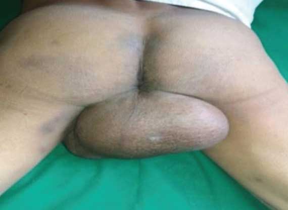
On physical examination, a single large ovoid mass of 13 cm × 11 cm size in the scrotal region. Firm in consistency, non-tender, local temperature not raised, fluctuation and transillumination (negative), not fixed to overlying skin and underlying tissue with bilateral testis palpable and clinically normal. The mass extended up to the anal verge [Table/Fig-1]. No rectal invasion was felt on per rectal examination. No regional lymphadenopathy was found on general physical examination.
Clinical picture of the scrotal swelling.
Ultrasonography showed a heterogeneous mass of size 12 cm × 11 cm in midline at inferior aspect of scrotal sac, with mild vascularity suggestive of a malignant mass. Laboratory studies were unremarkable. Fine-needle aspiration cytology showed benign spindle cell lesion of leiomyoma with aggressive nuclear morphology. No cytologically malignant cells were seen.
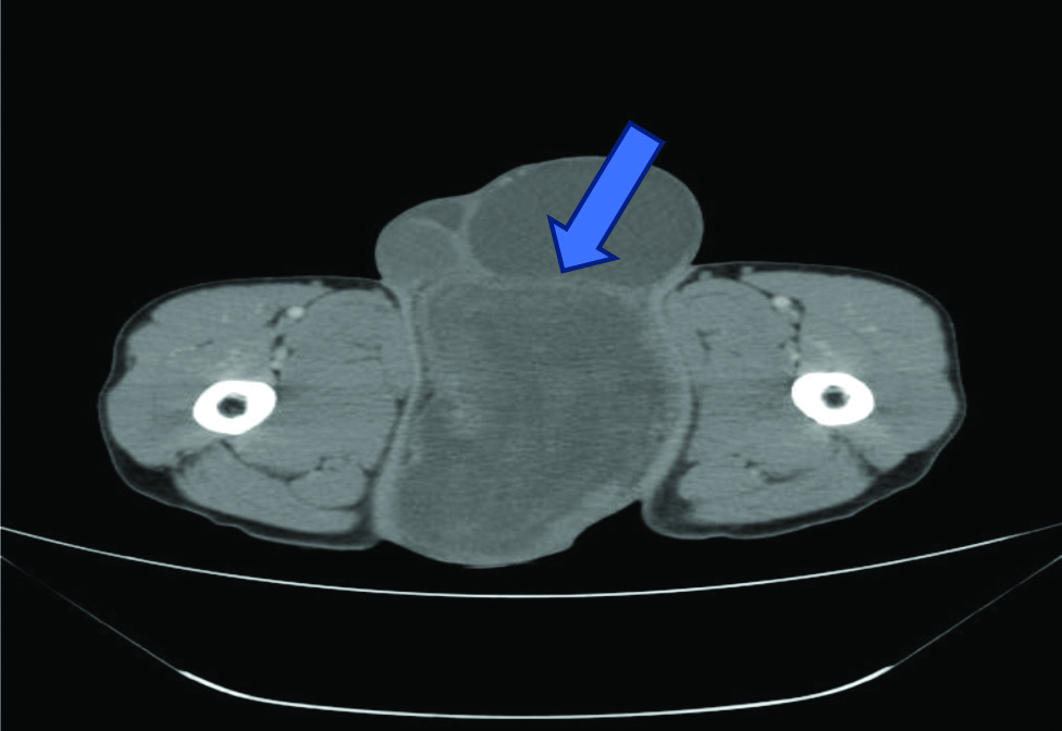
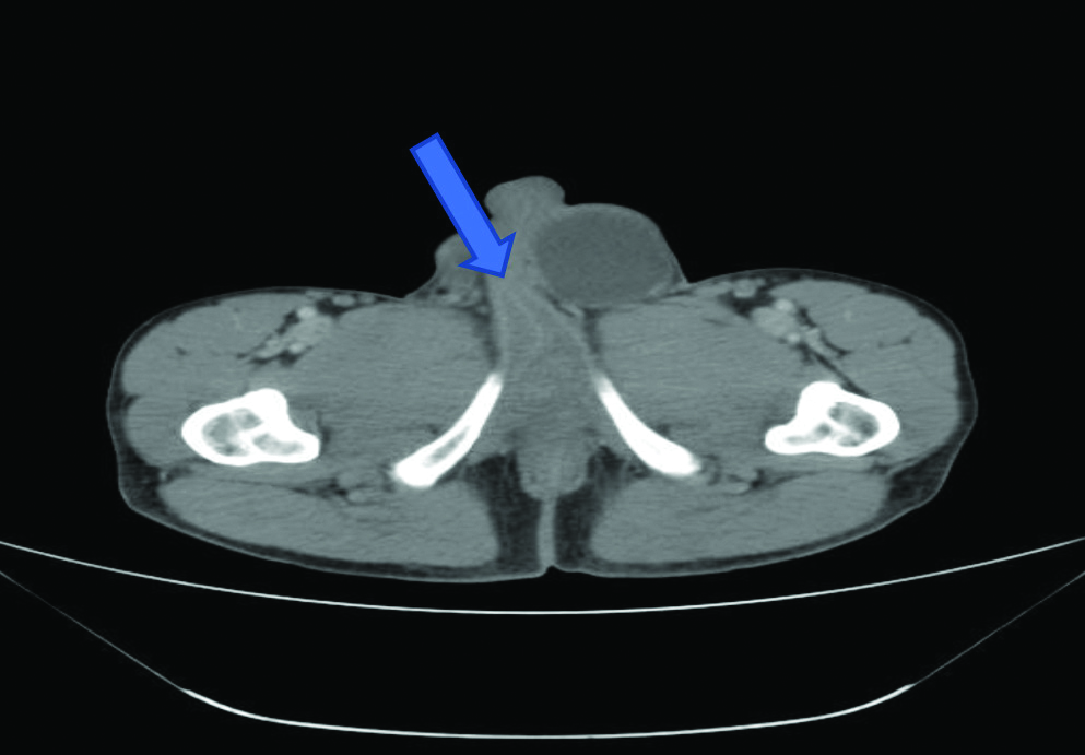
Contrast Enhanced Computed Tomography (CECT) showed well defined, oval hypodense mass measuring 14.5 cm x 10.8 cm, nodular, heterogenous enhancement within it, extending superiorly up to base of penis, displacing testis and scrotal sac and posteriorly compressing anal canal, no involvement of levator ani muscle [Table/Fig-2,3] and no involvement of anal canal which is suggestive of soft tissue tumour.
CECT image showing the extent of scrotal swelling. Arrow shows the line separating scrotal mass from both scrotal sacs.
CECT image showing the extent of scrotal swelling. Arrow shows the extension upto base of penis.
With the diagnosis of soft tissue tumour of scrotum, the patient was scheduled for surgery. A scrotal incision was performed and a large oval and firm mass were detected occupying a large portion in scrotum. Testes and epididymis were free. The mass was completely excised off the surrounding tissue by scrotal approach under spinal anaesthesia. Patient had an uneventful postoperative period.
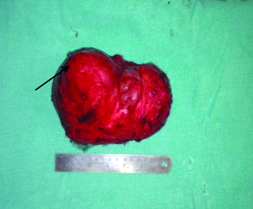
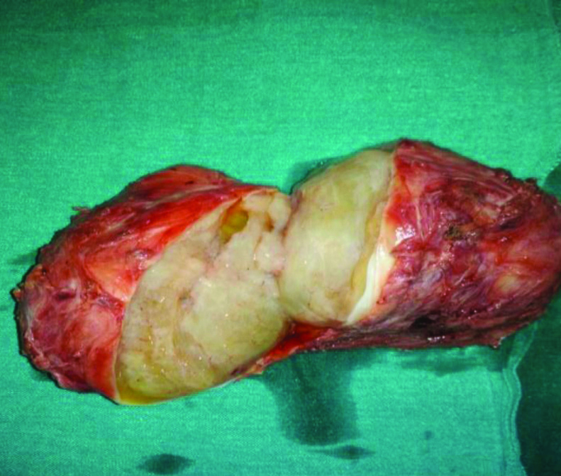
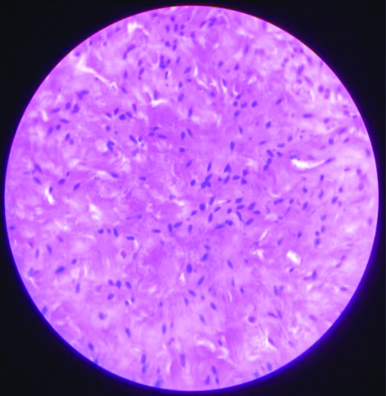
Pathologic examination of the tumour showed a large well-circumscribed smooth mass measuring 17 cm × 11 cm × 9 cm [Table/Fig-4]. The excised mass on cut section showed homogenous, gelatinous, dirty white/ creamy in colour, minimally vascular, no area of haemorrhage [Table/Fig-5]. Microscopic examination was suggestive of studies neurofibroma-composed of uniformly distributed spindle cells with inconspicuous nucleoli, dispersed chromatin and wavy nuclei with no atypia or mitosis [Table/Fig-6].
Gross image of resected scrotal mass.
Cut section of resected scrotal mass.
Microscopic view of the mass diagnosed as neurofibroma (H and E, 40x).
Discussion
Neurofibromatosis is an autosomal dominant condition wherein a tumour arises from nervous tissue. Clinical features that are diagnostic for neurofibromatosis are Cafe-au-lait spots, axillary and inguinal freckels, multiple neurofibromas in the various parts of the body [1-4]. Although it can be encountered anywhere within the central or peripheral nervous system, cranium, retroperitoneum, thorax, especially in the neck, and flexor surfaces of the extremities, localisation within the scrotum is the rarest [1,2,5-7]. The risk of developing cancer in a person with Neurofibromatosis is around 15% [8].
Scrotal tumours are mostly extratesticular, either originating from spermatic cord or epididymis. Benign scrotal tumours include leiomyoma, fibroma, lipoma, hemangioma, and epidermoid cysts [2]. Yamamoto M et al., reported the first case of solitary neurofibroma in the scrotum in 1982 [4]. Neurofibroma can be solitary or multiple and can present in any age group ranged between 8 and 77 years [3,5,7,9].
Few solitary neurofibroma cases, which involved the external genitalia in the absence of von Recklinghausen’s disease, were reported previously but only in one case, intratesticular tumour was reported [10]. In this case, exact anatomic structural origin of the tumour was not clear. However, mass was separate from the testis, vas deferens, and epididymis. So, as published by Issa MM et al., [11], reported a case of solitary neurofibroma in the scrotum which must have originated from the genital branch of the genitofemoral nerve lying posteriorly to the spermatic cord. This nerve innervates cremasteric muscle and distributes branches to the skin of the scrotum and adjacent thigh.
The surgical treatment of testicular and para-testicular tumours is performed by inguinal exploration [6,10]. The mass, in the index case, could be palpated outside the testis, had smooth contours. Radiographically, there were no lymphadenopathies, testis and paratesticular tissues were normal. These confirmed the diagnosis. In cases, where the tumour involves the testicle, orchidectomy is inevitable. But as testis were not involved in this case, so only complete surgical excision of the mass was done. Postoperative recovery was uneventful.
Conclusion(s)
Solitary Scrotal Neurofibromas not associated with Neuro-fibromatosis Type I (NF I) is a rare presentation. A neurofibroma in the scrotal region is a rare site of occurrence. Treatment of choice is complete surgical excision of the mass.
Author Declaration:
Financial or Other Competing Interests: None
Was informed consent obtained from the subjects involved in the study? Yes
For any images presented appropriate consent has been obtained from the subjects. Yes
Plagiarism Checking Methods: [Jain H et al.]
Plagiarism X-checker: Jan 16, 2020
Manual Googling: Mar 25, 2020
iThenticate Software: May 27, 2020 (17%)
[1]. Milathianakis KN, Karamanolakis DK, Mpogdanos IM, Trihia-Spyrou EI, Solitary Neurofibroma of theSpermatic CordUrol Int 2004 72(3):271-74.10.1159/00007713015084777 [Google Scholar] [CrossRef] [PubMed]
[2]. Türkyılmaz Z, Sönmez K, Karabulut R, Dursun A, Işık I, Başaklar C, A childhood case of intrascrotal neurofibroma with a brief review of the literatureJ Pediatr Surg 2004 39(8):1261-63.10.1016/j.jpedsurg.2004.04.02615300541 [Google Scholar] [CrossRef] [PubMed]
[3]. Geramizadeh B, Shakeri S, Karimi M, Hosseini M, Intrascrotal solitary neurofibroma: A case report and review of the literatureUrol Ann 2012 4(2):11910.4103/0974-7796.9556922629013 [Google Scholar] [CrossRef] [PubMed]
[4]. Yamamoto M, Miyake K, Mitsuya H, Intrascrotalextratesticular neurofibromaUrology 1982 20(2):200-01.10.1016/0090-4295(82)90364-8 [Google Scholar] [CrossRef]
[5]. Mishra VC, Kumar R, Cooksey G, Intrascrotal neurofibromaScand J Urol Nephrol 2002 36(5):385-86.10.1080/00365590232078392612487746 [Google Scholar] [CrossRef] [PubMed]
[6]. Agarwal PK, Palmer JS, Testicular and paratesticular neoplasms in prepubertal malesJ Urol 2006 176(3):875-81.10.1016/j.juro.2006.04.02116890643 [Google Scholar] [CrossRef] [PubMed]
[7]. Gupta S, Gupta R, Singh S, Pant L, Solitary intrascrotal neurofibroma: A case diagnosed on aspiration cytologyDiagn Cytopathol 2011 39(11):843-46.10.1002/dc.2155821994196 [Google Scholar] [CrossRef] [PubMed]
[8]. Miettinen MM, Antonescu CR, Fletcher CDM, Kim A, Lazar AJ, Quezado MM, Histopathologic evaluation of atypical neurofibromatoustumours and their transformation into malignant peripheral nerve sheath tumour in patients with neurofibromatosis 1-a consensus overviewHum Pathol 2017 67:01-10.10.1016/j.humpath.2017.05.01028551330 [Google Scholar] [CrossRef] [PubMed]
[9]. Erdemir F, Parlaktas BS, Uluocak N, Filiz NO, Acu B, Intrascrotal extratesticular neurofibroma: A case reportMarmara Med J 2008 21:064-67. [Google Scholar]
[10]. Livolsi VA, Schiff M, Myxoid Neurofibroma of the TestisJ Urol 1977 118(2):341-42.10.1016/S0022-5347(17)58002-7 [Google Scholar] [CrossRef]
[11]. Issa MM, Yagol R, Tsang D, Intrascrotal neurofibromasUrology 1993 41(4):350-52.10.1016/0090-4295(93)90594-Z [Google Scholar] [CrossRef]