The adhesion of bonding agents and their longevity are imperative in the dental field. No system can block microleakage, which significantly increases in cementum areas as evaluated by many studies. The hybrid layer is said to be the key mechanism of bonding between conditioned dentin and adhesive system [2].
The collagen network is infiltrated by monomers that are polymerized to form the hybrid layer, thus reinforcing the demineralized dentin [3]. The leakage pathway allows oral fluid and microflora to permeate the resin/dentin interface which can cause degradation of the bond area and pulp inflammation. The main cause of pulpal irritation from dental restorative materials is the bacteria at the restoration/dental interface [4]. Certain procedures need to be adopted to reduce microleakage; such as maintaining a wet dentin, applying the adhesive according to manufacturer’s instructions and following the incremental technique while restoring with resin composite. Hence, treating dentin with acids leads to collapse of exposed collagen fibres by eliminating hyrodxyapatite and/or dentauring collagen [5]. To resolve this issue Self-etching adhesive systems were designed. Off late many new adhesive systems have been designed and their pre-treatment and adhesion promoters are being explored to better the clinical results [6].
Materials and Methods
An in-vitro study was conducted in School of Dental Sciences, Krishna Institute of Medical Sciences, Karad, Maharashtra, India in February 2019 with Ethics Committee approval (0110/2018-2019).
Sixty extracted human premolars for orthodontic treatment purpose that were free of caries, attrition, abrasion, erosion, restorations and craze lines were selected for the this study. After mechanical debridement and enamel integrity evaluation using a magnifying glass (4X), all teeth were cleaned with pumice water slurry. Teeth were cleaned of any calculus, stains, soft tissue and other debris and stored in distilled water till cavity preparation.
Class V cavity was prepared on the facial and lingual surfaces of all teeth with a cylindrical diamond bur. All cavities were prepared 3-mm-wide mesio-distally paralleling the cemento-enamel junction, 2.5-mm-wide occluso-gingivaly and 1.5 mm in depth approx. While the gingival half was extended 1mm below CEJ [7]. Dimensions of these preparations were measured with a periodontal probe.
Group 1: Fifteen teeth (30 cavity preparations) used the 5th generation bonding agent Adper Single Bond 2 (3M ESPE-3M)
Group 2: Fifteen teeth (30 cavity preparations) used the 6th generation bonding agent Adper SE Plus (3M ESPE-3M)
Group 3: Fifteen teeth (30 cavity preparations) used the 7th generation bonding agent Adper Easy One (3M ESPE-3M)
The dentin bonding systems were applied following the manufacturer’s instructions and the cavities of Group I, II and III were restored with resin composite (Z-100).
Group 4: Fifteen teeth (30 cavity preparations) were restored with Resin modified glass ionomer (Vitremer) as per manufacturer’s instructions.
The restored teeth were thermocycled for 200 cycles between temperatures of 5°C and 55°C with a dwell time of 60 seconds [7,11].
The specimens were subjected to dye-leakage tests. Root apices were sealed with epoxy resin. Two coats of nail varnish were applied to the entire tooth surface to within 1 mm of the restoration. The teeth were then immersed in 2% methylene blue dye for 24 hours at room temperature, removed and thoroughly rinsed with distilled water and dried for 10 minutes [7].
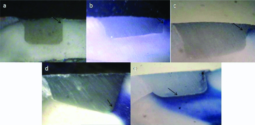
The teeth were sectioned with a thin diamond disc (DFS, Germany) such that two sections were obtained from each tooth. Sectioning was done through the center of the restoration from the facial to the lingual surface. The degree of dye penetration in each tooth was assessed using a stereomicroscope (Olympus SZ40) under 20X magnification [Table/Fig-1,2].
Dye penetration scores at enamel margin: a) score 0- no microleakage; b) score 1- die penetration within 1/3rd of cavity wall; c) score 2: within 2/3rd of cavity wall; d) score 3- within last 1/3rd of cavity upto axial wall; e) score 4- spreading along the axial wall [12,13].
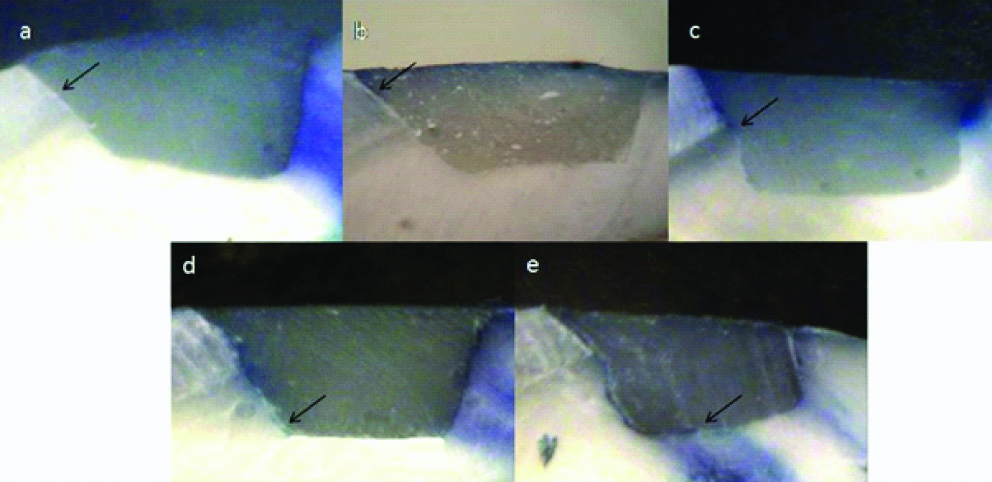
Dye penetration scores at dentinal margin: a) score 0- no microleakage; b) score 1- die penetration within 1/3rd of cavity wall; c) score 2: within 2/3rd of cavity wall; d) score 3- within last 1/3rd of cavity upto axial wall; e) score 4- spreading along the axial wall [12,13].
Based on the ordinal ranking system, the degree of dye leakage was determined as follows:
0-No microleakage
1-Dye penetration within 1/3 of cavity wall
2- Dye penetration within 2/3 of cavity wall.
3- Dye penetration within last 1/3 of cavity wall upto axial wall
4- Dye penetration spreading along axial wall
Dye penetration at the restoration-tooth interface was scored for both occlusal and cervical margins and on both sides buccal and lingual [12,13].
Statistical Analysis
Statistical analysis was done using Kruskall Wallis test and Mann- Whitney non-parametric analysis with significant p-value being p<0.05 and result was expressed through sum of ranks.
Results
[Table/Fig-3] depicts the microleakage scores at the coronal margins(enamel) for all the groups. [Table/Fig-4] depicts the microleakage scores at apical margins(cementum) for all the groups. [Table/Fig-5] depicts comparison amongst microleakage scores between enamel and cementum.
Distribution of microleakage scores at the coronal margins (Enamel).
| Score | | |
|---|
| Study group | 0 | 1 | 2 | 3 | 4 | Mean score | Standard deviation |
|---|
| Group 1 (adper single bond 2) (N=30) | 17 | 9 | 4 | 0 | 0 | 0.57 | 0.73 |
| Group 2 (adper SE plus) (N=30) | 6 | 4 | 14 | 4 | 2 | 1.73 | 1.14 |
| Group 3 (adper easy one) (n=30) | 4 | 7 | 12 | 4 | 3 | 1.83 | 1.15 |
| Group 4 (vitremer) (n=30) | 19 | 9 | 2 | 0 | 0 | 0.43 | 0.63 |
Distribution of microleakage scores at the apical margins (cementum).
| Score | | |
|---|
| Study group | 0 | 1 | 2 | 3 | 4 | Mean score | Standard deviation |
|---|
| Group 1 (adper single bond 2) (n=30) | 2 | 2 | 4 | 9 | 13 | 2.97 | 1.22 |
| Group 2 (adper SE plus) (N=30) | 3 | 3 | 7 | 12 | 5 | 2.43 | 1.19 |
| Group 3 (adper easy one) (N=30) | 3 | 2 | 10 | 11 | 4 | 2.37 | 1.13 |
| Group 4 (vitremer) (n=30) | 18 | 10 | 2 | 0 | 0 | 0.47 | 0.63 |
Comparison between microleakage scores in the enamel and cementum, The statistical analysis was done using Kruskall Wallis test and Mann-Whitney non-parametric analysis.
| Groups | p-value | Significance |
|---|
| Group 1 | 0.001 | Significant |
| Group 2 | 0.001 | Significant |
| Group 3 | 0.002 | Significant |
| Group 4 | 0.833 | Non- Significant |
At the enamel margins, Adper Single Bond 2 and Vitremer showed the lowest mean leakage. The mean leakage score of Adper Single Bond 2 and Vitremer were 0.57±0.73 and 0.43+0.63, respectively. Adper Easy One showed the highest mean leakage score of 1.83±1.15. At the cementum margins, Vitremer showed the lowest mean leakage score of 0.47±0.63 and highest mean leakage score shown by Adper Single Bond 2, the mean score being 2.97±1.22.
Statistically significant leakage (p<0.05) was found at dentin/cementum margins for all bonding agents when compared to the modified glass ionomer. In Groups I, II, III, difference in microleakage scores in the enamel and cementum margins is very significant (p<0.01), whereas in Group IV, the difference was not significant (p=0.833).
Discussion
All the resin composites are associated with polymerisation shrinkage resulting in volumetric contraction and formation of a gap at the periphery of the restoration. Other factors which play a role in marginal gap formation are the physical characteristics of resin composites (filler loading, modulus of elasticity, water sorption, coefficient of thermal expansion), C-factor of the cavity, occlusion components, tooth flexure, finishing and polishing effects etc. It is the adhesive that provides bonding of the resin composite with the cavity walls and counteracts the formation of marginal gap [14].
Marginal leakage has been defined as the “marginal permeability to bacterial, chemical and molecular invasion at the tooth/material interface”. It is a result of a breakdown of the tooth-restoration interface which can lead to discoloration, recurrent caries, pulpal inflammation and may require restoration replacement [15].
A standardised Class V cavity preparation design was chosen in this study as Class V cavities can be most challenging to the adhesive systems used owing to the high C-factor. The C-factor is the ratio between the bonded and unbonded surfaces. An increase in C-factor leads to an increase in the shrinkage stresses at the adhesive interface, thus reducing the sealing ability [16,17].
This study used aging by thermocycling to simulate degradation of bond over a period of time due to changes of temperature in oral cavity. All the teeth in this study were thermally cycled between 5°C and 55°C with a dwell time of 60 seconds [7].
Teeth were assessed for microleakage by dye penetration method using 2% methylene blue, since it is the most employed method owing to its simplicity and cost effectiveness.
The result of this in-vitro study indicate that none of the dentinal adhesive tested completely eliminated microleakage, both at the enamel and cementum (dentin) margins. All the three adhesives showed more microleakage at the cementum (dentin) margin when compared to enamel margin [18]. The difference in leakage in this study can be related to the composition of enamel and dentin substrate. Enamel is primarily inorganic in nature with the inorganic component constituting 95%. Hence, etching of enamel causes demineralisation of the inorganic surfaces creates microporosities, which result in the penetration of adhesive resin into these microporosities, forming a strong bond. Dentin has a higher organic content, tubular structure, odontoblastic processes and a moist surface which precludes ideal bonding. Therefore, the bonding to enamel is more stable and efficient than that obtained with dentin, resulting in greater microleakage at dentin interface [19].
Analysis of data for leakage at enamel margin revealed significantly lower microleakage for the total-etch adhesive systems Adper Single Bond 2 as compared to both the self-etch systems viz., Adper SE Plus and Adper Easy One (p<0.05). This finding was in agreement with studies by Koliniotou-Koumpia E et al., Pradelle-Plasse N et al., and Gagliardo RM and Avelar RP who also reported lower leakage associated with total-etch systems at enamel margins [20-22]. Scanning Electron Microscopy (SEM) studies have depicted that the use of phosphoric acid as an enamel etchant enhances enamel penetration and the subsequent attachment of adhesive monomers [23]. Bis-GMA, HEMA and polyalkenoic acid are the main chemical components of the Adper Single Bond 2. Vitrebond copolymer in composition of Adper Single Bond 2 form a chemical bond to the hydroxyapatite by forming a complex with the calcium ions
At cementum margin, Adper Single Bond 2 showed highest microleakage. These results are in accordance with findings by Amaral CM et al., Tenniswood CA et al., and Deliperi S et al., found significantly greater microleakage with a total-etch adhesive than self-etch adhesive in their study [24-26]. Probable reason for greater microleakage is the depth of demineralisation and the depth of penetration of the primer is not the same, which may leave exposed and unprotected collagen fibers. The Phosphoric acid etching of dentin completely removes the smear layer and opens the dentinal tubules. This increases the permeability of dentin resulting in dentinal fluid flowing to the dentinal surface which may result in interference with adhesion [27].
Self-etch adhesives Adper SE Plus and Adper Easy One showed greater microleakage at enamel margin can be attributed to incomplete etching of enamel by the acidic monomers present in the self-etch adhesive [28].
The seventh generation, ’all-in-one’ adhesive, Adper Easy One showed the highest mean leakage score at enamel margin. This finding can be related with the observations of Weerasinghe DD et al., who in a scanning electron microscope micro-analysis study observed that seventh generation, ‘all-in-one’ adhesive produced only a mild etching pattern of ground enamel [29]. The reason can be ascribed to Adper Easy One being a ‘mild’ self-etch adhesive with a pH of about 3.5, as compared to phosphoric acid etchants having a pH of 0.5-1 and Adper SE Plus with a pH below 1.
At the dentin interface both the self-etch adhesives, Adper Easy One and Adper SE Plus showed significantly lesser microleakage than Adper Single Bond 2 (p≤0.05). These results are in accordance with Tenniswood CA et al., [25]. He observed that two 7th generation self-etch systems showed significantly lesser dye penetration than a total-etch system. The etching depth and the depth of penetration of the adhesive are identical in case of self-etch adhesives which thus prevents the collagen fibers from collapsing. Therefore, unlike the 5th generation adhesive systems, they do not depend on “moist bonding”. This is important because technique sensitivity associated with bonding systems requiring a “moist bonding” technique may be associated with postoperative sensitivity. Lesser leakage scores associated with self-etch systems can be attributed to one or more of the following reasons; self-etch systems which are composed of aqueous mixture of acidic functional monomers do not require a separate acid etch component and subsequent rinsing procedures. In addition, they do not require application of the primer to a particular condition of wetness of dentin due to the self-etch adhesive water content. This greatly reduces the technique sensitivity and thus the risk of errors during application and manipulation becomes minimal. In the self-etch approach, as the infiltration of resin occurs simultaneously with the self-etching process, the risk of discrepancy between both processes is low or non-existant [30].
Vitremer is used in this study as a control group. Resin modified glass ionomer Vitremer showed less microleakage in enamel and cementum margin than total-etch and self-etch adhesive.
The underlying mechanism of adhesion for conventional glass-ionomers is based on a dynamic ion-exchange process, in which the polyalkenoic acid softens and infiltrates the hydroxyapatite structure, where it displaces calcium and phosphate ions out of the substrate, forming an intermediate adsorption layer of calcium and aluminum-phosphates and polyacrylates at the glass-ionomer hydroxyapatite interface. Resin-modified glass ionomers probably adhere through a combination of the latter mechanism and a micro-mechanical bonding mechanism that has been described for resin-based adhesives [31].
Two surfaces with equivalent surface energies should be brought together to achieve a good adhesive bond. Also the adherend should wet the substrate intimately. Vitremer tri-cure glass ionomer system is composed of the Vitrebond copolymer, HEMA (2-hydroxyethylmethacrylate), ethanol and photoinitiators. The acidic low viscosity primer modifies smear layer and wets the tooth structure to provide a surface receptive to glass ionomer mix. The acidic polymer of the primer is attracted to dentin and enamel surfaces. Photocuring the primer crosslinks the methycrylate groups of the polymer and provides a surface for placing glass ionomer mix. Once the Vitremer mix is placed on the primer, the polyacid of the primer reacts with the fluoroaluminosilsicate glass of the glass ionomer mix, hence making the primer a part of the overall glass ionomer restoration [33].
Vitremer tri-cure glass ionomer system has three distinct curing reactions; acid-base glass ionomer reaction (initiated when powder and liquid are mixed and can proceed in the dark), photoinitiated free radical methacrylate cure (initiated when the powder/liquid mix is exposed to light and occurs only where light penetrates), dark cure free radical methacrylate cure (initiated when powder and liquid are mixed and can proceed in the dark). A diferentiating feature of 3M tri-cure-based materials is the mechansim of dark methacrylate cure. This leads to uniform curing throughout the glass ionomer restorations, hence enhancing the physical properties even if placed in bulk. Vitremer offers various benefits over composite; such that it directly bonds to tooth structure and does not require separate adhesive, bulk placement of material is possible so there is no need to place in increments like composite, fluoride release for caries inhibition and moisture compatibility [34].
Limitation(s)
For this study, dye penetration is used for evaluating microleakage; using advanced and more reliable methods can be helpful for better result bonding agents routinely used.
Conclusion(s)
Among the materials used in the study, Vitremer exhibits the best marginal sealing ability (least microleakage) at both enamel and cementum margins and Adper Easy One shows the least sealing ability at the enamel margin, while Adper Single Bond 2 at the cementum margin. At the enamel margin, Adper Single Bond 2 and Vitremer shows significantly lesser microleakage than Adper Easy One and Adper SE Plus while that at the cementum margin, Vitremer shows significantly lesser microleakage than Adper Single Bond 2 and Adper SE Plus. Adper Single Bond 2, Adper SE Plus and Adper Easy One shows significantly greater dye penetration along the cementum (dentin) margin than at enamel margin. Vitremer shows similar leakage at both enamel margin and cementum margin. Resin modified glass ionomers show promising results compared to 5th, 6th as well as 7th generation dentin bonding agents when it comes to minimal microleakage and thus, can be advised for improved adhesive dentistry after considering all other properties as well.