“Resorption is associated with either a physiologic or a pathologic process resulting in a loss of dentin, cementum and/or bone” [1]. The process of physiologic resorption is completed when the deciduous teeth are exfoliated followed by the eruption of permanent teeth. This sequential process of resorption of deciduous teeth reflects the path of eruption of permanent teeth. Pattern of resorption of deciduous teeth may be affected by the position and size of the succeeding permanent teeth [2].
Root resorption of deciduous teeth is traditionally been studied radiographically. However, this method may not provide accurate information as radiographs are two dimensional representation of a three dimensional process [3]. Moreover, presence of overlap in the region of maxillary and mandibular anterior teeth would further obscure the visibility thereby leading to inaccurate observations [4,5].
More recently, micro-computerised tomography has been used for further understanding the process of resorption; however the process requiring specialised equipment and trained personnel limits the use of this procedure. Also, most of the studies are conducted on the dry skull of children and hence, it may not be possible to assess the continuous process and may restrict to a particular time frame of process of resorption [6,7].
Hence, this study was conducted to assess the pattern, surfaces and extent of physiologic root resorption in deciduous incisors, canines and molars at different stages of root resorption.
Materials and Methods
This observational study was conducted in JSS Dental College and Hospital, JSS Academy of Higher Education and Research, Mysuru, Karnataka. This study was conducted as a part of extensive research studying the changes occurring in the primary teeth during the different stages of root resorption. The duration of the study was 2.5 years (June 2015 to December 2017) which was required for the collection of primary teeth that would provide accurate information. The primary teeth were extracted from healthy individuals (3-10-year-old) undergoing interceptive orthodontic procedures or from the individuals who have undergone dental trauma demanding the extraction of primary teeth. Primary teeth thus collected were stored in 10% formalin until further use. Ethical clearance (JSS/ACP/Ethical/2012-13) was obtained from the Institutional Ethical Board (IEB) for performing the study. Since, it was observational trial, saturation sampling was done. Saturation sampling is done in qualitative research. In which, the procurement of samples is discontinued after a point beyond which no additional information could be obtained from the data, due to lack of prior information on measurements under Indian context. Hence, at 80% power of study and 5% level of significance the overall sample size was 138 with at least minimum of 10 samples in each sub-group.
The study consisted of 4 groups with 3 sub-groups in each. The sub grouping was done based on the RRL [Table/Fig-1,2]. The percentage RRL was used to group the deciduous teeth into stages of root resorption. Each sample tooth was radiographed using Radiovisiography (RVG) to rule out any internal pathology like internal resorption.
Criteria for grouping of teeth based on the Remaining Root Length (RRL).
| Tooth | % Remaining root length | Stage of resorption | Clinical observation |
|---|
 | 67% to 100% | Stage 1 | Starting of resorption where in resorption is seen only in the apical third |
 | 34% to 66% | Stage 2 | Moderately resorbed root with resorbing front in the middle third of the root |
 | 0% to 33% | Stage 3 | Severe resorption with resorption front in the cervical third of the root |
Grouping of the samples. Sub-groups divided on basis of stages of root resorption as determined by RRL.
| Groups | Sub-groups | Sample size (n) |
|---|
| Incisors | Stage 1 | 12 |
| Stage 2 | 12 |
| Stage 3 | 18 |
| Canines | Stage 1 | 12 |
| Stage 2 | 12 |
| Stage 3 | 12 |
| Maxillary molars | Stage 1 | 10 |
| Stage 2 | 10 |
| Stage 3 | 10 |
| Mandibular molars | Stage 1 | 10 |
| Stage 2 | 10 |
| Stage 3 | 10 |
| Total number of samples studied (n) | 138 |
Measurement of Remaining Root Length (RRL)
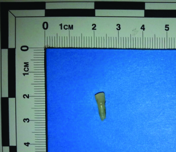
For the purpose of the study, the extracted teeth were grouped into four: Incisors, Canines, Maxillary Molars and Mandibular Molars. Each group was further divided into 3 sub-groups based on the stages of root resorption (as explained in [Table/Fig-2]) which was determined based on the RRL measurement. The sub grouping based on RRL helped in assessing resorption pattern. Maxillary and mandibular incisors were grouped together in Incisors group. Similarly, maxillary and mandibular canines were also grouped together as canines. For the measurement of RRL, teeth were photographed on blue background along No. 2 photomacrographic scale designed by American Board of Forensic Odontology (ABFO) [Table/Fig-3] [8]. The teeth were positioned with the help of modelling wax (1 mm by 1 mm) such that the long axis of the tooth was parallel to base [Table/Fig-4]. Teeth were photographed to capture the buccal, mesial, distal and palatal/lingual aspects. The images thus obtained were used for assessing the pattern of resorption and measurement of the RRL.
Measurement of Root Length using ABFO scale for digital calibration -single rooted teeth.
Tooth positioned such that the long axis of the tooth is perpendicular to the line of exposure.
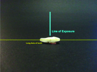
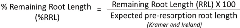
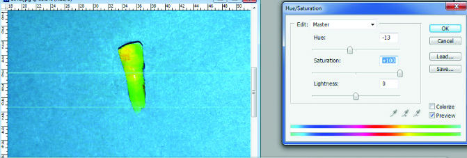
Digital calibration and measurement of the RRL was done using the ADOBE Photoshop CS3 Extended (Version 10.0) software. After importing the photograph of the tooth into the software, the ruler was set and unit of measurement was set to millimeter (mm) scale. The gradations on ABFO scale was used for calibration of the photograph to actual size. After the image calibration, the digital measurement of the RRL was recorded. The RRL was measured in mm as the distance between the trough of the cervical line and the deepest point of resorption on the root surface [Table/Fig-5]. The percentage of RRL was derived using the following formula [9]. The expected pre-resorption root length for primary teeth was obtained from Kramer WS and Ireland RI study [10].
Remaining Root Length (RRL)-Distance between the trough of the cervical line and the deepest point of resorption on the root surface- single rooted teeth.
Statistical Analysis
The data thus obtained was submitted to statistical analysis using SPSS software version 22.0. The following statistical tests were applied. The p-value <0.05 was considered to be statistically significant. Descriptive statistics (Mean and standard deviation) was calculated and Repeated measure ANOVA was used for determining the significance within the groups. The observations were obtained by the same researcher three times on four tooth surfaces. Intra-class correlation analysis using two way mixed models resulted in coefficients ranging between 0.996-0.999 indicating extremely high reliability of observations.
Results
[Table/Fig-6] provides the comparison of root resorption across different surfaces stage wise among incisors. The mean length of resorption for stage 3 was significantly different across different surface with a p-value 0.0015 using repeated measures of ANOVA. The mesial surface of the root showed the least mean length in the stage 3. The mean length of resorption was insignificantly different across different surfaces for stage 1 and stage 2 with a p-value of 0.0532 and 0.0946, respectively.
Comparison of root resorption across different surfaces according to stages-Incisors.
| Incisors | Stage1 (n=12) | Stage 2 (n=12) | Stage 3 (n=18) |
|---|
| Mean (mm) | SD | Mean (mm) | SD | Mean (mm) | SD |
|---|
| Buccal/Labial | 11.01 | 1.61 | 7.54 | 1.21 | 4.43 | 0.77 |
| Distal | 10.44 | 1.85 | 6.50 | 1.13 | 1.51 | 0.69 |
| Lingual | 10.43 | 1.94 | 5.32 | 1.97 | 1.54 | 0.22 |
| Mesial | 10.47 | 1.83 | 5.43 | 1.59 | 1.21 | 0.75 |
| p-value* | 0.0532 | 0.0946 | 0.0015 |
*Obtained using repeated measures of ANOVA; p-value < 0.05 considered significant
[Table/Fig-7] provides the comparison of root resorption across different surfaces stage wise among canines. The mean length of resorption for canines in stage 2 was significantly different across different surface with a p-value 0.0453 using repeated measures of ANOVA. The mean length was least for lingual surface of the root. The mean length of resorption for canines was insignificantly different across different surfaces for stage 1 and stage 3 with a p-value of 0.596 and 0.0872, respectively.
Comparison of root resorption across different surfaces according to stages-canines.
| Canines | Stage 1 (n=12) | Stage 2 (n=12) | Stage 3 (n=12) |
|---|
| Mean (mm) | SD | Mean (mm) | SD | Mean (mm) | SD |
|---|
| Buccal/Labial | 12.12 | 1.29 | 6.54 | 1.67 | 1.96 | 0.02 |
| Distal | 11.59 | 1.25 | 5.90 | 1.18 | 0.98 | 0.08 |
| Lingual | 11.03 | 1.61 | 5.38 | 1.35 | 0.75 | 0.06 |
| Mesial | 11.44 | 1.41 | 5.63 | 1.39 | 1.35 | 0.07 |
| p-value* | 0.596 | 0.0453 | 0.0872 |
*Obtained using repeated measures of ANOVA; p-value <0.05 considered significant
[Table/Fig-8] provides the comparison of root resorption across different surfaces according to stages of maxillary molar. The mean length of resorption for maxillary molar in stage 1 was significantly different across different surface with a p-value 0.0232 using repeated measures of ANOVA. The mean length was the least for the disto-buccal root. The mean length of resorption for maxillary molar was insignificantly different across different surfaces for stage 2 and stage 3 with a p-value of 0.3402 and 0.4626, respectively.
Comparison of root resorption across different surfaces according to stages-Maxillary molar.
| Maxillary molar | Stage 1 (n=10) | Stage 2 (n=10) | Stage 3 (n=10) |
|---|
| Mean (mm) | SD | Mean (mm) | SD | Mean (mm) | SD |
|---|
| Disto buccal | 7.69 | 1.34 | 5.13 | 1.59 | 0.79 | 0.04 |
| Mesio-buccal | 8.88 | 1.44 | 6.39 | 1.57 | 1.12 | 0.05 |
| Palatal | 8.39 | 1.06 | 6.02 | 0.98 | 1.13 | 0.02 |
| p-value* | 0.0232 | 0.3402 | 0.4626 |
*Obtained using repeated measures of ANOVA; p-value <0.05 considered significant
[Table/Fig-9] provides the comparison of root resorption between two different surfaces according to stages of mandibular molar. The mean length of resorption for mandibular molar in stage 1 and stage 2 was significantly different between two surfaces with a p-value 0.0005 and < 0.0001, respectively with distal root showing lesser mean length suggesting greater resorption comparatively. The mean length of resorption for mandibular molar was insignificantly different between both surfaces for stage 3 with a p-value of 0.755.
Comparison of root resorption between two surfaces according to stages-Mandibular molar.
| Mandibular molar | Stage 1 (n=10) | Stage 2 (n=10) | Stage 3 (n=10) |
|---|
| Mean (mm) | SD | Mean (mm) | SD | Mean (mm) | SD |
|---|
| Distal | 7.18 | 0.74 | 5.48 | 0.39 | 1.23 | 0.07 |
| Mesial | 8.17 | 1.06 | 7.85 | 1.35 | 1.10 | 0.09 |
| p-value* | 0.0005 | <0.0001 | 0.755 |
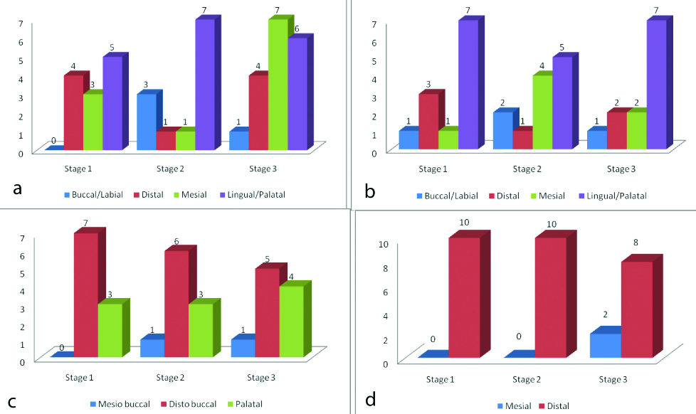
The frequency distribution graphs represented that among incisors [Table/Fig-10a], the lingual surfaces showed maximum resorption in stage 1 and 2 with mesial surface predominating in stage 3. Similarly, among canines [Table/Fig-10b] the lingual surface showed predominantly more resorption among all the stages. [Table/Fig-10c] shows that among the maxillary molars the pattern of resorption indicated that the distobuccal roots undergoes consistently more resorption among all the stages. [Table/Fig-10d] shows that among mandibular molars, consistently among all the stages, distal root shows more resorption when compared to the mesial root.
a) Comparison of number of samples with maximum resorption along different surfaces in various stages of resorption among Incisors; b) Comparison of number of samples with maximum resorption along different surfaces in various stages of resorption among Canines; c) Comparison of number of samples with maximum resorption along different surfaces in various stages of resorption among Maxillary Molars; d) Comparison of number of samples with maximum resorption along different surfaces in various stages of resorption among Mandibular Molars.
Discussion
The present study aimed to assess the pattern, surfaces and extent of physiologic root resorption in primary teeth. The pattern of resorption was assessed irrespective of age and gender. As per the literature, there are various factors which can influence the eruption of teeth such as gender, genetic control, craniofacial morphology, socio-economic status, systemic factors and so on [11]. As these factors influence the eruption of teeth, they may definitely have an influence on the timing of resorption and may not influence the pattern of resorption. Irrespective of the age and gender, the anatomic pattern has been the prime objective of this study hence, the collection of teeth specifically for the study was not undertaken rather extracted teeth were preserved in 10% formalin prior to recording the observations. Also, there is lack of sufficient literature which would suggest that factors such as age and gender may have an influence on the pattern of resorption. Hence, this study aimed to assess only the pattern of resorption irrespective of the age, gender and various other factors. However, this can be considered as the extended scope of the study.
Pattern of resorption was assessed by photographically measuring the RRL from all the aspects of the tooth. This method would accurately depict the pattern of resorption as it would provide the three dimensional assessment of the same when compared to radiographic method which is a two dimensional representation of the ongoing event of resorption. As mentioned previously, the photographs of the teeth were taken with ABFO scale and distance between the trough of the cervical line and the deepest point of resorption on the root surface was measured from buccal, mesial, distal and palatal/lingual aspect. This method would provide the amount of resorption occurring on each aspect, thereby clearly depicting the pattern of resorption.
The assessment of pattern of resorption of the incisors based on the above mentioned method indicates during the stage 1 of root resorption, among the buccal and palatal aspects, the palatal aspect predominantly showed more resorption. Among the proximal surfaces, mesial aspect showed more resorption in the later stages of resorption. As the resorption proceeds, growth of the jaws favours the labial positioning of the permanent successor, thereby leading to involvement of the labial surface in the advanced stages and showing more uniform pattern of resorption [12]. Literature also explains axio-palatal/lingual pattern of resorption of incisors [13]. However, this study describes mesial aspect of the root undergoing resorption along with palatal/lingual aspect in the later stages. Pattern of resorption among canines have not been discussed separately in the literature. The present study describes that the pattern of resorption in canines closely follows that of incisors with palatal/lingual aspect exhibiting more resorption. As the resorption proceeds towards stage 2 and stage 3 the resorption shows more uniformity with proximal surfaces following palatal/lingual pattern of resorption. However, buccal/labial aspect shows less resorption compared to the other surfaces. Comparison with other studies is not possible as literature does not directly assess the pattern of resorption of canines.
In the maxillary molars, the disto-buccal root shows early resorption followed by the palatal root and the mesio-buccal counterpart in sequence. The inner aspects of all the roots were advancing in the resorptive process. The same pattern of resorption continues as root proceeds to stage 2 of resorption. Prove SA et al., describes the pattern of resorption of the maxillary molars with distobuccal aspect resorbing earlier. However, this study does not describe the resorption pattern of palatal aspect along with distobuccal aspect. The difference in the findings may have been as a result of difference in the methodology of studying the resorption pattern as study conducted by Prove SA et al., was based on radiographic findings which would provide assessment based on two dimensional findings [14].
Among the mandibular molars, the distal root seems to be resorbing earlier especially in the lingual aspects followed by the mesial aspect [15]. Some of the cases showed a uniform resorption of both the mesial and distal roots, with the progressive resorptive end in the inner aspect and towards lingual surface. However, as the resorption proceeds to stage 3, the developing permanent successor occupies more apical position thereby bringing about uniformity in the pattern of mesial and distal roots of resorption. However, distal roots show more preponderance of resorption compared to the mesial root. Study conducted by Peretz B et al., reported 55% of samples showing resorption of distal root [16].
Limitation(s)
This study aims to assess the pattern of resorption irrespective of the various other confounding factors such as age and gender. However, the scope of the study can be further extended to correlate the pattern of resorption with that of the age of the subjects. This would further clarify the age wise resorptive pattern and also may be a useful adjunct in age estimation.
Conclusion(s)
Different deciduous teeth follow a different pattern of resorption. Incisors and canines however follow similar pattern with lingual surface resorbing predominantly more than other surfaces. The maxillary molars indicated that the distobuccal and palatal roots resorb earlier than the mesiobuccal root however at stage 3, the pattern of resorption regularises with mesial root resorbing almost at the same rate as distobuccal and palatal. Similarly mandibular molars indicated predominantly distal pattern of resorption.
*Obtained using repeated measures of ANOVA; p-value < 0.05 considered significant
*Obtained using repeated measures of ANOVA; p-value <0.05 considered significant
*Obtained using repeated measures of ANOVA; p-value <0.05 considered significant