Management of Bilateral Dental Agenesis with Aesthetic Rehabilitation by Groper’s Appliance- A Six-year Follow-up
Zohra Jabin1, Pooja Dudeja2, Krishan Dudeja3
1 Associate Professor, Department of Pedodontics and Preventive Dentistry, IDST, Modinagar, Noida, Uttar Pradesh, India.
2 Professor, Department of Conservative Dentistry, ESIC, Rohini, Delhi, India.
3 Professor, Department of Prosthodontics, Krishna Dental College, Noida, Uttar Pradesh, India.
NAME, ADDRESS, E-MAIL ID OF THE CORRESPONDING AUTHOR: Zohra Jabin, D-716, Jalvayu Towers, Sector-47, Noida-201301, Uttar Pradesh, India.
E-mail: drzohrajabin@gmail.com
Tooth agenesis is the most clearly recognised dental anomaly in humans. Tooth agenesis may be single or multiple and may involve single or both arches. However, agenesis of permanent tooth is more commonly seen than primary tooth agenesis. Various studies reported 2.6-11.3% incidence of missing permanent teeth, out of which 0.4-0.9% occurs in the primary dentition and most commonly lateral incisors are involved. Women are usually more affected and the male-to-female ratio is about 2:3. The clinical features evident in children with missing teeth are compromised growth of alveolar ridge, speech discrepancies and reduced lower facial height resulting in deep bite. Missing anterior compromises aesthetics and affects the psychological and social well being of the child. Absence of teeth results in speech defects and unbalanced jaw growth which predisposes the patient to dental malocclusion. Thus, the condition requires careful treatment planning and subsequent management. Limited treatment options are available for the management of this condition due to child’s tender age and co-operation levels. Among these Groper’s appliance presents a promising treatment modality. This appliance has proven to be compliant functionally as well as aesthetically. Agenesis of multiple anterior teeth in both the arches and involving primary as well as permanent dentition is a rare presentation and literature shows paucity of data pertaining to this anomaly. Thus, there is a need for the documentation of such cases in literature. The author reported a case of nonsyndromic bilateral agenesis of primary and permanent interiors in a healthy three-year-old Indian male patient and a brief overview on its clinical implications and management.
Central incisor, Nonsyndromic, Oligodontia, Primary dentition, Sibling, Succedaneous teeth
Case Report
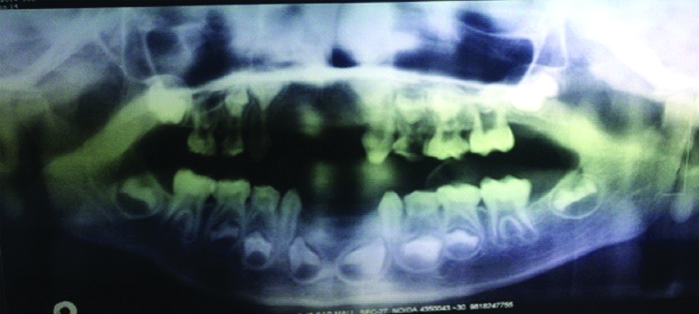
Parents of a three-year-old Indian male patient reported to the Dental Wellness Centre, Noida with a complain of non eruption of upper as well as lower anterior teeth of their child. The patient did not have any difficulty in mastication and speech. The child was born to non-consanguineous parents. The pregnancy and delivery were uneventful. There was no history of any trauma, severe systemic diseases or infections to the anterior region. Family history revealed similar finding in patient’s younger sibling [Table/Fig-1]. Extraoral features like patient height, weight, built and general appearance was normal. Facial profile was bilaterally symmetrical. Patient was well nourished and responded actively to conversation. on examination inntraorally 51,52,61,62,71,72,81 and 82 were found to be missing. Primary canines and molars were present in both arches. The erupted teeth were in normal shape, size and colour. Gingiva, tongue and oral mucosa was normal in colour and appearance. Investigations done were panoramic examination [Table/Fig-2] which reveals congenital agenesis of primary central and lateral incisors in maxillary and mandibular arches. It also showed absence of succedaneous tooth buds of all the missing primary teeth, though only mandibular permanent central incisor tooth buds were evident. Thus, after considering all facts the final diagnosis was Nonsyndromic Dental Agenesis.
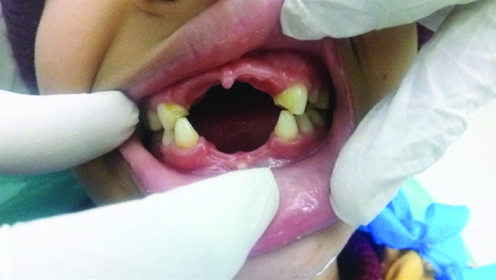
Intraoral view showing multiple primary and permanent tooth agenesis in both arches.
Preoperative radiographic evaluation of the patient. Panoramic radiograph revealing congenital absence of primary maxillary and mandibular incisors along with permanent tooth buds. Presence of permanent central incisor tooth bud evident in mandibular arch.
Treatment Plan
Replacement of primary anteriors was done by placing Groper’s appliance in maxillary as well as mandibular arch. In this appliance primary molars were used to support the appliance through bands. This appliance was chosen owing to its benefits like preserving an open space to prevent arch length loss, restoration of function and aesthetics, allows maxillary growth and prevents improper tongue function. The strength and durability of the appliance is due to its construction each prosthetic tooth is individually attached to a metal pad that is welded to the palatal wire. A written consent form obtained from the patient.
First Appointment
Band adaptation (band size: 0.005″×0.180″) was done on 55, 65. After band adaptation impressions were taken with irreversible hydrocolloid material -Alginate (Dentsply Zelgan).
Lab Procedure
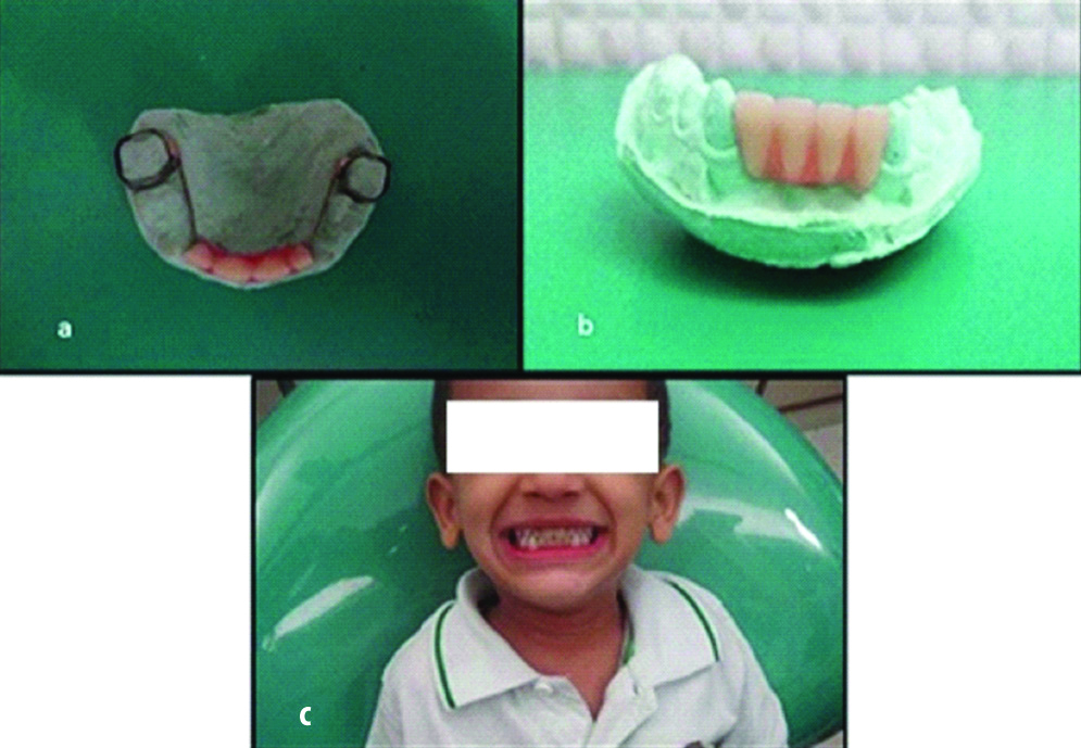
Casts were poured with Type III gypsum product dental stone (Neelkanth). Since the available teeth are too big in size and by trimming it were difficult to achieve the desired morphology, thus composite teeth were custom made by using the mould. These teeth were contained by means of a wire (21 gauge 0.9 mm) bearing gingival coloured acrylic flange. It was soldered to the bands and its anterior part was embedded into acrylic (DPI-RR) covering the vestibule. The final appliance was trimmed, polished and was ready for a try in [Table/Fig-3a-c].
(a) Maxillary appliance on the study model; (b) Mandibular appliance on the study model; (c) Patient with maxillary and mandibular groper’s appliance.
Second Appointment
Wax-up trial was done and occlusion was checked with lower teeth. Later separating media was applied in the cast and then denture base was fabricated using cold cure acrylic. The appliance was seated by means of molar band and checked for accurate fit, any premature contacts and occlusal discrepancies. The banded appliance was then cemented using Glass Ionomer Cement (GC Corporation, Tokyo, Japan). Post-insertion instructions were given to the patient which included proper oral hygiene maintenance specially cleaning of acrylic flange area after every meal. The patient was instructed to visit the clinic every month for follow-up and report immediately in case of breakage of appliance. Patient appliance was re-fabricated several times during the follow-up period of 6 years in order to accommodate the growth changes occurring in the maxilla and mandible. After changing the appliance several times, the decalcification of molars was noticed due to repeated debanding and replacement of bands with the new ones during every change. Decalcification was taken care by application of Acidulated Phosphate Fluoride (APF) gel (Dentbay Pascal) at regular intervals and in order to prevent further damage to permanent molars, the Groper’s appliance was converted into bonded rather than banded.
Design of Bonded Groper’s Appliance
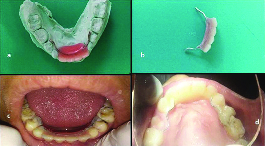
The wire extensions were bonded on the palatal surfaces of canines and first primary molars bilaterally. In the mean course the first primary molar of right side showed mobility and got exfoliated. Thus, the wire extension on this side needed to be extended till second primary molar [Table/Fig-4a-d].
(a) Mandibular bonded Appliance on model; (b) Maxillary bonded Appliance; (c) Mandibular appliance after cementation; (d) Maxillary appliance after cementation.
Follow-Up
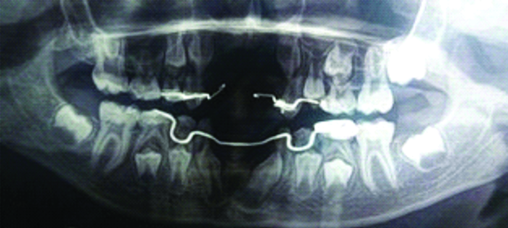
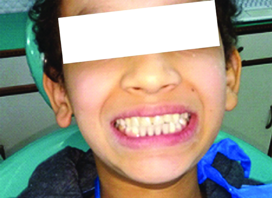
The patient follow-up was done for periodic adjustments and replacements of the appliance to accommodate for the growing arch. The appliances was repaired and reconstructed several times during the 6 year duration as patient reached mixed dentition phase and several teeth erupted and exfoliated. Panoramic radiograph [Table/Fig-5] showing erupting canines and maintained arch length after 6 year follow-up. Patient with maxillary and mandibular appliance after 6 year follow-up is depicted in [Table/Fig-6].
Panoromic view after 6 year follow-up period.
Maxillary-Mandibular appliance in occlusion in patient aged 9 years.
Discussion
Though absence of all teeth is a rare finding but congenital absence of one or more teeth is seen often during dental examination. It can be termed hypodontia when there is absence of 1 to 6 teeth whereas Oligodontia is a condition in which more than six teeth are missing [1]. Tooth agenesis is largely idiopathic and its exact aetiology is unknown. Though, several factors like localised infection or inflammation, tooth bud anomalies, orofacial trauma during odontogenesis, exposure to radiation therapy in the head and neck region and health conditions like metabolic disorders and disturbance of the endocrine system serve as predisposing risk factors [2,3]. Heredity or familial distribution also plays a role. Brooks AH proposed that agenesis or hypodontia arises as a result of combined effects of genetic and environmental factors [4]. Present case showed bilateral absence of central and lateral incisors involving both the dentitions. Venkataraghavan K et al., have reported a similar case of multiple missing primary teeth (n=18) with congenital absence of all primary teeth except the maxillary right and left primary second molars with the presence of all the developing permanent tooth buds showing defective formation of dentin, roots of the permanent molars and lower centrals [5]. Although, thorough history and examination did not indicate presence of any syndrome in this patient neither any associated tooth discrepancies.
In the present case, replacement of teeth was planned considering several factors like perseverance of space, maintenance of aesthetics, prevention from deleterious oral habits and to facilitate the child’s speech development. Timely tooth replacement prevented inappropriate speech compensations and inability to articulate certain speech sounds, since many sounds are made with the tongue touching the lingual side of the maxillary incisors [6]. However, Gable TO et al., found loss of incisors doesn’t result in speech derangement in children who are over 4 years of age [6]. This patient was three-year-old and had no complaints with speech, it was stipulated that timely teeth replacement with groper’s appliance would aid in his speech development at later ages [7]. Patient’s general physical examination was done thoroughly. No hair thinning and sparcity was observed, nails did not show brittleness, there were no signs of glaucoma or any eye ailment and skin was normal in texture and appearance. These findings ruled out presence of ectodermal dysplasia and other related syndromes. Studies suggest that there is no strong evidence that early loss of maxillary incisors will have any significant, long-lasting effect on the growth and development of the child [8].
On the contrary, some opine that agenesis of anterior teeth may affect development of smaller cranial base length and leads to shorter maxilla, prognathic mandible, shorter lower anterior facial height, retroclination of maxillary and mandibular incisors [9]. Out of these, loss of intercanine width is of utmost concern. In the present case, the timely placement of appliance prevented the loss of intercanine width and facilitated its increase with the growing maxilla [Table/Fig-5]. Normal denture was not taken into consideration as a treatment option due to its non-compliance in young patients and its retention issues in children. Absence of anterior teeth in children has a strong influence on the psyche of the children. Kapur A et al., reported that the child’s self esteem can be guarded by aesthetic rehabilitation of the primary dentition [10]. At an age as early as 5 years, children formulate a mental image of their appearance. Being able to maintain this image facilitate their acceptance among their peer group [11]. In the present case aesthetics was the primary concern for the child’s parents and they demanded aesthetic rehabilitation in order to boost self confidence of the child. The patient did not have any complain with mastication and speech. In the Groper’s appliance preformed bands were not used and bands were custom made which provided snug fit and positioning and stability. The wire incorporated was rigid enough to withstand the masticatory load and it was positioned laterally to avoid its interference with the tongue positioning. In the present case, younger sibling of patient also presented with hypodontia but the expression was dissimilar. Kagitha PK et al., reported a case of concordant expression of hypodontia in two girl siblings aged 11 and 13 years [12].
Jasmin JR and Groper JN, were the first ones to document groper’s appliance. Their design lacked the acrylic flange and teeth were directly attached to metal cleats that were soldered to the palatal wire bar [13]. Similarly, Shanmugaavel AK et al., fabricated grasce appliance using the palatal wire to attach the teeth directly [14]. They placed stainless steel crowns on molars instead of molar bands. In the present case, acrylic flange was used as it is able to stabilise the teeth better and also prevents development of gap between teeth and alveolus due to improper fit. In this case, molar bands were preferred as it is less cumbersome to remove and replace them for periodic modifications in the appliance, to accommodate growth changes than stainless steel crown. Moreover, the patient had healthy vital molars and it was preferred to avoid unnecessary reduction and crown placement in them. In the present case, successful replacement of teeth ensured the stability of alveolar bone volume due to physiological stimulation of the periodontal ligament. Implant treatment is postponed until the completion of jaw growth in adolescence.
Conclusion(s)
Absence of tooth at younger age may result in psychological trauma to the child. The restoration of anterior aesthetics with this appliance gave an essential psychological boost to the child and his parents. Groper’s appliance enhanced aesthetics, assisted in proper speech development and restored normal functioning; Above all, it provided self-confidence to the child and improved his social wellbeing.
Author Declaration:
Financial or Other Competing Interests: None
Was informed consent obtained from the subjects involved in the study? Yes
For any images presented appropriate consent has been obtained from the subjects. Yes
Plagiarism Checking Methods: [Jain H et al.]
Plagiarism X-checker: Feb 13, 2020
Manual Googling: Apr 20, 2020
iThenticate Software: May 09, 2020 (10%)
[1]. Tavajohi-Kermani H, Kapur R, Sciote JJ, Tooth agenesis and craniofacial morphology in an orthodontic populationAm J Orthod Dentofacial Orthop 2002 122:39-47.10.1067/mod.2002.12394812142896 [Google Scholar] [CrossRef] [PubMed]
[2]. Galluccio G, Castellano M, La Monaca C, Genetic basis of nonsyndromic anomalies of human tooth numberArch Oral Biol 2012 57(7):918-30.10.1016/j.archoralbio.2012.01.00522325622 [Google Scholar] [CrossRef] [PubMed]
[3]. De Coster PJ, Marks LA, Martens LC, Huysseune A, Dental agenesis: Genetic and clinical perspectivesJ Oral Pathol Med 2009 38(1):01-17.10.1111/j.1600-0714.2008.00699.x18771513 [Google Scholar] [CrossRef] [PubMed]
[4]. Brooks AH, A unifying aetiological explanation or anomalies of human tooth number and sizeArch Oral Biol 1984 29:373-78.10.1016/0003-9969(84)90163-8 [Google Scholar] [CrossRef]
[5]. Venkataraghavan K, Anantharaj A, Prasanna P, Sudhir R, Oligodontia in the primary dentition: Report of a caseJ Dent Child (Chic) 2007 74:154-56. [Google Scholar]
[6]. Gable TO, Kummer AW, Lee L, Creaghead NA, Moore LJ, Premature loss of the primary maxilllary incisors: Effects on speech productionJ Dent Child 1995 62:173-79. [Google Scholar]
[7]. Waggoner WF, Kupietzky A, Anterior esthetic fixed appliances for the preschooler: Considerations and a technique for placementPediatr Dent 2001 23:147-50. [Google Scholar]
[8]. Kirzioglu Z, Erturk MS, Success of reinforced fiber material space maintainersJ Dent Child 2004 71:158-62. [Google Scholar]
[9]. Endo T, Yoshino S, Ozoe R, Kojima K, Shimooka S, Association of advanced hypodontia and craniofacial morphology in Japanese orthodontic patientsOdontology 2004 92:48-53.10.1007/s10266-004-0034-515490305 [Google Scholar] [CrossRef] [PubMed]
[10]. Kapur A, Chawla HS, Goyal A, Gaube K, An esthetic point of view in very young childrenJ Clin Pediatr Dent 2005 30(2):99-103.10.17796/jcpd.30.2.360k2j445277341816491961 [Google Scholar] [CrossRef] [PubMed]
[11]. Wilson CFG, Management of trauma to primary and developing teethDent Clin N Am 1995 39:133-67. [Google Scholar]
[12]. Kagitha PK, Namineni S, Tupalli A, Challa S, Agenesis of permanent mandibular central incisors: A concordant condition in siblingsInt J Clin Pediatr Dent 2016 9(1):74-77.10.5005/jp-journals-10005-133727274160 [Google Scholar] [CrossRef] [PubMed]
[13]. Jasmin JR, Groper JN, Fabrication of a more durable fixed anterior esthetic applianceJ Dent Child 1984 51:124-27. [Google Scholar]
[14]. Shanmugaavel AK, Deepa G, Lavanya S, Smile reconstruction for the preschoolers using GRASCE appliance -Two case reportsClin Diagn Res 2016 10(8):19-22.10.7860/JCDR/2016/19817.825127656579 [Google Scholar] [CrossRef] [PubMed]