JCDR - Register at Journal of Clinical and Diagnostic Research
Journal of Clinical and Diagnostic Research, ISSN - 0973 - 709X
Microbiology Section DOI : 10.7860/JCDR/2020/43642.13703
Year : 2020 | Month : May | Volume : 14 | Issue : 05 Full Version Page : DC04 - DC07

Comparison of Manual and Automated Method for Speciation and Antifungal Susceptibility of Candida Species Causing Blood Stream Infection in Critically ill Patients

Swati Jain1, Bichitrananda Swain2, Sunita Kabi3

1 Assistant Professor, Department of Microbiology, IMS and SUM Hospital, S"O"A University, Bhubaneswar, Odisha, India.
2 Associate Professor, Department of Microbiology, SLN Medical College and Hospital, Koraput, Odisha, India.
3 Assistant Professor, Department of Microbiology, IMS and SUM Hospital, S"O"A University, Bhubaneswar, Odisha, India.


NAME, ADDRESS, E-MAIL ID OF THE CORRESPONDING AUTHOR: Dr. Sunita Kabi, 144, Sri Satyasai Enclave, Kolathia, Bhubaneswar-751020, Odisha, India.
E-mail: sunitakabi@gmail.com
Abstract

Introduction

Globally, the incidence Blood Stream Infection (BSI) by Candida species is increasing. It is associated with high mortality rates especially in immunocompromised critically ill patients. The epidemiology has been shifted from Candida species to Non-albicans Candida (NAC) in recent decades. Speciation of Candida spp. can help to improve the outcome of patients as few Candida species are intrinsically resistant to antifungal agents.

Aim

To identify the spectrum of Candida species causing BSI, study their sensitivity pattern to antifungal agents and comparison of conventional (germ tube test, Dalmau plate culture and Chromagar) and automated (Vitek-2) methods for the same.

Materials and Methods

Blood specimens of the clinically suspected cases of septicaemic patients were cultured by BacT/ALERT before administration of antibiotics or antifungals during the study period of one and half year. The isolated Candida were identified to the species level along with antifungal susceptibility testing done by both conventional and automated (Vitek-2) methods. Statistical analysis was done using Chi-square test.

Results

Of the total 100 Candida isolated, the most common Candida species was C.parapsilosis (26%), followed by C.tropicalis (23%), C.albicans (21%) and C.auris (15%). Total concordance between manual and Vitek-2 identification was 74%. Significant risk factors involved in patients with candidemia were malignancy, diabetes mellitus and chronic kidney disease. Antifungal susceptibility rate for amphotericin B, caspofungin, Fluconazole, Flucytosine, Micafungin and Voriconazole was 94%, 90.5%, 73.8%, 96.4%, 98.8% and 95.2%, respectively. Mortality rate was 45% due to sepsis and associated complications.

Conclusion

Early isolation, speciation and antifungal susceptibility is the key for management of candidemia cases to get the better outcome as C.auris, the important multidrug resistant variant has caused a major epidemiologic shift in candidemia. Molecular studies are required for accurate speciation of Candida species like C. auris, C. haemulonii, C. famata, etc.

Keywords

Introduction

A global increase in incidence of Candida infection has been reported by several authors over the past two decades showing remarkable shift in prevalence of different Candida species [1]. BSI by Candida, also known as candidemia, is defined as the isolation of Candida species from at least one blood culture in patients with symptoms or signs of a systemic infection [2]. Invasive candidiasis includes a spectrum of clinical conditions, the most frequent of which is candidemia, has become a remarkable threat to public health, a life-threatening infection which is associated with morbidity and mortality to be as high as 40% [3]. It represents 10% of nosocomial infections in hospitalised patients [4] and is the fourth most common blood stream pathogen [5]. The incidence of candidemia expressed as cases per 100,000 inhabitants has been reported to range from 1-8 cases [6] and the prevalence was found to be 6.9 per 1000 cases according to a survey of Intensive Care Units (ICUs) worldwide [7]. Risk factors associated with candidemia are premature birth, advanced age, long term hospitalisation, prolonged exposure to antimicrobial drugs, invasive procedures such as using intravascular catheters, multiple interventions and the immunosuppressive conditions like diabetes, HIV and malignancy [8]. Although Candidaalbicans continues to be the predominant species to cause candidemia, recent studies have detected a growing proportion of BSI by NAC species like C. tropicalis, C. parapsilosis, C. glabrata, C. krusei [9]. Ninety percent of all Candida BSI globally are attributed by these species [10]. Importantly, many NAC spp. Like C. krusei and C. glabrata are shown to have intrinsic resistance against Fluconazole [7]. There has been a recent epidemiologic shift in candidemia driven by C.auris, a novel Candida spp., which is a multidrug-resistant pathogen causing protracted healthcare-associated outbreaks [8,11]. It is now essential to do the species-level identification as antifungal agent can be selected by the clinicians for the better patient care [12,13]. Continuous surveillance is needed to know the incidence, species distribution and antifungal drug susceptibility profile of Candida causing BSI.

The present study was designed with an aim to identify the spectrum of Candida species causing BSI, study their sensitivity pattern to antifungal agents and comparison of conventional (germ tube, dalmau plate culture and CHROMagar) and automated (Vitek-2) methods for the same.

Materials and Methods

This hospital based prospective study was carried out in the Department of Microbiology, IMS & SUM Hospital, Bhubaneswar, Odisha, India over a period of one and half year (January 2018-June 2019) after obtaining ethical clearance from institutional ethical committee (Ref No/DMR/IMS-SH/SOA/16075). After taking informed consent, the blood samples from 8156 ICU patients, clinically suspected to have septicaemia, were collected following all the aseptic precautions and before administration of antibiotics or antifungals and were screened for candidemia. All the Candida isolates obtained as a single pathogen from blood cultures were included in the study. Patients other than those of ICU admitted and also those who were under antibiotics or antifungals even from ICU admission status were excluded from this study.

A total of 8156 specimens were inoculated into the BacT/ALERT (BioMerieux) blood culture bottles and incubated at 37°C for a maximum period of seven days in BacT/ALERT 3D system, a fully automated blood culture system for detection of aerobic growth in blood samples and if there was no growth, the result was read as negative. From culture bottles which were flagged positive in the system, Gram stain was performed. If yeast cells were observed on staining, blood was subcultured on two Sabouraud’s Dextrose Agar (SDA) and blood agar plates and incubated at 37°C. SDA and blood agar plates were examined for white to cream coloured, smooth and pasty colonies after 24-48 hours of incubation [14].

Gram staining was performed from the colony and the morphology of yeast cells was noted. All the isolates were processed by both conventional and automated methods in parallel.

Colonies from the SDA were then plated onto cornmeal agar with Tween 80 (Dalmau plate culture) for chlamydospores production [15]. Germ tube test was performed from colonies for presumptive identification of C. albicans [16].

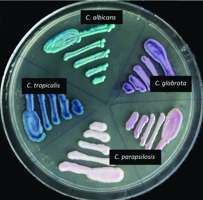
Colonies from SDA were plated onto CHROM agar (HiCrome Candida differential agar- Himedia), a differential culture medium that is claimed to facilitate the isolation by colorimetric presumptive identification [17] and were incubated at 37°C for 48 hours. The results were interpreted according to the colour of the colony formed and as per manufacturer’s guidelines [Table/Fig-1,2]. All the isolates that gave doubtful morphology or not identified by conventional methods were taken as “Candida spp.”

Appearance of various Candida species on CHROMagar.

Candida speciesMorphology on CHROM agar
C. albicansApple green
C. dubliniensisDark green
C. tropicalisMetallic blue with a pink halo
C. parapsilosisWhite to pale pink
C. glabrataPale pink to violet
C. kruseiFuzzy pink coloured colonies with matt surface and white edges

Colour production of various Candida species on CHROMagar.

In parallel to conventional methods listed above, for all the isolates identification and antifungal susceptibility testing were also done by Vitek-2 (BioMerieux) with 0.5% McFarland suspension from the colonies from SDA using the Identification Card-Yeast (ID-YST) and Antifungal Susceptibility Testing card for yeast (AST YS01) as per Clinical and Laboratory Standards Institute (CLSI) guidelines and manufacturer’s instructions [12]. Finally, the results of the two methods (conventional and Vitek-2) were compared with each other. Candida albicans ATCC 90028 strain was used as control for evaluation of various methods [18].

Statistical Analysis

It was done as per Chi-square test to compare the conventional and the automated method.

Results

A total of 100 non-duplicate isolates of Candida species were obtained from 2202 positive blood culture cases as per inclusion criteria during the study period.

Male patients (55/100, 55%) predominated the female patients (45/100, 45%) in terms of isolation frequency. Maximum number of candidemia patients were in the elderly age group (>60 years, mean age- 71), i.e., 43%, followed by 41-60 years (mean age- 51.3) (33%); 14% and 10% patients were seen in the age groups of 21-40 years (mean age- 32) and up to 20 years (mean age- 8.4), respectively.

Overall 60% of cases had associated co-morbid conditions like malignancy (29/100), kidney disease (40/100), indwelling vascular catheters (47/100), surgical interventions (26/100) and diabetes mellitus (52/100), etc.

Candida parapsilosis (26/100, 26%) was the predominant species causing candidemia followed by Candida tropicalis (23/100, 23%), Candida albicans (21/100, 21%), Candida auris (15/100, 15%), Candida glabrata (5/100, 5%) and others (10/100, 10%) [Table/Fig-3].

Comparison of Candida spp. identification by conventional and automated methods.

Manual identificationVitek 2 identification
C. albicansC. tropicalisC. parapsilosisC. glabrataC. aurisC. guillermondiC. utilisC. famataC. pelliculosaC. haemuloniiC. rugosaTotal
C. albicans21000000000021
C. tropicalis02300000000023
C. parapsilosis00261800000035
C. glabrata000400000004
Candida spp.0000723112117
Total212326515231121100

NB-concordance rate between two methods- 74/100


For speciation, of the total 100 Candida isolates concordance between conventional and Vitek-2 method was 74 (74%). Of the total C.parapsilosis identified by conventional method, 1 isolate as C. glabrata and 8 isolates as C. auris was established by Vitek-2 method. 17 isolates of Candida which could not be identified by conventional chromogenic method were interpreted as Candida spp. but final identification was considered as per Vitek-2 results. Vitek-2 is the better method (p<0.001) than the conventional method for Candida species identification.

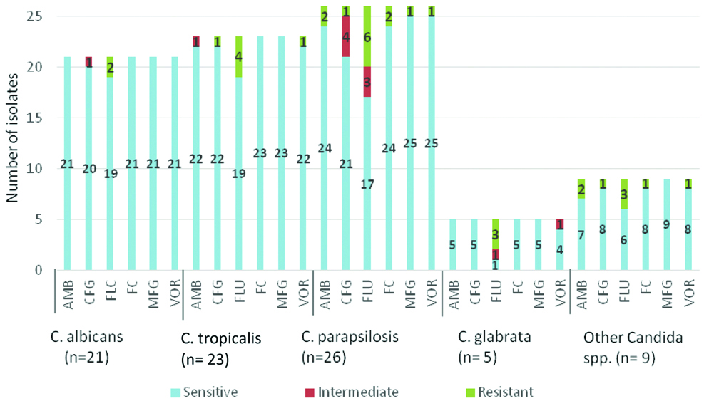
Vitek-2 Compact system does not give Antifungal susceptibility test results for C.auris and C.famata, so Antifungal susceptibility rates were calculated for 84 isolates (n=84). Antifungal susceptibility rate for Amphotericin B, Caspofungin, Fluconazole, Flucytosine, Micafungin and Voriconazole was 94%, 90.5%, 73.8%, 96.4%, 98.8% and 95.2%, respectively [Table/Fig-4].

Antifungal susceptibility testing rate of various antifungals.

AntifungalsSensitivePercentage
Amphotericin B79/8494%
Caspofungin76/8490.5%
Fluconazole62/8473.8%
Flucytosine81/8496.4%
Micafungin83/8498.8%
Voriconazole80/8495.2%

Different Candida species also showed variable sensitivity result to different antifungal drugs used [Table/Fig-5].

Antifungal susceptibility testing result for different Candida spp.

Mortality rate was found to be 45% due to sepsis and associated complications, 11% of patients were lost for follow-up and rest of the cases were treated successfully.

Discussion

Candidemia is an emerging problem in healthcare settings, particularly among those in ICU’s. Early isolation, speciation and antifungal susceptibility is essential for the choice of the best therapeutic approach for the patient, thereby, decreasing morbidity and mortality. Therefore, knowledge of epidemiology of candidemia can help to salvage patients. In this study, an overview of candidemia was obtained, including Candida species identification, antifungal susceptibility pattern, epidemiological characteristics and patient outcome.

In this study, candidemia was most commonly seen in patients >60 years of age (43%) which is in concordance with other studies [19,20]. On the contrary, a study by Thomas T and Dias M shows a higher preponderance in middle age groups (18-50 years) [9]. This was possibly because present study mainly focused on the critically ill patients in ICU’s. In this study, the common risk factors for candidemia were malignancy, diabetes mellitus and chronic kidney disease which is similar to a study by Thomas T and Dias M [9].

The isolation rate of candidemia in the present study was 4.54% which is in concordance with many other studies from India [Table/Fig-6] [5,14,18,21-23].

Studies showing incidence of candidemia from different parts of India [5,14,18,21-23].

AuthorYearAreaCandidemia rate
Verma AK et al., [5]2003North India1.61%
Giri S et al., [18]2013South India5.76%
Chander J et al., [21]2013North India5.79%
Sudan SS et al., [22]2016J&K4.48
Bhattacharjee P [23]2016Kolkata4.03%
Gandham NR et al., [14]2016Pune14.8%

In the present global scenario, there is a changing trend of Candida species, with predominance of NAC spp. In the present study C. parapsilosis (26%) was isolated as the predominant isolate which is similar to a study by Van Schalkwyk E et al., [8], while some other studies had C. tropicalis [9,24] or C. albicans [2] as the predominant pathogen.

C. auris has surpassed the number of cases caused by C. albicans or NAC for the last 10 years and is a major cause of candidemia. In this study also, it was the fourth most common isolated species comprising 15% of candidemia cases. It was identified by Vtek-2 method though which has certain limitation in identification. C.auris identification is confirmed if the initial identification of C.auris is made and C.auris identification is possible if the initial identification of C.haemulonii, C.duobushaemulonii, C.famata or C.lusitaniae is made [25]. There have been reports of C. famata being misdiagnosed by Vitek-2 [26]. So, the ideal method of identification of such species is by Matrix Assisted Laser Desorption Ionisation-Time Of Flight Mass Spectrometry (MALDI-TOFMS). This multidrug resistant pathogen is known to cause protracted healthcare associated outbreaks. This possibly happens due to its ability to persist on surfaces, form biofilms and resist routinely used environmental cleansing agents. Similarly, in this study, C.auris was associated with micro-outbreaks. Presently, disinfectants having sporicidal activity or hydrogen peroxide- based products has been recommended to clean the surfaces in rooms of patients infected or colonised with C.auris because of its high efficiency in eliminating this species from inert surfaces [27,28].

Candida species differ in their susceptibility to antifungal agents. For instance, C.glabrata is not very sensitive to Fluconazole, C.krusei is intrinsically resistance to Fluconazole and C.lusitaniae is resistant to amphotericin B [29]. In this study, all antifungal agents except Fluconazole demonstrated excellent activity against all Candida species. Fluconazole being a cheaper drug has been used as main drug against Candida infections. So, the use of this drug should be as per susceptibility result. Similar findings were published in a study from Southern India [30].

Death rate by candidemia is quite high. According to some studies, invasive infection with Candida is associated with mortality rates of 35-80% in ICU settings [31-33]. The mortality rate of patients with candidemia in this study was 45% which is well within the range reported by other studies and even more concern is about auris which showed 100% mortality.

Candida speciation by conventional methods has long been used as benchmark identification procedures. However, these methods are time consuming, laborious and not reliable in identifying broad spectrum of species and require additional tests like assimilation and fermentation. Several commercial systems were developed which can produce rapid yeast identification and their antifungal susceptibility in at least 15 hour. Their use is limited because these systems are not reliable for identification of some species and some are also misidentified. So, now-a-days, molecular strategies, PCR or non-PCR based methods, are being used as complementary to conventional methods for providing more accurate results in less time.

Limitation(s)

Limitation of present study is that we have not done fermentation and assimilation tests which are the crucial means of species identification by conventional method, also considered as reference method. Although Vitek-2 is a reliable method for identification of common fungal species, for accurate speciation of C.auris, C.guilliermondii, C.haemulonii, C.famata, etc., molecular studies like nucleic acid sequencing may be required as researchers are now claiming Vitek-2 as the inaccurate method for identification of such species.

Conclusion(s)

Even though CHROMagar helps with identification at a lower cost as compared to Vitek-2, which is useful in countries having low resources, but it takes longer duration for complete identification, which is the main drawback of this method. Vitek-2 is considered as a reliable technique for antifungal susceptibility of yeast species, it also has the added advantage of being more rapid and easier than the alternative procedure developed by CLSI, broth microdilution method which is cumbersome and expensive. So, a fast and accurate technique for yeast identification is very important for microbiological laboratories. Accordingly, Vitek-2 can be applied for early identification and antifungal susceptibility testing. Local epidemiological data and antifungal susceptibility profile should be taken into consideration when establishing antifungal treatment strategies. Infection control measures like hand and personal hygiene by healthcare workers, proper catheter care, frequent clinical examination of patients who are weaned off invasive device to be practiced to reduce nosocomial transmission as candidemia represents 10% of nosocomial infections in hospitalised patients and also antibiotic stewardship must be emphasised.

NB-concordance rate between two methods- 74/100

References

[1]Antinori S, Milazzo L, Sollima S, Galli M, Corbellino M, Candidemia and invasive candidiasis in adults: A narrative review Europ J Int Med 2016 34:21-28.10.1016/j.ejim.2016.06.02927394927  [Google Scholar]  [CrossRef]  [PubMed]

[2]Xiao Z, Wang Q, Zhu F, An Y, Epidemiology, species distribution, antifungal susceptibility and mortality risk factors of candidemia among critically ill patients: Aretrospective study from 2011 to 2017 in a teaching hospital in China Antimicrob Resist Infect Control 2019 8:8910.1186/s13756-019-0534-231161036  [Google Scholar]  [CrossRef]  [PubMed]

[3]Wisplinghoff H, Bischoff T, Tallent SM, Seifert H, Wenzel RP, Edmond MB, Nosocomial bloodstream infections in US hospitals: Analysis of 24,179 cases from a prospective nationwide surveillance study Clin Infect Dis Off Publ Infect Dis Soc Am 2004 39(3):309-17.10.1086/42194615306996  [Google Scholar]  [CrossRef]  [PubMed]

[4]Ghrenassia E, Mokart D, Mayaux J, Demoule A, Rezine I, Kerhuel L, Candidemia in critically ill immunocompromised patients: Report of a retrospective multicenter cohort study Ann Intensive Care 2019 9:6210.1186/s13613-019-0539-231161475  [Google Scholar]  [CrossRef]  [PubMed]

[5]Verma AK, Prasad KN, Singh M, Dixit AK, Ayyagari A, Candidemia in patients of a tertiary health care hospital from North India Ind J Med Res 2003 177:122-28.  [Google Scholar]

[6]Neofytos D, Lu K, Hatfield-Seung A, Blackford A, Marr KA, Treadway S, Epidemiology, outcomes, and risk factors of invasive fungal infections in adult patients with acute myelogenous leukemia after induction chemotherapy Diagn Microbiol Infect Dis 2013 75(2):144-49.10.1016/j.diagmicrobio.2012.10.00123142166  [Google Scholar]  [CrossRef]  [PubMed]

[7]Kett DH, Azoulay E, Echeverria PM, Vincent JL, Extended prevalence of infection in ICU study (EPIC II) group of investigators. Candida bloodstream infections in intensive care units: Analysis of the extended prevalence of infection in intensive care unit study Crit. Care Med 2011 39(4):665-70.10.1097/CCM.0b013e318206c1ca21169817  [Google Scholar]  [CrossRef]  [PubMed]

[8]Van Schalkwyk E, Mpembe RS, Thomas J, Shuping L, Ismail H, Lowman W, Epidemiologic shift in candidemia driven by Candida auris, South Africa, 2016-2017 Emerg Infect Dis 2019 25(9):1698-707.10.3201/eid2509.19004031441749  [Google Scholar]  [CrossRef]  [PubMed]

[9]Thomas T, Dias M, Epidemiological profile of Candida isolated from septicaemic patients Indian J Microbiol Res 2018 5(4):508-11.10.18231/2394-5478.2018.0103  [Google Scholar]  [CrossRef]

[10]Giri S, Kindo AJ, A review of Candida species causing blood stream infection Indian J Med Microbiol 2012 30(3):270-78.10.4103/0255-0857.9948422885191  [Google Scholar]  [CrossRef]  [PubMed]

[11]Ruiz-Gaitan A, Moret AM, Tasias-Pitarch M, Aleixandre-Lopez AI, Martinez-Morel H, Calabuig E, An outbreak due to Candida auris with prolonged colonisation and candidaemia in a tertiary care European hospital Mycoses 2018 61:498-505.10.1111/myc.1278129655180  [Google Scholar]  [CrossRef]  [PubMed]

[12]Pahwa N, Kumar R, Nirkhiwale S, Bandi A, Species distribution and drug susceptibility of Candida in clinical isolates from a tertiary care centre at Indore Indian J Med Microbiol 2014 32(1):44-48.10.4103/0255-0857.12430024399387  [Google Scholar]  [CrossRef]  [PubMed]

[13]Souza MN, Ortiz SO, Mello MM, Oliveira FM, Severo LC, Goebel CS, Comparison between four usual methods of identification of Candida species Rev Inst Med Trop Sao Paulo 2015 57(4):281-87.10.1590/S0036-4665201500040000226422150  [Google Scholar]  [CrossRef]  [PubMed]

[14]Gandham NR, Vyawahare CR, Jadhav SV, Misra RN, Candidemia: Speciation and antifungal susceptibility testing from a tertiary care hospital in Maharashtra, India Med J DY Patil Univ 2016 9:596-99.10.4103/0975-2870.192165  [Google Scholar]  [CrossRef]

[15]Kurtzman CP, Fell JW, Boekhout T, Robert V, Methods for Isolation, Phenotypic Characterization and Maintenance of Yeasts. In: Kurtzman, C.P., Fell, J.W. and Boekhout, T., Eds The Yeasts 2011 5th EditionElsevierAmsterdam:87-110.10.1016/B978-0-444-52149-1.00007-0  [Google Scholar]  [CrossRef]

[16]Taschdjian CL, Burchall JJ, Kozinn PJ, Rapid identification of Candida albicans by filamentation on serum and serum substitutes AMA Am J Dis Child 1960 99:212-15.10.1001/archpedi.1960.0207003021401113836995  [Google Scholar]  [CrossRef]  [PubMed]

[17]Vijaya D, Harsha TR, Nagaratamma T, Candida speciation using Chrom agar J Clin Diag Res 2011 5(4):755-58.  [Google Scholar]

[18]Giri S, Kindo AJ, Kalyani J, Candidemia in intensive care unit patients: A one year study from a tertiary care center in South India J Postgrad Med 2013 59(3):190-95.10.4103/0022-3859.11803624029196  [Google Scholar]  [CrossRef]  [PubMed]

[19]Shivaprakasha S, Radhakrishnan K, Karim PMS, Candida spp. other than Candida albicans: A major cause of fungaemia in a tertiary care centre Indian J Med Microbiol 2007 25(4):405-07.10.4103/0255-0857.3735018087096  [Google Scholar]  [CrossRef]  [PubMed]

[20]Dewan E, Biswas D, Kakati B, Verma S, Kotwal A, Oberoi A, Epidemiological and mycological characteristics of candidemia in patients with hematological malignancies attending a tertiary-care center in India Hematol Oncol Stem Cell Therapy 2015 8(3):99-105.10.1016/j.hemonc.2015.06.00626173033  [Google Scholar]  [CrossRef]  [PubMed]

[21]Chander J, Singla N, Sidhu SK, Gombar S, Epidemiology of Candida blood stream infections: Experience of a tertiary care centre in North india J Infect Dev Ctries 2013 7(9):670-75.10.3855/jidc.262324042103  [Google Scholar]  [CrossRef]  [PubMed]

[22]Sudan SS, Sharma P, Sharma M, Sambyal SS, Candidemia in a tertiary care hospital in Jammu (J&K)- A comparison of conventional methods and chromagar techniques for speciation IJSR 2016 5(8):451-54.  [Google Scholar]

[23]Bhattacharjee P, Epidemiology and antifungal susceptibility of Candida species in a tertiary care hospital, Kolkata, India Curr Med Mycol 2016 2(2):20-27.10.18869/acadpub.cmm.2.2.528681016  [Google Scholar]  [CrossRef]  [PubMed]

[24]Jose LR, Savio J, Candidemia isolates, identification and antifungal susceptibility testing: A study from a tertiary care centre Indian J Microbiol Res 2017 4(4):464-67.  [Google Scholar]

[25]Algorithm to identify Candida auris based on phenotypic laboratory method and initial species identification. National center for emerging and zoonotic infectious diseases. CDC 2019. https://www.cdc.gov/fungal/diseases/candidiasis/pdf/Testing-algorithm-by-Method-temp.pdf  [Google Scholar]

[26]Rajkumari N, Mathur P, Xess I, Misra MC, Distribution of different yeasts isolates among trauma patients andcomparison of accuracy in identification of yeasts by automated method versus conventional methods for better use in low rei countries Indian J Med Microbiol 2014 32(4):391-97.10.4103/0255-0857.14224325297023  [Google Scholar]  [CrossRef]  [PubMed]

[27]Cadnum JL, Shaikh AA, Piedrahita CT, Sankar T, Jencson AL, Larkin EL, Effectiveness of disinfectants against Candida auris and other Candida species Infect Control Hosp Epidemiol 2017 38(10):1240-43.10.1017/ice.2017.16228793937  [Google Scholar]  [CrossRef]  [PubMed]

[28]Biswal M, Rudramurthy SM, Jain N, Shamanth AS, Sharma D, Jain K, Controlling a possible outbreak of Candida auris infection: Lessons learnt from multiple interventions J Hosp Infect 2017 97(4):363-70.10.1016/j.jhin.2017.09.00928939316  [Google Scholar]  [CrossRef]  [PubMed]

[29]Ruiz L, Sugizaki M, Montelli A, Matsumoto F, Pires M, da Silva B, Fungemia by yeasts in Brazil: Occurrence and phenotypic study of strains isolated at the Public Hospital, Botucatu, São Paulo J Mycol Med 2005 15(1):13-21.10.1016/j.mycmed.2004.12.002  [Google Scholar]  [CrossRef]

[30]Adhikary R, Joshi S, Species distribution and antifungal susceptibility of candidemia at a multi super specialty centre in Southern India Indian J Med Microbiol 2011 29(3):309-11.10.4103/0255-0857.8392021860117  [Google Scholar]  [CrossRef]  [PubMed]

[31]Mean M, Marchetti O, Calandra T, Bench-to-bedside review: Candida infections in the intensive care unit Crit Care 2008 12:20410.1186/cc621218279532  [Google Scholar]  [CrossRef]  [PubMed]

[32]Falagas ME, Roussos N, Vardakas KZ, Relative frequency of albicans and the various non-albicans Candida spp among candidemia isolates from inpatients in various parts of the world: A systematic review Int J Infect Dis 2010 14(11):954-66.10.1016/j.ijid.2010.04.00620797887  [Google Scholar]  [CrossRef]  [PubMed]

[33]Horn DL, Neofytos D, Anaissie EJ, Fishman JA, Steinbach WJ, Olyaei AJ, Epidemiology and outcomes of candidemia in 2019 patients: Data from the prospective antifungal therapy alliance registry Clin Infect Dis 2009 48(12):1695-703.10.1086/59903919441981  [Google Scholar]  [CrossRef]  [PubMed]