Threatened abortion is defined as pregnancy with gestational age <20 weeks with closed cervical os, having complaints of pain in abdomen and vaginal bleeding or spotting [1]. A 5-25% of women with threatened abortion carries the risk of spontaneous abortion [2], and these women are 2.6 times more prone to abort in comparison to healthy pregnancy [3].
Progesterone has a pivotal role in the maintenance of pregnancy. Estimation of serum progesterone level in the early gestation can be of immense help for predicting the prognosis of patients with threatened abortion.
The non-invasive investigation of the maternal and fetal circulation has been possible because of the discovery of Colour Doppler USG [4]. In the 1st trimester, colour Doppler USG of the uterine arteries will not have any issues for safety of the fetus as the fetus or embryo does not lie in the range of colour Doppler USG beam [5]. Therapeutic value of progesterone in threatened abortion is still a matter of argument. Also, different studies have shown wide variation in cutoff values for serum progesterone in early pregnancy from 9.9 ng/mL to 32.7 ng/mL [6,7]. This study was done to evaluate the hypothesis that early pregnancy failure and adverse pregnancy outcome is associated with unusual patterns of blood flow in the feto-maternal circulation. Also, an attempt was made to determine the levels of serum progesterone in threatened abortion patients who aborted and also those whose pregnancy continued up to period of viability (28 weeks). Progesterone is considered as essential for sustenance of pregnancy, but progesterone levels show wide variations in early pregnancy, Hence in addition to determination of serum progesterone levels in early pregnancy, this study aimed to analyse blood flow through uterine artery by transvaginal colour Doppler in women presenting with signs and symptoms of threatened abortion in early pregnancy.
Materials and Methods
It was a cross-sectional study conducted in the Department of Obstetrics and Gynaecology, Acharya VinobaBhave Rural Hospital, JNMC, Sawangi (Meghe) Wardha, Maharashtra. Study duration was 2 years from September 2017 to August 2019. Study population were all pregnant women reporting with signs and symptoms of threatened abortion in early pregnancy (6-12 weeks). Ethical clearance was taken from Institutional Ethics Committee of Datta Meghe Institute of Medical Sciences (DMIMS) Sawangi, (Meghe), Wardha. (Ref No. DMIMS (DU)/IEC/ 2017-18/6666). All patients included in study were explained about the methodology and purpose of the study. Informed valid written consent in local language was taken from all patients included in this study.
Total population (N=120) patients of threatened abortions attending in AVBRH, Sawangi, Meghe, Wardha during period of two years were included in the study.
Estimated sample size for the study was 101 cases based on standard formula.
χ2=Chi-square value for 1 degrees at some desired probability level. This is 3.84 at 5% level of significance.
P=50% proportion; Q=100-p=50; C=Confidence interval of the one choice (95% CI)=0.05.
By assuming 10% non-response rate, total sample size would be =92+9.2=101.2=101.
Selection Criteria of Subjects under Study
Inclusion criteria: Pregnant women with 6-12 weeks GA presenting with pain in abdomen or with bleeding per vagina. In this institute, majority of the cases of threatened abortion were in the first trimester. Also, because the study was a comparative study with serum progesterone and at this institute, the supplementation with serum progesterone is limited to the first trimester. In second trimester, abortions were less frequent and the most common cause in them was incompetent os.
Only viable pregnancies were included as detected by ultrasound B scan (showing fetal heart activity).
Exclusion criteria: Multi-fetal pregnancy, Molar pregnancy, Ectopic pregnancy, Missed abortion/Blighted ovum, those patients who have already received progesterone and those not giving consent were excluded.
All women were assessed with pre-designed proforma related to relevant history, clinical examination and investigations at the time of admission. The data was kept confidential at all levels of the study. Serum progesterone level estimation was done by collecting 2 mL of blood sample and its levels were determined using the Advia Centaur CP machine. The principle of the assay was a combination of enzyme immunoassay competition method with final fluorescent detection. TVS Colour Doppler of uterine arteries were done by using machine Hitachi Aloka Arietta S70 with 5-7.5 MHZ trans-vaginal probe. Methodology for the calculation of the three most commonly used indices to calculate vascular impedance:
RI=(S-D)/S
PI=(S-D)/M
S=Systolic peak velocity
D=Diastolic peak velocity
M=Mean velocity
Follow-up: Then the patients were followed-up to 28 weeks of gestation to note the continuation of pregnancy.
Statistical Analysis
Statistical analysis was done by using descriptive and inferential statistics using student’s unpaired t-test, sensitivity, specificity, PPV, NPV, ROC analysis and software used in the analysis were SPSS 22.0 version and Graph Pad Prism 7.0 version and p-value <0.05 was considered as level of significance.
Results
After samples were analysed and it was found that women who continued their pregnancy had a mean serum progesterone level of 24.21±4.48 ng/mL and those who aborted had a mean of 20.49±3.06 ng/mL. The p-value was 0.004 and it was statistically significant [Table/Fig-1]. In this study, RI was 0.48±0.08 in women who continued their pregnancy and RI was 0.63±0.10 in women who aborted. The p-value was 0.0001 and it was statistically significant [Table/Fig-2]. On the other hand, Pulsatility Index (PI) was 0.94±0.45 in patients whose pregnancy was continued up to period of viability. The p-value was 0.40 and it was statistically not significant [Table/Fig-3]. S/D ratio was 2.05±0.56 in women who continued their pregnancy and 2.56±1.52 in women who aborted. The p-value was 0.023 and it was found statistically significant [Table/Fig-4].
Correlation of Serum Progesterone (ng/mL) with pregnancy outcome.
| Pregnancy outcome | N | Mean | Standard deviation | Standard error mean | t-value |
|---|
| Continued | 87 | 24.21 | 4.48 | 0.48 | 2.98 p=0.004 |
| Aborted | 14 | 20.49 | 3.06 | 0.82 |
Student’s unpaired t-test
Correlation of uterine artery RI with pregnancy outcome (p-value was 0.0001).
| Pregnancy outcome | N | Mean | Std. deviation | Std. error mean | t-value |
|---|
| Continued | 87 | 0.48 | 0.08 | 0.009 | 5.31p<0.001 |
| Aborted | 14 | 0.63 | 0.10 | 0.028 |
Correlation of uterine artery PI with pregnancy outcome.
| Pregnancy outcome | N | Mean | Std. deviation | Std. error mean | t-value |
|---|
| Continued | 87 | 0.94 | 0.45 | 0.04 | 0.83 p=0.40 |
| Aborted | 14 | 1.05 | 0.44 | 0.11 |
Correlation of S/D ratio with pregnancy outcome.
| Pregnancy outcome | N | Mean | Std. deviation | Std. error mean | t-value |
|---|
| Continued | 87 | 2.05 | 0.56 | 0.06 | 2.31 p=0.023 |
| Aborted | 14 | 2.56 | 1.52 | 0.40 |
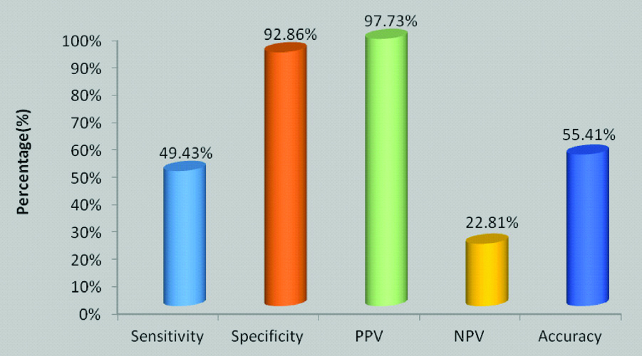
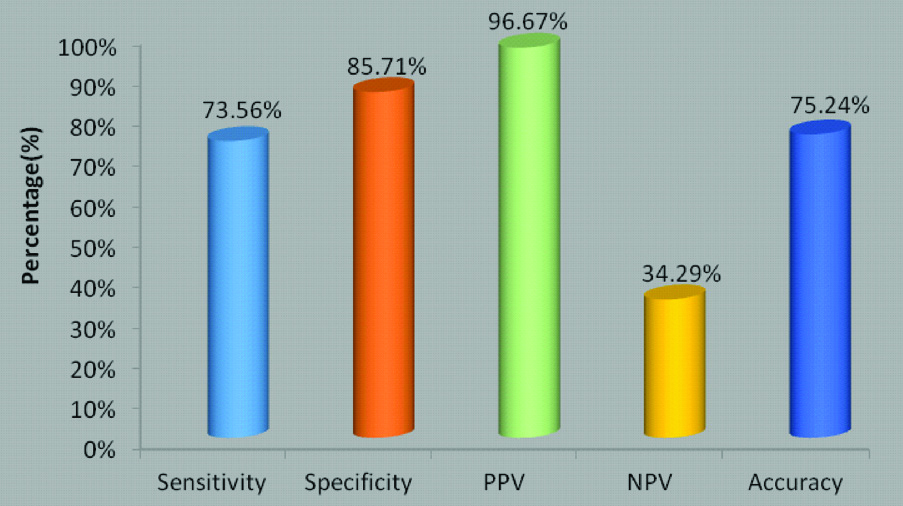
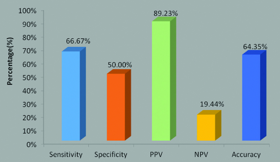
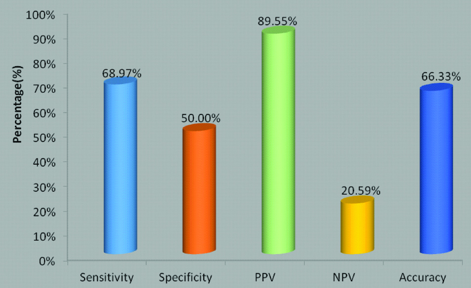
The sensitivity and specificity of the serum progesterone level was calculated as 49.43% and 92.86% with PPV, NPV as 97.73%, 22.81%, respectively [Table/Fig-5]. Similarly, 73.56%, 85.71% was sensitivity and specificity, respectively, of uterine artery resistance index [Table/Fig-6], whereas for the pulsatility index specificity was 50.00% and sensitivity was 66.67% [Table/Fig-7]. Sensitivity and specificity for S/D ratio was also calculated and shown in [Table/Fig-8].
Sensitivity and specificity of Serum Progesterone level.
Sensitivity and specificity of uterine artery RI.
Sensitivity and specificity of uterine artery PI.
Sensitivity and specificity of S/D ratio.
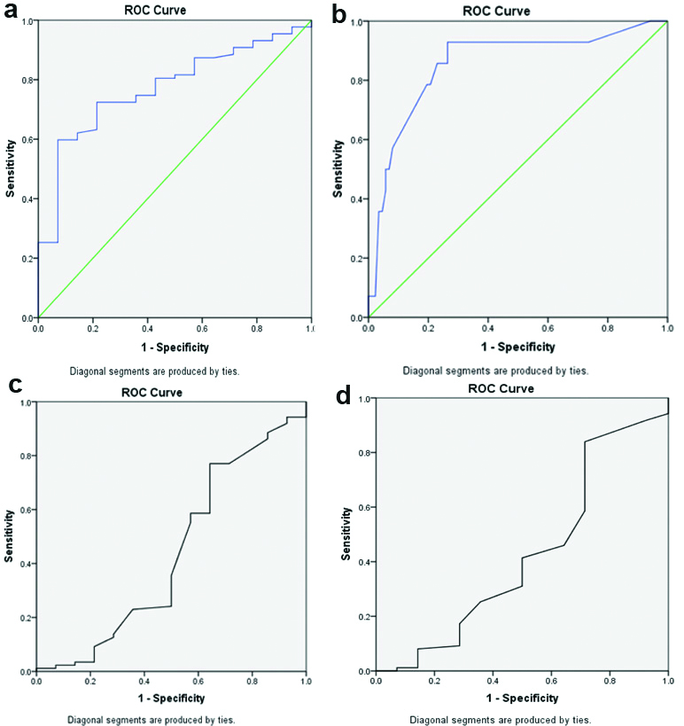
In general, the area under an ROC curve measures about the usefulness of a test, i.e. a greater area denotes a more useful test. Above ROC curves show that area under curve for Progesterone is 0.773, for Uterine artery RI is 0.855, for PI is 0.437 and S/D ratio is 0.423. It means serum progesterone and Uterine artery RI can play significant role in the evaluation of the outcome of threatened abortion in early pregnancy, whereas Uterine artery PI and S/D ratio has no significant role. It was noted that AUC of uterine artery RI is more than serum progesterone. Hence, Uterine artery RI can be better tool than serum progesterone for prediction of outcome of threatened abortion in early pregnancy [Table/Fig-9a-d].
ROC for Serum Progesterone, RI, PI, S/D ratio. a) Serum progesterone. Area under the curve=0.773; b) Uterine artery RI. Area under the curve=0.855; c) Uterine artery PI. Area under the curve=0.437; d) S/D/Ratio. Area under the curve=0.423
Few cases showing TVS colour Doppler of uterine arteries in pregnant women depicting values of RI, PI and S/D ratio [Table/Fig-10a-c].
Photographs of TVS colour doppler of uterine arteries showing values of RI, PI and S/D ratio. a) Pregnancy continued. b) Pregnancy continued. c) Pregnancy aborted before reaching upto period of viability.

Discussion
Early pregnancy losses can have a lot of emotional, family, social and economic issues associated with them. Ultrasonography is an investigation that is routinely done in the first trimester for knowing fetal viability and dating of the pregnancy. Adding Doppler study to the initial ultrasound assessment can be more convenient and cost-effective to the patient. In the present study of 101 women with threatened abortion, 87 (86.14%) women continued their pregnancy and 14 (13.86%) aborted. The 87 women who continued their pregnancy, were followed-up to 28 weeks of GA which was considered as period of viability.
In the present study, it was found that mean age of women were 26.37±4.73 years. Dave A et al., in their study had mean age of women as 25.7±4.35 years [8].
Serum Progesterone
Serum progesterone concentrations <5 ng/mL suggest a dying pregnancy, whereas values >20 ng/mL support the diagnosis of a healthy pregnancy [9]. According to past literatures, the viable pregnancy had serum progesterone value 5.1 ng/mL at the lowest range while serum progesterone level of 25 ng/mL had 97% probability for viable pregnancy [10].
In this study, women who continued their pregnancy had a mean serum progesterone level of 24.21±4.48 ng/mL. those who aborted had a mean of 20.49±3.06 ng/mL. (Level of significance was taken as 0.05 throughout the study). Kant RH et al., conducted a study of 100 women with threatened abortion in Jaipur <12 weeks GA found mean serum progesterone level of 21.5±10.4 ng/mL in those who aborted and patients who continued their pregnancy had mean serum progesterone 41.6±10.8 ng/mL [11]. Abdelazim IA et al., in their study on 260 women with threatened abortion in Ahmadi and Al-Rashid hospital, Kuwait had higher values of mean serum progesterone in viable pregnancy 46.7±7.4 ng/mL and mean serum progesterone was 9.9±4.8 ng/mL in non-viable pregnancies [12]. Hanita O and Hanisah AH, in a study of 95 women with threatened abortion at <13 weeks GA and followed-up to 22 weeks of GA to know the viability status. They showed that patient who continued their pregnancy had progesterone level 89.7±33.2 ng/mL and who aborted had progesterone level 23.3±12 ng/mL [7]. Liu X et al., in a study on 48 women with threatened abortion had mean serum progesterone 15.0±4.9 ng/mL at 12 weeks and 20.4±3.0 ng/mL at 28 weeks GA [13]. Pandey VP et al., conducted a study in Jaipur over 100 pregnant women with threatened abortion. They showed that patients who continued their pregnancy had mean serum progesterone level 18.18±6.09 ng/mL and the mean serum progesterone was 6.56±2.94 ng/mL in patients who aborted. Considering 9.9 ng/mL as cut-off value, it had 94.9% sensitivity and 92.7%, specificity [6].
Resistance Index
In the present study, mean RI of total 101 women was 0.52. Mohamed MA et al., in a study with 100 women with threatened abortion in Sudan. They had RI 0.83±0.3, in women who aborted and RI of 0.47±0.2 in women who continued their pregnancy [14].
Romero-Guiterrez RG et al., in their study conducted at Leon, Mexico with 223 women with threatened abortion found a RI 0.79±0.07 in women who aborted and 0.76±0.09 in women who continued their pregnancy [15]. Kurjak A et al., in their study of 20 women with threatened abortion and 131 normal pregnancy found that threatened abortion patients had 0.81±0.06 uterine artery RI whereas normal pregnancy had 0.78±0.10 [16].
Pulsatility Index
In the present study, Pulsatility Index (PI) was 0.94±0.45 in patients whose pregnancy was continued up to period of viability and was 1.05±0.44 in women who aborted. The p-value was 0.40 and it was statistically not significant. Mohamed MA et al., conducted a study with 100 women with threatened abortion at Sudan. Their study had PI 2.3±0.8 in women who aborted and PI of 1.66±0.8 in women who continued their pregnancy [14]. Kurjak A et al., in their study conducted in Croatia (university of Zagreb) had 20 patients with threatened abortion and 131 women with healthy pregnancy; showed PI of 2.13±0.48 in threatened abortion and PI of 1.91±0.61 in normal pregnancy [16].
S/D Ratio
In the present study, S/D ratio was 2.05±0.56 in women who continued their pregnancy and 2.56±1.52 in women who aborted. Al-Waeely AJ, conducted a study with 25 women with threatened abortion at Basrah Maternity and child hospital and they got S/D ratio of 5.45±3.02 [17].
ROC Curves
In the present study ROC curves showed that area under curve for Progesterone is 0.773, for Uterine artery RI is 0.855, for PI is 0.437 and S/D ratio is 0.423. This shows that serum progesterone and Uterine artery RI can play significant role in predicting outcome of threatened abortion in early pregnancy, whereas Uterine artery PI and S/D ratio has no significant role as area under curve was < 0.5. It was found that area under curve of uterine artery RI is more than that of serum progesterone. Hence, Uterine artery RI can be better tool than serum progesterone for predicting outcome of threatened abortion.
Dave A et al., in their study which was conducted at Indore included 100 women had AUC for progesterone was 0.971 [8]. Hanita O and Hanisah AH, conducted a study with 95 patients at Malaysia found that the AUC for progesterone was 0.95 [7]. Pandey VP et al., in a study from Jaipur had AUC for serum progesterone as 0.975±0.013 [6]. The present authors did not find any study that measured Doppler study of uterine arteries measuring the Area under curve of RI, PI and S/D Ratio.
There are only few studies in the literature which compare efficacy of transvaginal Colour Doppler ultrasound of uterine arteries with serum progesterone levels in predicting outcome of threatened abortion in early pregnancy.
Silwinska PA et al., conducted a study in 2001 with study population of 30 pregnant women with threatened abortion between 5-12 weeks GA. No correlation was found between maternal serum progesterone and doppler parameters in prediction of outcome of threatened abortion patients [18].
Hameed A and Hasan M, conducted a study at Zagazig University, Egypt with 143 pregnant females at 6-12 weeks of gestation; 93 cases with threatened abortion and 50 cases with normal pregnancy were enrolled in the study. They concluded that Serum β-HCG and progesterone level together had more sensitivity and specificity. Doppler study of Uterine artery did not play significant role in predicting the outcome of threatened abortion [19].
Limitation(s)
The sample size was small and the study was of short duration.
Conclusion(s)
In this study, it was found that uterine artery blood flow studies by trans vaginal colour doppler USG are useful for predicting the outcome of threatened abortion in early pregnancy. Serum progesterone and Uterine artery RI both play significant role in evaluation of the outcome of threatened abortion in early pregnancy. It was noted that AUC of uterine artery RI is more than serum progesterone. Hence, Uterine artery RI can be better tool than serum progesterone for prediction of outcome of threatened abortion in early pregnancy.
Further studies with larger sample size are necessary for corroborate the findings. Also, the present study was limited to first trimester pregnancy (6-12 weeks GA). Second trimester pregnancies up to 20 weeks GA with threatened abortion can also be studied.
Student’s unpaired t-test