Enamel plays a protective role for the tooth against external factors. But it undergoes some damage when acidic environment, caused by fermentation of carbohydrates by bacteria, acts on it. Despite various enamel surface treatments, enamel decay is still one of the major concerns world wide. Streptococcus mutans is an important bacterium responsible for the development of dental caries. Various forms of fluoride are the most effective way to prevent tooth decay [1].
Due to the simplicity of its use, low risk of being swallowed and patient preference, fluoride varnish is most preferred by all dentists [2]. The long-term prophylactic effect of varnishes depends on sustained release of fluoride.
Remineralisation effect of fluoride is its ability to reduce acid formation by fermentation of carbohydrates by S. mutans in dental plaques or reduction in S. mutans count in saliva [3]. Since only use of fluoride varnishes had minimal effect on the total salivary levels of S. mutans, they are suggested to be used in combination with other active agents which enhance the overall effect of the varnish. CPP-ACP is a novel agent for the prevention and arresting the progression of dental caries [4]. Applications of CPP-ACP paste along with standard oral hygiene reduced the size and degree of demineralisation of white spot lesions in clinical study [5]. Xylitol intake leads to favourable results like low incidence of caries and S. mutans levels in oral flora due to the inefficacy of five-carbon sugar xylitol to be fermented by S. mutans [6]. Xylitol incorporated varnishes have shown to be promising alternatives to increase enamel remineralisation [7].
Comparison of two fluoride varnishes with active ingredients like CPP-ACP and Xylitol coated calcium phosphate has never been done to assess their antimicrobial and enamel surface change potential. Keeping this in mind the present In-vitro study was performed to probe into the antibacterial effectiveness of two fluoride varnishes on S. mutans and to compare the enamel microhardness after treating the enamel with these two varnishes.
Materials and Methods
Antibacterial efficacy: The study was carried out in School of Dental Sciences, Krishna Institute of Medical Sciences, Karad, Maharashtra from November 2019 to January 2020. The ethical clearance was obtained for the study (Protocol No:277/2019-2020). In this in-vitro study, the antibacterial effects of two sodium fluoride varnishes, namely MI Varnish (GC Dental) containing CPP-ACP and Embrace Varnish (Pulpdent) containing Xylitol coated calcium and phosphate ions designated as sample A and B respectively were evaluated against S. mutans. For the study, to obtain statistically significant results, the sample size of 10 was considered significant, the power of the study was 90% at 5% significance. antimicrobial efficacy was checked in 10 wells of each group using agar well diffusion method. The bacteria required for the study were isolated clinically from saliva of patients in the age group of 5-9 years. Three patients with high caries index were selected for saliva collection. The patients were informed about the procedure and written informed consent was obtained from the parents. The patients were instructed to avoid eating 1 hour before the saliva collection. The procedure was carried out in the morning session. The patients were asked to spit in sterile containers and unstimulated saliva was collected as the influence of stimulants like citric acid or paraffin wax was to be eliminated from the study. This saliva was serially diluted with isotonic saline (0.95% NS).
Fourth serial dilution was spread evenly on Tryptone Yeast Extract Cystine with M1975 Sucrose and without Bacitracin (TYCSB) Agar Base in a petridish. The dish was incubated anaerobically for 48 hours at 37°C. After 48 hours, colonies were taken from the dish with the help of sterile inoculating loop and suspension was prepared with 50% optical density in normal saline. Sterilised TYCSB media was cooled at 45°C and prepared inoculum was added to it. The medium was mixed thoroughly and allowed to cool at room temperature. Two wells were bore in the agar plate using sterile borer and 100 microlitre of sample A and sample B was dispensed using micropipette in each well. Ten such petridishes were incubated anaerobically for 48 hours at 37°C. Subsequently, the sensitivity of S. mutans to fluoride varnish was evaluated by measuring the maximum diameter of the inhibition zone using digital Vernier calliper. The mean and standard deviation were calculated and data was analysed using unpaired t-test.
Enamel surface microhardness: This in-vitro experimental study was carried on 30 human premolar teeth which were extracted due to orthodontic purposes. The inclusion criteria for the study samples was clinically healthy teeth with absence of caries, cracks and hypoplastic lesions. The samples were decoronated at cemento-enamel junction. The buccal enamel surface of these samples was flattened using 1000 and 800 grit sand paper serially to obtain a flat surface for testing. Square label with dimensions of 4×4 mm was placed on the buccal surfaces of flattened samples. The samples were embedded in acrylic discs leaving the labelled portion exposed. The exposed enamel surface was coated with clear nail varnish. The labels were then removed to obtain an enamel window of 4×4 mm. Thirty samples were then divided randomly in 3 groups of 10 each. Samples in group A (n=10) were coated with MI Varnish according to the manufacturer’s instructions. Similarly, samples in group B (n=10) were coated with Pulpdent Embrace Varnish. The samples in group C (n=10) did not receive any fluoride treatment. All the samples were stored in artificial saliva for 24 hours at 37°C. Varnishes were slowly cleaned from the surface of treated samples with periodontal curette after 24 hours to synchronise the loss of varnish from the enamel surface in the oral cavity [8]. The samples were entered into pH cycle given by AlAmoudi SA et al., so as to create laboratory conditions similar to the mouth [2]. Each cycle was performed for 24 hours. Initially, samples were placed separately in a demineralisation solution for three hours. The demineralising solution consisted of 2.2 mM CaCl2, 2.2 mM NaHPO4, 0.05 M acetic acid, adjusted with 1 ml KOH at pH=4.5. The samples were then immersed in distilled water for 30 minutes.
Thereafter, samples were placed in a remineralisation solution for 20 hours and were again washed and immersed in distilled water for 30 minutes. The remineralising solution contained 1.5 mM CaCl2, 0.9 mM NaHPO4, KCl 0.15 mM with pH=7.0. This cycle was repeated for 10 days [2].
At the end of the pH cycling for 10 days, microhardness of enamel was measured using microhardness tester machine (Mitutoyo HM-210). Microhardness of each sample was assessed by making an indentation on enamel by applying 100 gm force for a dwell time of 10 seconds as shown in [Table/Fig-1] [9]. The readings were obtained on two points in each sample and their mean was taken.
Experimental sample being tested under Universal Testing Machine for surface hardness.

Statistical Analysis
Data obtained was tabulated and subjected to statistical analysis using one-way ANOVA. p≤0.05 was considered statistically significant. The data was tabulated and results were statistically analysed in Graph Pad Instat Software version 3.06.
Results
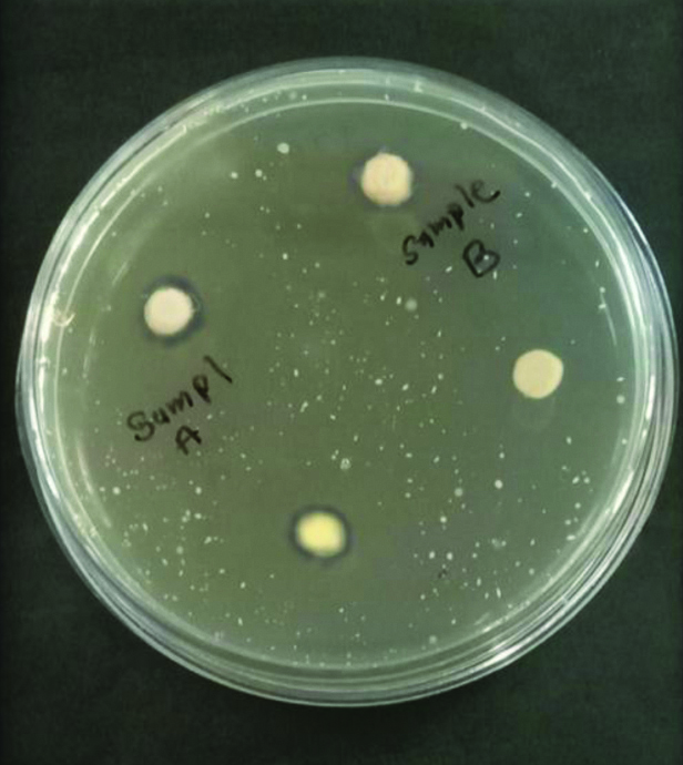
[Table/Fig-2] shows comparison of mean zone of inhibition around the discs coated with fluoride varnishes (mm). Both the varnishes showed antibacterial effects against S. mutans. However, 4 out of 10 samples showed no zone of inhibition in group B. MI Varnish had a greater diameter of no growth around the sample wells as shown in [Table/Fig-3]. In [Table/Fig-4] on statistical comparison by unpaired t-test, MI Varnish showed significantly higher antibacterial effect, compared to Pulpdent Embrace Varnish (p≤0.05).
Comparison of mean zone of inhibition (mm) of two varnishes.

TYCSB-agar plate showing zone of inhibition around the sample of group A.
Results of unpaired t-test comparing mean diameter of inhibition zone around the wells containing fluoride varnish (mm).
| Varnish | Mean | SD | p-value |
|---|
| Grp A (MI Varnish) | 1.120 | 0.3293 | 0.0290 |
| Grp B (Pulpdent Embrace Varnish) | 0.160 | 0.1506 | |
[Table/Fig-5] compares the mean microhardness Vickers Hardness Number (VHN) of the three groups tested.
Comparison of mean VHN of three groups.
| Sample No. | Group A (MI Varnish) | Group B (Pulpdent Embrace) | Group C (Control) |
|---|
| 01 | 128.9 | 118.0 | 106.5 |
| 02 | 128.6 | 112.6 | 109.3 |
| 03 | 127.7 | 109.5 | 103.2 |
| 04 | 126.4 | 116.4 | 105.5 |
| 05 | 133.4 | 115.5 | 102.5 |
| 06 | 129.1 | 112.1 | 105.9 |
| 07 | 131.0 | 118.2 | 104.4 |
| 08 | 133.3 | 119.1 | 104.0 |
| 09 | 139.4 | 117.5 | 101.3 |
| 10 | 129.6 | 112.7 | 103.4 |
| MEAN | 130.74 | 115.16 | 104.6 |
A significant difference regarding the means of enamel microhardness was found between the groups (p<0.05) by ANOVA after the application of fluoride varnish [Table/Fig-6]. Group A (MI Varnish) showed the highest microhardness as compared to Group B (Pulpdent Embrace Varnish) and Control.
Results of ANOVA between groups comparing the VHN of the samples.
| Mean | SD | p-value |
|---|
| Grp A (MI Varnish) | 130.74 | 3.782 | |
| Grp B (Pulpdent Embrace) | 115.16 | 3.231 | 0.001 |
| Grp C (Control) | 104.60 | 2.292 | |
Discussion
Fluoride varnish is often preferred by most dentists because of the ease and speed of use, lower risk of being swallowed leading to toxicity and patient compliance. The long-term prophylactic effect of varnishes depends on sustained fluoride release.
The most significant anticaries effects of fluoride is the inhibition of demineralisation and augmentation of remineralisation of early caries lesions as well as fluoride can inhibit acid production by bacteria and reduce the number of S. mutans [10].
Using only fluoride varnishes has minimal effect on the total salivary levels of S. mutans, they should be used along with other active agents which increase the overall effect of the varnish. CPP-ACP is a novel agent for the prevention as well as arrest of dental caries progression. This milk product facilitates remineralisation, inhibits demineralisation, and prevents dental caries by forming a calcium phosphate ion reservoir for enamel to resist acid attacks [11].
Also, Calcium Phosphate fuses into the salivary pellicle and decreases the adhesion of S. mutans to the tooth surface. The combination of CPP-ACP with fluoride results in localisation of calcium and phosphate ions with fluoride ions at the enamel surface [3]. The advantage of CPP-ACP is the availability of calcium, phosphate, and fluoride in one product.
Each molecule of CPP can bind up to 25 calcium ions, 15 phosphate ions, and 5 fluoride ions. The calcium phosphate in these complexes is biologically available for remineralisation of sub-surface lesions in tooth enamel [12]. Formation of a remineralisation reservoir on the surface by CPP-ACP could be the attributing factor in its higher remineralisation potential in the current study.
Xylitol is a sugar alcohol derived primarily from the forest and agricultural materials which acts as a sweetening agent. Various studies have reported that xylitol intake leads to favorable results like reduced incidence of caries and S. mutans levels in oral flora [3]. This could be due to the inability of five-carbon sugar xylitol to be fermented by S. mutans to produce acids. Addition of xylitol is known to increase the patient compliance. Xylitol incorporated varnishes have also proven to be a favourable replacement to enhance enamel remineralisation.
Studies assessing various varnishes for their antimicrobial action and change in enamel surface properties have been conducted in the past as discussed in [Table/Fig-7] [3,13-17].
Studies conducted to assess the efficacy of varnishes [3,13-17].
| Author | Year | Conclusion |
|---|
| Llena C et al., [13] | 2015 | At 4 weeks, CPP-ACFP is superior to fluoride varnish at remineralising smooth-surface white spot lesions. |
| Cardoso CA et al., [14] | 2014 | Enamel surface remineralisation was significantly increased by Duraphat™, 10% xylitol plus F and 20% xylitol plus F formulations, while significant subsurface mineral remineralisation could be seen only for enamel treated with Duraphat™, Duofluorid™ and 20% xylitol formulations.20% xylitol varnishes seem to be promising alternatives to increase remineralisation of artificial caries lesions. |
| KL Girish Babu et al., [15] | 2018 | There was no significant difference in the remineralising potential of varnish containing CPP-ACP and fluoride and varnish containing only fluoride. |
| Jafari K et al., [3] | 2018 | According to the results, Polimo showed the highest antibacterial effects, compared to the other three varnishes (p≤0.05). Growth inhibition zones were not observed in V-varnish and Preventa. The mean diameter of inhibition zone around the MI varnish was significantly higher, compared to those of the V varnish and Preventa (p≤0.05). |
| Yadav S et al., [16] | 2019 | Fluoride vanish with CPP-ACP resulted in reduction of more number of S. mutans than xylitol-containing fluoride varnish and Fluor protector®. |
| Varma V et al., [17] | 2019 | As compared to clinpro fluoride releasing varnish, MI varnish with CPP-ACP had the highest fluroide release.MI varnish is a 5% NaF varnish with CPP-ACP that releases high amount of bioavailable fluoride, calcium and phosphate and hence is successful in reversing early caries. |
| Current study | 2020 | MI Varnish showed higher anti-bacterial efficacy as compared to Pulpdent Embrace Varnish against S. mutans by disc diffusion method on TYCSB agar. The surface microhardness of enamel group treated with MI Varnish was significantly greater than Pulpdent embrace and control groups. |
In the current study, we used two fluoride varnishes containing CPP-ACP and Xylitol coated calcium and phosphate ions as active ingredients.
Different methods to assess the antibacterial activity of restorative materials are well diffusion, disc diffusion, flow cytometry and qPCR. However, owing to its feasibility and reliability, the well diffusion method was chosen for the current study to check the antimicrobial efficacy of the varnishes. MI varnish showed a significantly higher antimicrobial activity against S. mutans as compared to Pulpdent Embrace Varnish. This disparity could be because of the composition of the varnishes and their mechanisms of action.
Vongsavan K et al., demonstrated that the combination of fluoride and xylitol varnishes was favourable for preventing enamel demineralisation however, the addition of xylitol to fluoride varnish showed no significant difference than fluoride varnish alone in-vitro [1].
In a study done by Jafari K et al., all fluoride varnishes, except for V-varnish and Preventa, had antimicrobial effect. Among the four evaluated fluoride varnishes, Polimo MI varnish showed the highest antibacterial effect against S. mutans. Furthermore, the application of Polimo and MI varnish not only inhibited demineralisation and increased remineralisation but also lowered the level of S. mutans in the oral cavity [3].
In the current study, Vickers Hardness method was used to compare micro hardness of tooth enamel by using two fluoride varnishes after simulating the oral environment in the lab. The pH cycling method by creating acidic challenges helps in the simulation of the oral environment in the laboratory. However, 100% simulation cannot be achieved because of the key role of the factors associated with remineralisation process like speed and flow rate of saliva and composition and buffering capacity of saliva [12].
Evaluation of changes in the enamel surface area has great importance because surface layer has a vital role in decay process. The calculation of surface’s micro hardness is a suitable method which was carried out using Vickers Hardness Measuring method. The benefit of this method is accuracy and quantitative measurement capability. Also, the option of applying force with various sizes and the feasibility of re-measuring hardness of specimens within a specific time frame. The microhardness was measured at two points in each sample and mean of the values was taken.
The present study showed higher values of microhardness in fluoride treated groups as compared to the control group. Similar to the results obtained in this study Nalbantgil D et al., concluded that microhardness after the application of two fluoride varnish was more than the control group [18].
Navabi B et al., concluded that the difference between microhardness was observed in enamel before and after the application of fluoride varnish [19]. In a study done by Tavassoli-Hojjati S et al., Iranian APF gel (Kimia) increased the microhardness of tooth enamel compared to control group which is in accordance to the present study [20]. Majithia U et al., conluded that MI Varnish showed slightly more surface microhardness as compared to Flor-Opal® Varnish and Premier® Enamel Pro® Varnish [21]. MI Varnish increased the enamel microhardness slightly higher than Pulpdent Embrace Varnish in this study.
This study depicts that the two varnishes increased the enamel micro hardness. However, remineralisation observed in in-vitro conditions could be slightly inconsistent in comparison to the oral conditions.
Limitation(s)
One limitation of this study includes using only S. mutans while the other pathogenic bacteria associated with the dental biofilms causing caries were not considered.
Conclusion(s)
MI Varnish showed higher anti-bacterial efficacy as compared to Pulpdent Embrace Varnish against S. mutans by disc diffusion method on TYCSB agar. The surface microhardness value of enamel samples treated with MI Varnish were significantly greater than other two groups. The enamel surface microhardness of control group was the least suggesting improvement in the microhardness on application of fluoride varnishes.