Spontaneous Spleen Rupture: Rare But Not Exceptional
Atef Mejri1, Mohamed Firas Ayadi2, Khaoula Arfaoui3
1 Visceral Surgeon, Department of General Surgery, Regional Hospital of Jendouba, Jendouba, Tunisia.
2 Faculty, Department of General Surgery, Regional Hospital of Jendouba, Jendouba, Tunisia.
3 Visceral Surgeon, Department of General Surgery, Regional Hospital of Jendouba, Jendouba, Tunisia.
NAME, ADDRESS, E-MAIL ID OF THE CORRESPONDING AUTHOR: Atef Mejri, BP 296, Jendouba, Bou Salem, Tunisia.
E-mail: atef.mejri@hotmail.com
Spontaneous splenic rupture is a rare life threatening condition that occurs in 0.1% to 0.5% of all the spleen rupture cases. In case of a young patient with no history who consults for acute abdominal pain, the diagnosis of spontaneous spleen rupture is often forgotten or ignored in favour of other more frequent diagnoses. However, it has the highest mortality rate so it should always be kept in mind in front of signs of internal haemorrhage especially if it is associated with haemodynamic instability. Emergency Splenectomy is often performed with good reported outcomes. We present a case of a spontaneous idiopathic splenic rupture, diagnosed early and managed successfully.
Atraumatic, Emergency, Idiopatic, Laparotomy
Case Report
A 36-year-old male presented to the emergency for vomiting, pallor and left-sided abdominal pain of sudden onset, evolving for two hours. No history of recent trivial abdominal trauma or fever was reported. The patient did not work out too hard and did not make a hard effort of coughing or sneezing recently and he did not smoke. There was no significant medical history and he was not on any regular medications.
Clinical examination revealed- BP: 122/55 mmHg, pulse: 110 BPM, pallor and no jaundice. He had epigastric and left upper quadrant guarding. The rest of the examination was unremarkable. Haematological investigation revealed, WBC: 16.000/μL; Haemoblobin level at 12.4 g/dL; Blood group: B positive, no other abnormality was found in the biological assessment. The only radiological examination that was done was the abdominal CT scan. Given the availability and the high diagnostic accuracy of this imaging modality and the emergency situation, we immediately decided to perform it. In fact, it helps establish a quicker and more precise diagnosis than an abdominal ultrasound. The latter is certainly a noninvasive examination but it is still subjective as it relies on the skill of the practitioner.
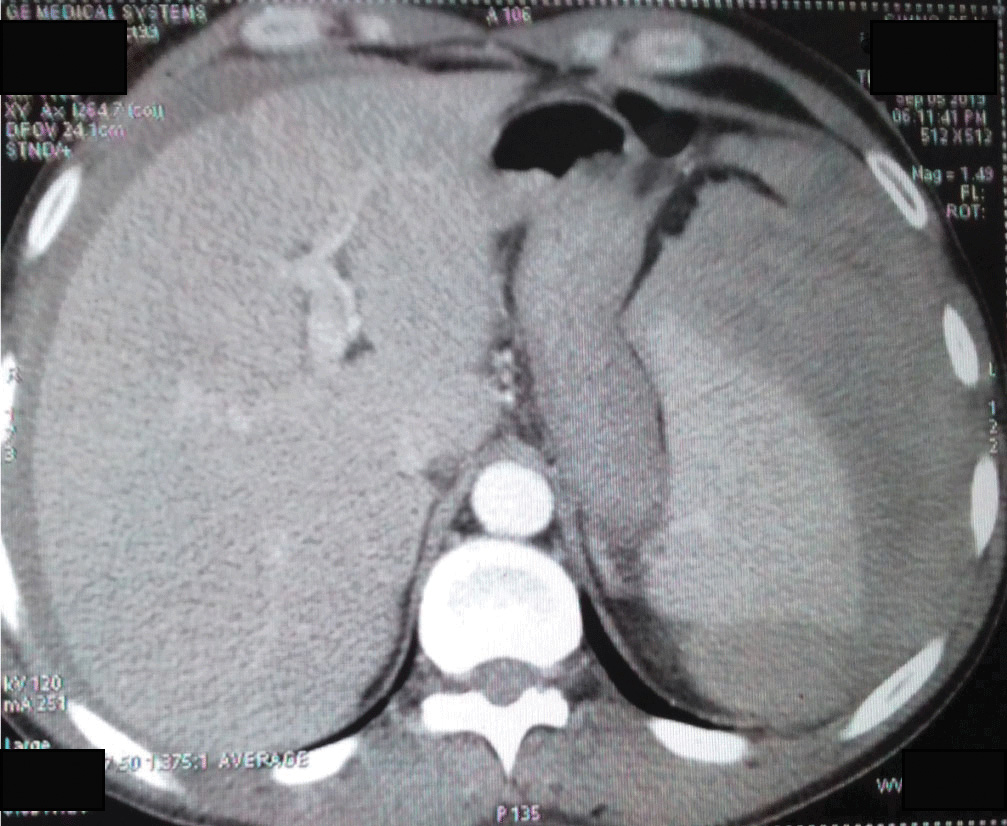
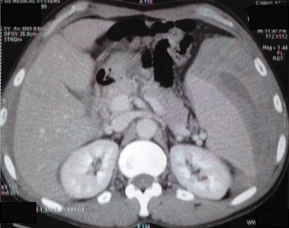
The CT scan revealed a 5 cm spleen haematoma, and free intra-peritoneal fluid, suggesting a non-traumatic splenic rupture [Table/Fig-1,2], while realising the CT scan, BP dropped to 82/36 mmHG and the pulse increased to 140 BPM, the oxygen saturation was 92%. Blood test was repeated and it showed- Hb: 7.6 g/dL; PT=78%; a normal platelet count; no blood electrolyte disorder; and a normal renal function. The arterial blood gas revealed a metabolic acidosis: pH=7,2.
CT scan: arterial enhancement: 5 cm spleen haematoma with free intraperitonial fluid staged grade III.

CT scan: portal enhancement: perisplenic hematoma.

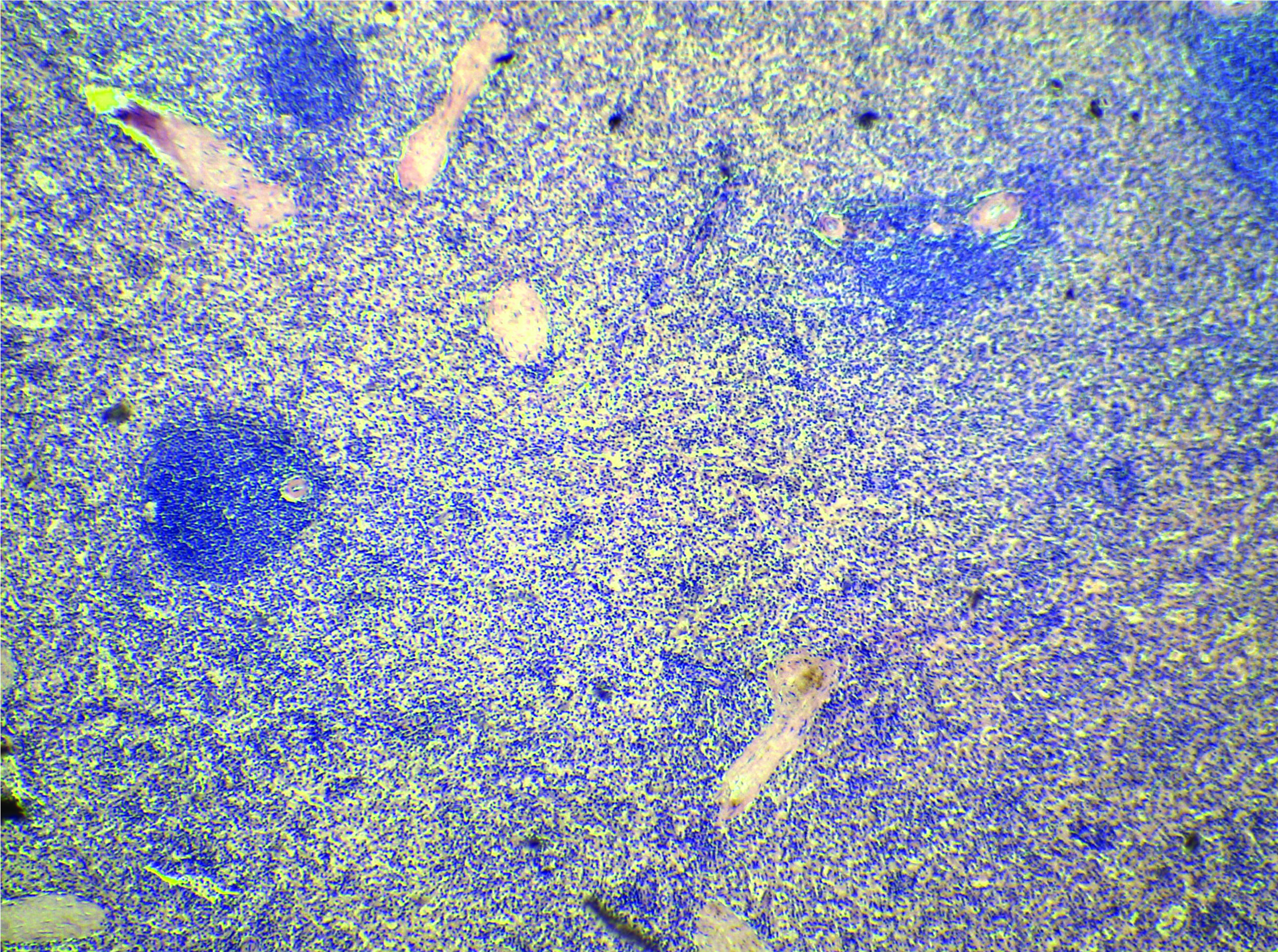
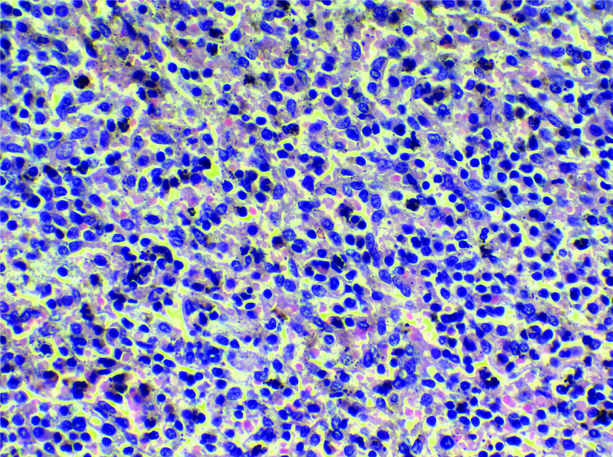
After a quick fluid resuscitation, it was decided to proceed for splenectomy. Postoperatively, the patient received pneumococcal, vaccination and penicillin prophylaxis was initiated. All of the aetiological tests were negative, the histological examination showed an engorgement of the splenic tissue with no other underlying pathology [Table/Fig-3,4]. The diagnosis of atraumatic-idiopathic splenic rupture was considered.
Splenic tissue (magnification X4) showing an atrophic red pulp dominated by a hyperplastic white pulp.

Splenic tissue (magnification X40) showing a cell population richin histiocytes sometimes engorged with haemosiderin.

Discussion
Spontaneous Splenic Rupture (SSR) is a rare clinical presentation with potentially grave medical outcome. It occurs in 0.1 to 0.5% of patient with no history of recent trauma [1]. It is documented since the 19th century [2]. However, the reason for its rupture is still not understood yet. One of the main mechanisms that have been reported in literature and the most likely responsible for SSR in the index case is the increased intra-splenic tension caused by cellular hyperplasia and engorgement [3].
It should also be kept in mind that simple physiological acts, like sneezing or coughing or other daily harmless activities or moves may lead to the compression of the spleen by the abdominal musculature and then turn fatal. According to Renzulli P et al., the aetiologies of SSR are divided into two major categories: the underlying pathology-related rupture is predominant (93%) and it includes neoplasic, infectious, inflammatory causes and drug or treatment correlated causes. In 7% of cases, the cause is said to be idiopathic [4]. This can be explained by misdiagnosis due to its non-specific signs, rare occurrence and the absence of recent trauma [5].
In fact, the main symptom is left upper quadrant abdominal pain [1], which can be generalised with rigidity and signs of haemorrhagic shock in later stages. In 20% of the cases, a sharp radiating pain to the left shoulder (Kehr’s sign) was observed [6]. CT scan is often essential to make the diagnosis and grade the splenic injury on a scale for I to V [2]. Regarding the important role of the spleen as a lymphoid organ, conservative low-grade injuries (I–II) can be managed conservatively in the condition of a haemodynamically stable patient, regular clinical and biological supervision. This attitude could not be held in the case of this patient due to the sudden haemodynamic instability and deglobulisation occurring in the radiology department. While in case of grade IV-V injuries usually splenectomy is done. Thapar PM et al., reported a similar case of 23-year-old male who underwent an emergency laparoscopic splenectomy for a SSR, that was safely accomplished. Although the laparoscopic approach is an emerging surgical tool that was proven to offer a faster recovery and a shorter hospital stay duration, it should be considered wisely without compromising the patient’ life [7]. The clinical situation is the most important factor in guiding the decisions.
Conclusion(s)
Spontaneous rupture of the spleen is an extremely rare and dangerous pathology. The non-specific signs and the absence of a blunt abdominal trauma can be responsible of a diagnosis delay that can be hazardous and responsible for the high mortality. The CT scan is the key examination permitting to make the diagnosis and grade the splenic injury, which guide the therapeutic management and helps to make the choice between splenectomy and a conservative management.
Author Declaration:
Financial or Other Competing Interests: None
Was informed consent obtained from the subjects involved in the study? Yes
For any images presented appropriate consent has been obtained from the subjects. Yes
Plagiarism Checking Methods: [Jain H et al.]
Plagiarism X-checker: Nov 14, 2019
Manual Googling: Feb 08, 2020
iThenticate Software: Feb 28, 2020 (7%)
[1]. Gedik E, Girgin S, Aldemir M, Keles C, Tuncer MC, Aktas A, Non-traumatic splenic rupture: Report of seven cases and review of the literatureWorld J Gastroenterol WJG 2008 14(43):6711-16.10.3748/wjg.14.671119034976 [Google Scholar] [CrossRef] [PubMed]
[2]. Weaver H, Kumar V, Spencer K, Maatouk M, Malik S, Spontaneous splenic rupture: A rare life-threatening condition; Diagnosed early and managed successfullyAm J Case Rep 2013 14:13-15.10.12659/AJCR.88373923569554 [Google Scholar] [CrossRef] [PubMed]
[3]. Samuk I, Seguier-Lipszyc E, Baazov A, Tamary H, Nahum E, Steinberg R, Emergency or urgent splenectomy in children for non-traumatic reasonsEur J Pediatr 2019 178(9):1363-67.10.1007/s00431-019-03424-631312939 [Google Scholar] [CrossRef] [PubMed]
[4]. Renzulli P, Hostettler A, Schoepfer AM, Gloor B, Candinas D, Systematic review of atraumatic splenic ruptureBr J Surg 2009 96(10):1114-21.10.1002/bjs.673719787754 [Google Scholar] [CrossRef] [PubMed]
[5]. Walker AM, Foley EF, Surgical management of atraumatic splenic ruptureInt Surg J 2016 3(4):2280-88.10.18203/2349-2902.isj20163613 [Google Scholar] [CrossRef]
[6]. Kaniappan K, Lim CTS, Chin PW, Non-traumatic splenic rupture-a rare first presentation of diffuse large B-cell lymphoma and a review of the literatureBMC Cancer 2018 18(1):779PubMed-NCBI. https://www.ncbi.nlm.nih.gov/pubmed/30068299 (accessed 24 Dec2019)10.1186/s12885-018-4702-130068299 [Google Scholar] [CrossRef] [PubMed]
[7]. Thapar PM, Philip R, Masurkar VG, Khadse PL, Randive NU, Laparoscopic splenectomy for spontaneous rupture of the spleenJ Minimal Access Surg 2016 12(1):75-78.10.4103/0972-9941.15895026917926 [Google Scholar] [CrossRef] [PubMed]