Entrance Skin Dose Measurement for Diagnostic Spinal Radiographic Examinations in King Khalid Hospital, Saudi Arabia: A Prospective Study
Yousif Mohamed Abdallah1, Nouf Hussain Abuhadi2
1 Associate Professor, Department of Radiological Science and Medical Imaging, College of Applied Medical Science, Majmaah University, Majmaah, Riyadh, Saudi Arabia.
2 Assistant Professor, Department of Diagnostic Radiology, College of Applied Medical Science, Jazan University, Jazan, Saudi Arabia.
NAME, ADDRESS, E-MAIL ID OF THE CORRESPONDING AUTHOR: Yousif Mohamed Abdallah, Majmaah-11952, Riyadh, Saudi Arabia.
E-mail: y.yousif@mu.edu.sa
Introduction
Radiographic examinations has necessary role in the identification of spine injuries and pathologies. There are many hazards associated with the radiation exposure which included the acute (radiation injury) and chronic exposure effects (cancer).
Aim
To measure the entrance skin dose of spine vertebra (cervical, thoracic, lumbar and sacral) in AP and Lateral Views.
Materials and Methods
A prospective study was conducted with a sample of 250 adults and 100 paediatrics patients. The imaging apparatus, which was used in this study was Siemens with pipe Filtration 2.0-3.0 mm of AL/70 KVp. The parameters of patients collected were patients’ characteristics and exposure factors. The dose was measured using Entrance Skin Dose (ESD) and International Atomic Energy Agency (IAEA) formula and compared nationally and internationally.
Results
The measured exposure parameters were 78.1±5.7 and 19.9±7.8 for the machine kVp and mAs, respectively. The measured ESD dose for cervical, thoracic, Lumbosacral (AP and LAT.) and sacral (AP) for adult population were 0.11±0.06 mGy and 0.15±0.07 mGy, (0.86±0.06 and 0.91±0.09 mGy, 0.88±0.07 and 0.92±0.09 mGy and 0.25±0.04 mGy, respectively. Similarly, measured ESD dose for cervical, thoracic, Lumbosacral (AP and LAT.) and sacral (AP) for paediatrics population were 0.09±0.01 mGy and 0.12±0.07 mGy, 0.32±0.03 mGy and 0.42±0.06 mGy, 0.38±0.06 and 0.74±0.08 mGy and 0.09±0.01, respectively.
Conclusion
The results of the study were within the range of permissible dose of the spine vertebrae dose (4.0-30.0 mGy). More studies are recommended to study radiation dose of the spine vertebrae with large patients’ data and more than one modalities to compare.
Radiation dose, Traumatic, Vertebra
Introduction
Radiation Exposure is main hazard in radiographic investigations. Now-a-days human organ imaging is performed by different systems and methods. As the new diagnostic methods including CT, MRI, and sonography are evolving but plain radiography is still a powerful tool with enough benefits to the patients undoubtedly. Therefore, patients’ exposure to radiation has been increased all over the world due to this diagnostic radiography [1]. The basic concept to reduce the radiation exposure is to use the minimum dose needed for good image quality by radiological tests. Radiation hazard arises from abuse of equipments, high exposure factors and exposure to different dose levels for the same clinical investigations [2-4]. Radiation exposure can cause severe injuries and possibly leads to cancer [5-6]. Trauma is a grievance which leads to emotional and physical impact [7]. In recent times, in Saudi Arabia, the amount of road traffic accidents and their effects has augmented considerably [8,9]. There is no perfect procedure to define radiation experience of patients during radiation examinations [10]. The normal radiation exposure differs between 10-100 mGy, which may rise the chance of cancer occurrence usually among population who are highly unprotected [11-13]. The traumatic X-rays imaging considered as one of the most common analytical tool used to study and identify the pathological circumstances [14]. Since the entitlements of traumatic radiology are growing speedily, it is critical subject to evaluate the radiation dosages during the examination and try to reduce them as much as possible [15-17]. The sacral realm (sacrum) is at the bottom of the spine and prevarication between the fifth segments of the lumbar. Conventional X-rays examination is an accepted modality in detection and identification of the different spine disorders in both paediatric and geriatric patients [18,19]. However, X-rays exposure considers dangerous especially in fledgling patients. Some studies showed that the irradiation in the early ages could increase the probability of having radiation sickness and malignant disorders due to tissues hypersensitivity. Therefore, the justified request and optimised protection measures should be applied especially in the younger patients [20-22]. The measurement of the radiation exposure in spine vertebrae x-rays is very crucial as the cutaneous and subcutaneous tissues exposed to large amount of the dose [23]. The measured dose can be used to formulate diagnostic reference levels at national level.
One of the variables of radiation protection is the dosage of patients. The dose is usually determined by evaluating the Entrance Skin Dose (ESD) for patients who are exposed to X-rays diagnosis. The ESD is defined as the dose of air absorbed by the patient’s entrance surface at the intersection of the beam axis, including the back dispersion. The surface dose entry is one of the critical quantities for estimating the patient dose and maximising the patient dose. This quantity is the fundamental requirement to be contrasted with other global dose levels of comparison, which are very important for safety against radiation. The ESD can generally be measured using two methods, either directly on patients’ skin using Thermoluminescent (TLD) measurements, or indirectly using the template quality estimates of the X-ray machine [24-28]. The aim of the study is to measure the entrance skin dose of spine vertebra (cervical, thoracic, lumbar and sacral) in AP and Lateral Views.
Materials and Methods
A prospective study was conducted with a sample of 350 patients who undergone radiological examinations at King Khaled Hospital, Majmaah, Saudi Arabia’s radiological department, between October 2018 to June 2019. The research was approved by IRB00010471 (H-01-R-012, OHRP/NIH (FWA00018774) licensing committee of King Abdelaziz City of Science and Technology (KACST). A MUREC-Nov.2l/COM-2018/9 permit is given from the Ethics Committee of the University of Majmaah.
The patient’s characteristics measured were age, Body Mass Index (BMI) and exposure factors, Focus-to-Skin Distance (FSD) and projections. The imaging system used in this study was Siemens, AXIOM imaging system made in Germany 2014, model: AlOIC with pipe Filtration 2.0-3.0 mm AL/70 KVp which was accompanied with dose controller (AEC). The machine output was measured and calibrated using reference dosimeter device with the high accuracy (±3%) and TLDs. The ESD was calculated as follows:
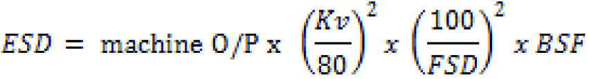
where:
(OP): the output of the machine
(mA) the product of the tube current
(FSD) the focus-to-skin distance (in cm).
(BSF) the backscatter factor,
Inclusion criteria: All traumatic patients who had conventional x-ray examination in area of the study were included.
Exclusion criteria: All traumatic patients who had other examinations rather than x-rays in area of the study.
Statistical Analysis
For statistical analysis SPSS version 23 was used. All data from this study are shown as mean plus standard range variability.
Results
The mean age of adult patients was 32.9±7.1 years with range of 16-65 years and mean age of paediatrics’ patients were 7.1±1.3 years with range of 1-15 years. Maximum number of adult patients were in the age group of 61-65 years [Table/Fig-1].
The age distribution for both gender among adult patients in the study sample.
| Age group (years) | Male n (%) | Female n (%) |
|---|
| 15-20 | 5 (2.3%) | 1 (2.7%) |
| 21-25 | 5 (2.3%) | 1 (2.7%) |
| 26-30 | 4 (1.9%) | 3 (8.1%) |
| 31-35 | 12 (5.7%) | 2 (5.4%) |
| 35-40 | 23 (10.9%) | 3 (8.1%) |
| 41-45 | 28 (13.2%) | 3 (8.1%) |
| 46-50 | 33 (15.4%) | 7 (18.9%) |
| 51-55 | 35 (16.4%) | 6 (16.2%) |
| 56-60 | 29 (13.6%) | 4 (10.9%) |
| 61-65 | 39 (18.3%) | 7 (18.9%) |
The registered exposure factors were 78.1±5.7 with range of (61.8-84.8) KVp, 19.9±7.8 with range of 1-48 mAs, 107.3±12.5 with range of 105-115 cm FSD and 0.11±0.06 with range of (0.05-2.01) mGy for tube potential, tube current, FSD and ESD Dose, respectively [Table/Fig-2].
Imaging parameters of the study.
| Variables | Tube potential (KVp) | Tube current×time (mAs) | Focus-to-skin distance (cm) | Entrance skin dose (mGy) |
|---|
| Mean | 78.1 | 19.9 | 107.3 | 0.11 |
| Median | 74.8 | 17.15 | 106.1 | 0.10 |
| Standard deviation | 5.7 | 7.8 | 12.5 | 0.06 |
| Minimum | 61.8 | 1 | 105 | 0.05 |
| Maximum | 84.8 | 48 | 115 | 2.01 |
KVp: Kilovoltage peak; mAs: Milliampere-seconds; cm: centimeters; mGy: megagray (106 gray)
The exposure factors of the spine vertebrae cervical, thoracic, Lumbar and sacral Imaging is shown in [Table/Fig-3].
Exposure factors of the cervical, thoracic, Lumbar and sacral vertebrae imaging.
| Variables | Cervical spine | Thoracic spine | Lumbar spine | Sacral |
|---|
| KVp | mAs | KVp | mAs | KVp | mAs | KVp | mAs |
|---|
| Mean | 77.9 | 22.8 | 70.8 | 6.67 | 80.2 | 21.3 | 73.14 | 5.12 |
| Median | 73.7 | 21.1 | 68.1 | 6.15 | 78.3 | 20.9 | 72.9 | 4.7 |
| Standard deviation | 8.1 | 7.2 | 5.6 | 2.4 | 8.91 | 6.2 | 7.7 | 1.93 |
| Minimum | 60.1 | 29 | 61.8 | 2.1 | 72.9 | 3.2 | 72.9 | 0.4 |
| Maximum | 83.8 | 1 | 76.8 | 18.7 | 84.8 | 48 | 76.8 | 20.4 |
KVp: Kilovoltage peak; mAs: Milliampere-seconds
The measured ESD dose for cervical, thoracic, Lumbosacral (AP and LAT.) and sacral (AP) for adult population were 0.11±0.06 mGy and 0.15±0.07 mGy, (0.86±0.06 and 0.91±0.09 mGy, 0.88±0.07 and 0.92±0.09 mGy and 0.25±0.04 mGy, respectively Similarly measured ESD dose for cervical, thoracic, Lumbosacral (AP and LAT.) and sacral (AP) for paediatrics population were 0.09±0.01 mGy and 0.12±0.07 mGy, 0.32±0.03 mGy and 0.42±0.06 mGy, 0.38±0.06 and 0.74±0.08 mGy and 0.09±0.01, respectively [Table/Fig-4,5].
Entrance skin dose measured for adult population.
| Examination | Number | Mean | Minimum | Maximum |
|---|
| Cervical vertebrae |
| AP | 60 | 0.11±0.06 | 0.08 | 0.31 |
| Lateral | 0.15±0.07 | 0.09 | 0.39 |
| Thoracic vertebrae |
| AP | 40 | 0.86±0.06 | 0.01 | 0.99 |
| Lateral | 0.91±0.09 | 0.02 | 0.81 |
| Lumbar vertebrae |
| AP | 120 | 0.88±0.07 | 0.05 | 1.95 |
| Lateral | 0.92±0.09 | 0.06 | 2.01 |
| Sacral vertebrae |
| AP | 30 | 0.25±0.04 | 0.01 | 0.99 |
Entrance skin dose measured for paediatrics population.
| Examination | Number | Mean | Minimum | Maximum |
|---|
| Cervical vertebrae |
| AP | 35 | 0.09±0.01 | 0.06 | 0.23 |
| Lateral | 0.12±0.07 | 0.04 | 0.45 |
| Thoracic vertebrae |
| AP | 20 | 0.32±0.03 | 0.01 | 0.92 |
| Lateral | 0.42±0.06 | 0.03 | 0.77 |
| Lumbar vertebrae |
| AP | 40 | 0.38±0.06 | 0.02 | 1.18 |
| Lateral | 0.74±0.08 | 0.05 | 1.01 |
| Sacral vertebrae |
| AP | 5 | 0.09±0.01 | 0.01 | 0.39 |
Discussion
This present study was performed to measure the ESD received in spine vertebrae (C/S, D/S, L/S, S/S) AP and lateral projections. A total 250 adults and 100 paediatric patients were examined in two radiology departments in king Khalid Hospital, Majmaah in which 85% of the patients were males and 15% were females. In a study conducted by Aliasgharzadeh A et al., the mean ESD values were 2.18 and 5.36 for lumbar AP and Lateral, respectively [1] which was much higher in comparison to the results of present study. The ESD obtained for this study was compared with other studies nationally and internationally in [Table/Fig-6]. In present study, the mean radiation dose for spine lumbar sacral in AP-OBL position was 0.88±0.07 mGy, the lowest radiation dose was 0.65 mGy in KKUH, the highest radiation dose was 40 mGy in KACST [11]. However, the mean radiation dose for spine lumbar sacral in LAT-OBL projection was 0.92±0.09 mGy in present study which is lower than KKUH (1.17 mGy) [11]. The dose amounts were different in this study comparing with other Saudi hospitals (KKUH, KACST and SFH) and international places (IAEA, UK, and Malaysia). As it was observed that ESD results obtained in different studies varies a lot. The reason for this might be due to different patient size, investigation method, medical situation as well as the expertise of the radiologist. Another reason could be different values of tube current, tube potential, beam field of view, distance to patient The results of present study paediatrics population cannot be compared as no such studies are conducted nationally and Internationally which have measured the ESD dose in spinal radiography. The forthcoming study should contain more patients and many imaging modalities and departments. This study will help the medical physicists and radiation protection investigators to discover the critical areas of medical exposures that many investigators were not able to explore.
The mean values of ESD (mGy) of Vertebrae spine examination of the study sample compared with other scientists results nationally and internationally.
| Examination | Present study | Abdelhalim A [11] | KACST [11] | Malaysia [11] | IAEA [11] | UK [11] | Abdelhalim M et al., [11] (SFH) |
|---|
| 0.11 | 2.280±1.56 | 10 | 1.02 | - | - | 0.67 |
| Cervical spine (AP) | 0.15 | 5.790±4.85 | 30 | 1.60 | - | - | 0.99 |
| Cervical spine (Lateral) | 0.86 | - | - | | | - | - |
| Thoracic spine (AP) | 0.91 | - | - | | | - | - |
| Thoracic spine (lateral) | 0.88 | 9.19±2.69 | 40 | 10.56 | 10.0 | 6.10 | 5.23 |
| L/S (AP) | 0.92 | 21.22±3.85 | 40 | 18.60 | 30 | 16.0 | 8.90 |
| L/S (LAT) | 0.25 | - | - | | - | - | - |
| Sacral (AP) | | | | | | | |
SFH: Security force hospital; KACST: King Abdelaziz City of Science and Technology; IAEA: International atomic energy agency
Limitation(s)
This study did not study the other radiological modalities’ exposures, which should be studied to check the radiation dose and to highlight the radiation hazard. The study did not link the ESDs with diagnostic image quality of spine vertebrae examination.
Conclusion(s)
The results of the study were within the range of permissible does of the spine vertebrae X-rays examination and lower than the most of other studies (4.0-30.0 mGy). The Computed radiology and exposure control could decrease the radiation dose sufficiently. The effective quality assurance should be applied in radiology department.
KVp: Kilovoltage peak; mAs: Milliampere-seconds; cm: centimeters; mGy: megagray (106 gray)
KVp: Kilovoltage peak; mAs: Milliampere-seconds
SFH: Security force hospital; KACST: King Abdelaziz City of Science and Technology; IAEA: International atomic energy agency
Author Declaration:
Financial or Other Competing Interests: As declared above
Was Ethics Committee Approval obtained for this study? Yes
Was informed consent obtained from the subjects involved in the study? Yes
For any images presented appropriate consent has been obtained from the subjects. NA
Plagiarism Checking Methods: [Jain H et al.]
Plagiarism X-checker: Sep 24, 2019
Manual Googling: Feb 01, 2020
iThenticate Software: Feb 20, 2020 (7%)
[1]. Aliasgharzadeh A, Mihandoost E, Masoumbeigi M, Salimian M, Mohseni M, Measurement of entrance skin dose and calculation of effective dose for common diagnostic X-Ray examinations in Kashan, IranGlob J Health Sci 2015 7(5):202-07.10.5539/gjhs.v7n5p20226156930 [Google Scholar] [CrossRef] [PubMed]
[2]. Bushberg T, Seibert A, Leidholdt M, Boone M, The Essential Physics of Medical Imaging 2003 2nd edPhiladelphia. USALippincott William and Wilkins10.1118/1.1585033 [Google Scholar] [CrossRef]
[3]. Dowd S, Tilson R, Practical Radiation Protection and Applied Radiobiology 1999 2nd edPennsylvaniaSunders Company [Google Scholar]
[4]. Compagnone M, Baleni G, Pagan L, Calzolaio F, Barozzi L, Ergamini C, Comparison of radiation doses to patients undergoing standard radiographic examinations with conventional screen-film radiography, computed radiography and direct digital radiographyBritish Journal of Radiology 2006 79:899-904.10.1259/bjr/5713858317065288 [Google Scholar] [CrossRef] [PubMed]
[5]. Hart D, Hillier M, Wall B, Dose to patients from medical x-ray/examinations in the UK-1995 review, NRPB-R289 2010 LondonHMSOHenner Anja, Radiographer students learning dose management of the patients, Proceedings of Third European IRPA Congress:14-18. [Google Scholar]
[6]. Helsin K, Henshaw PS, Hawkins JW, Incidence of leukemiain physiciansJ Natl Cancer Inst. 1944 2010 4:339-46. [Google Scholar]
[7]. Herrmann A, Bonél H, Stabler A, Kulinna C, Glaser C, Holzknecht N, Chest imaging with flat-panel detector at low and standard doses: comparison with storage phosphor technology in normal patientsEur Radiol 2002 2:385-90.10.1007/s00330-001-1166-411870439 [Google Scholar] [CrossRef] [PubMed]
[8]. Jones G, Stoddart J, Radiation use in the orthopaedic theater: A prospective auditAustralian and New Zealand Journal of Surgery 1998 68(11):782-84.10.1111/j.1445-2197.1998.tb04676.x9814741 [Google Scholar] [CrossRef] [PubMed]
[9]. Abu K, Loogane M, Rana M, Naidoo N, A quantitative analysis of ionizing radiation exposure to the hands, thyroid and whole body of orthopaedic registrars at King Edward VIII Hospital during fluoroscopic internal fixation of the lower limbsJ. Al-Aqsa Unv 2006 10(S.E.) [Google Scholar]
[10]. Abdelhalim M, The formulation of local diagnostic reference levels for several diagnostic X-ray examinations at Security Forces Hospital in Riyadh (A survey for the doses received by patients undergoing diagnostic X-ray at Security Forces Hospital in Riyadh and identifying the factors required for lowering the patient doses)Journal of American Science 2013 9:36-43. [Google Scholar]
[11]. Abdelhalim M, Darwish A, Ruqaya S, Al-Ayed M, Assessment of patient doses levels during X-ray diagnostic imaging using TL dosimeters and comparison with local and international levelsTrends in Medical Research 2008 3:72-81.10.3923/tmr.2008.72.81 [Google Scholar] [CrossRef]
[12]. Vañó E, Miller DL, Martin CJ, Rehani MM, Kang K, Rosenstein M, ICRP Publication 135: diagnostic reference levels in medical imagingAnn ICRP 2017 46(1):1-144.10.1177/014664531771720929065694 [Google Scholar] [CrossRef] [PubMed]
[13]. Vañó E, Rosenstein M, Liniecki J, Rehani MM, Martin CJ, Vetter RJ, ICRP Publication 113. Education and training in radiological protection for diagnostic and interventional proceduresAnn ICRP 2009 39(5):7-68.10.1016/j.icrp.2011.01.00221788173 [Google Scholar] [CrossRef] [PubMed]
[14]. ICRPAvoidance of radiation injuries from interventional procedures; international commission on radiation protection, ICRP Publication 85Annals of the ICRP 2000 30OxfordPergman Press:210.1016/S0146-6453(01)00004-5 [Google Scholar] [CrossRef]
[15]. Papadimitriou D, Perris A, Molfetas G, Panagiotakis N, Manetou A, Tsourouflis G, Patient dose, image quality and radiographic techniques for common X-ray examinations in two Greek hospitals and comparison with European guidelinesRadiat Port Dosimetry 2002 95(1):43-48.10.1093/oxfordjournals.rpd.a00652111468804 [Google Scholar] [CrossRef] [PubMed]
[16]. AAPMComprehensive methodology for the evaluation of radiation dose in X-ray computed tomography. AAPM report no. 111Report of AAPM Task Group 111: The future of CT dosimetry 2010 College Park [Google Scholar]
[17]. AAPMSite-specific dose estimates (SSDE) in paediatric and adult body CT examinations. AAPM Report No. 204Report of AAPM Task Group 204 of AAPM 2011 College Park [Google Scholar]
[18]. Descamps C, Gonzalez M, Garrigo E, Germanier A, Venencia D, Measurements of the dose delivered during CT exams using AAPM task group report No. 111Journal of Applied Clinical Medical Physics 2012 13(6)10.1120/jacmp.v13i6.393423149785 [Google Scholar] [CrossRef] [PubMed]
[19]. Inkoom S, Schandorf C, Boadu M, Emi-Reynolds G, Nkansah A, Adult medical X-ray dose assessments for computed tomography procedures in Ghana- A review paperJournal of Agricultural Science and Technology 2014 19(1&2):01-09. [Google Scholar]
[20]. UNSCEAR, Sources, effects and risks of ionization radiation (Report to the General Assembly. New York) 2013 [Google Scholar]
[21]. Nemtoi A, Czink C, Haba D, Gahleitner A, Cone beam CT: A current overview of devicesDentomaxillofac Radiol 2013 42(8)10.1259/dmfr.2012044323818529 [Google Scholar] [CrossRef] [PubMed]
[22]. Pauwels R, Araki K, Siewerdsen J, Thongvigitmanee S, Technical aspects of dental CBCT: State of the artDentomaxillofac Radiol 2015 44(1)10.1259/dmfr.2014022425263643 [Google Scholar] [CrossRef] [PubMed]
[23]. Kalender W, Dose in x-ray computed tomographyPhys Med Biol 2014 59:R129-50.10.1088/0031-9155/59/3/R12924434792 [Google Scholar] [CrossRef] [PubMed]
[24]. Tian X, Li X, Segars W, Paulson E, Frush D, Samei E, Pediatric chest and abdominopelvic CT: Organ dose estimation based on 42 patient modelsRadiology 2014 270:535-47.10.1148/radiol.1312261724126364 [Google Scholar] [CrossRef] [PubMed]
[25]. Al-Abdulsalam A, Brindhaban A, Occupational radiation exposure among the staff of departments of nuclear medicine and diagnostic radiology in Kuwait, Department of Radiologic Sciences, Kuwait UniversityMed Princ Pract 2014 23:129-33.10.1159/00035712324356092 [Google Scholar] [CrossRef] [PubMed]
[26]. Lukasiewicz A, Bhargavan-Chatfield M, Coombs L, Radiation dose index of renal colic protocol CT studies in the United States: A report from the American College of Radiology National Radiology Data RegistryRadiology 2014 271:445-51.10.1148/radiol.1413160124484064 [Google Scholar] [CrossRef] [PubMed]
[27]. Parakh A, Euler A, Szucs-Farkas Z, Schindera ST, Transatlantic comparison of CT radiation doses in the era of radiation dose-tracking softwareAJR Am J Roentgenol 2017 209:1302-07.10.2214/AJR.17.1808728898129 [Google Scholar] [CrossRef] [PubMed]
[28]. Malaysian Diagnostic Reference levels (DRLs) in Medical Imaging. 2010;220-80 [Google Scholar]