The goal of non-surgical endodontic retreatment is complete removal of gutta-percha from root canal walls, re-establish working length, promote disinfection and re-obturate the root canal to establish healthy periapical tissues following inadequate treatment or re-infection [1]. Complete removal of filling materials is essential for identification of the cause of treatment failure and rectification. The most common material used to obturate the root canal space is gutta-percha in combination with a Root Canal Sealer (RCS). Gutta-Percha (GP) can be removed using hand or nickel titanium rotary files, ultrasonic instruments aided by heated or chemical solvents [2]. However, residual gutta-percha and sealer have been observed to be left on canal walls by all the removal techniques [3,4].
Ultrasonics have been applied in endodontic retreatment procedures mainly as an adjunct to aid in the removal of filling materials. The technique is based on a combination of irrigation and ultrasonic vibration to loosen the Root Canal Filling (RCF) material [4]. Passive Ultrasonic Activation (PUA) involves ultrasonic activation of a size 15 (small sized) file or smooth wire freely in the root canal to induce acoustic microstreaming [5]. Several studies have used PUA in conjunction with irrigants like sodium hypochlorite (NaOCl), Ethylenediaminetetraacetic Acid (EDTA) and solvents to remove filling materials [6,7]. Passive ultrasonic irrigation after GP and sealer removal was seen to enhance the cleanliness of dentinal tubules though residual filling materials were observed on the canal walls [8]. Root canal sealers are difficult to eliminate from the canal ramifications during mechanical methods of RCF removal during retreatment procedures. During these circumstances, organic solvents may aid in penetrating these inaccessible areas and remove the residual filling materials [9]. Supplemental passive ultrasonic activation of Endosolv R, a GP solvent was found ineffective in removing filling residues from the canal walls [10]. However, it was found that it may aid in enhancing the dissolving properties of organic solvents [10]. To date, there are very few studies which have evaluated the effectiveness of hand or rotary systems when supplemented with PUA of xylene whilst removing obturating materials. Thus, the objective of this in-vitro study was to evaluate the efficacy of PUA of xylene assisted hand and rotary retreatment files in removing root canal filling materials during retreatment of teeth obturated with a hybrid warm gutta-percha obturation technique. The residual RCF was evaluated by a stereomicroscope after sectioning the teeth. The null hypothesis tested was that there would be no difference in the efficacy of PUA assisted hand and rotary retreatment files in the removal of root canal filling material during endodontic retreatment.
Materials and Methods
This experimental in-vitro study was carried out at the Department of Conservative Dentistry and Endodontics after obtaining the Institutional Ethics Committee approval (ethical clearance number: MGM/DCH/IERC/43/18), forty freshly extracted human single rooted permanent teeth with mature apices extracted for periodontal and orthodontic reasons were selected. The sample size was arrived at using the Pass sample size calculating software {PASS 16 Power analysis and sample size software (2018). NCSS, LLC. Kaysville, Utah, USA, ncss.com/software/pass}. The study was done over a period of 4 months (from October 2018 till February 2019). Teeth included were of similar anatomic form, with similar crown-root dimensions, roots with patent canals free from caries, cracks, previous endodontic treatment and resorption. Those with single canal and curvature ranging less than 10 degrees in accordance with Schneider technique, single apical foramen verified radiographically were selected [11]. The radiographs were analysed for the presence of canal complexities, calcifications, patency and previous endodontic treatment, such teeth were excluded from the study. The selected teeth were immersed in 5% NaOCl for an hour to remove any organic debris. Calculus and debris were further removed using hand scalers along with curettage of the root surface to remove any soft tissue remnants without causing undue damage to the root. Following which they were stored in phosphate buffered saline solution until further use. All the teeth were decoronated below the Cementoenamel Junction (CEJ) using a diamond disc to obtain a uniform root length of 16 mm for all the specimens.
Initial Endodontic Treatment
Access cavities were refined using high speed round carbide bur (Mani Inc, Japan). Working length was established at 15 mm, 1 mm shorter than the length at which the tip of a size 10 K-file was visible at the apical foramen. The canal orifices of all teeth were enlarged with size 1, 2, and 3 Gates Glidden drills (Dentsply, Maillefer, Ballaigues, Switzerland). Root canal preparation was accomplished using ProTaper Nextrotary files (PTN) (DentsplyMaillefer, Ballaigues, Switzerland) in a crown-down technique to an apical size of 30, 6% (X3). During instrumentation, the root canals were irrigated with 25 mL of 2.5% sodium hypochlorite (NaOCl), (Tripharma Pvt., Ltd.,). Post-instrumentation, the final irrigation was done with 2 mL of 17% EDTA solution (Dent Wash, Prime Dental Products, Mumbai, India) followed by 5 mL of normal saline, 5 mL of NaOCl and 5 mL of normal saline. Later the canals were dried with absorbent points (Sure Endo Paper Points, Sure Dent Corporation, Korea).
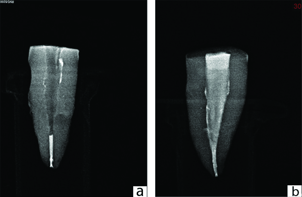
The root canal sealer AH-Plus sealer (DentsplyMaillefer, Ballaigues, Switzerland) was dispensed on a clean glass slab and mixed as per manufacturer’s recommendation. It has a working time of four hours and setting time of eight hours. The root canals were coated using the master gutta-percha point (DentsplyMaillefer, Ballaigues, Switzerland). The teeth were obturated by a hybrid warm gutta-percha obturation technique. Herein, the apical 5 mm of the root canal space was obturated by lateral compaction technique. The coronal gutta-percha was seared off using E&Q Master pen (Meta Biomed, Inc. Korea) and the GP was vertically condensed upto 10 mm length to obtain a 5 mm apical plug of GP [Table/Fig-1a]. The down pack was completed and further refined with pre adjusted hand pluggers. Backfill was done with thermoplasticised gutta-percha (Obtura Max system, Obturaspartan Endodontics, Algonquin, USA). Radiographs were taken to confirm the density of obturation [Table/Fig-1b]. The coronal 2 mm of gutta-percha was removed and cavity sealed with IRM (DentsplyMaillefer, Ballaigues, Switzerland). The specimens were stored in an incubator at 37°C and 100% humidity for 30 days to allow the sealer to set completely [12].
a) Radiograph of downpack done to obtain 5 mm apical plug of gutta-percha. b) Radiograph of backfill done with thermoplasticised gutta-percha.
Retreatment Procedures
After a period of 30 days, the teeth were randomly assigned to four groups (n=10) based on the retreatment procedure. During retreatment procedures, the roots in all the four groups were copiously irrigated with the same irrigation protocol as followed in initial endodontic treatment procedure both during and post instrumentation.
Group 1: Retreatment with H-file
Gutta-percha and sealer were removed using H-files (DentsplyMaillefer, Ballaigues, Switzerland) in a crown-down technique maintaining apical enlargement to size 30 H-file until no visual evidence of residual filling materials was seen on the file. No solvent was used in combination with H files while removing GP and sealer, in between instrumentation during retreatment procedures, the roots in all the four groups were copiously irrigated with the same irrigation protocol as followed in initial endodontic treatment procedure both during and post instrumentation. Root canal refinement was achieved using PTN X3 file at 300 rpm and #30 hand K-files. Later the canal was flooded with xylene (Fisher Scientific, Thermo Electron LLS India Pvt., Ltd., Mahape) and removed with absorbent points till they came out clean, white and dry (paper wicking) [13].
Group 2: Retreatment with H-file and PUA
Gutta-percha and sealer were removed similar to group 1, following which canal was flooded with 2 mL of xylene and PUA was done in three cycles of 20s each using #20 Irrisafe ultrasonic tip (Irrisafe tips, Acteon, Merignac, France) at a power setting of 3 for a total of 1 minute. The solvent was replenished after each cycle. This was followed by paper wicking.
Group 3: Retreatment with ProTaper Universal Retreatment Files
Gutta-percha and sealer were removed with ProTaper Universal Retreatment (PTUR) NiTi rotary instruments by a crown-down technique at a constant speed of 500 rpm for D1 instrument followed by D2 at 400 rpm and torque at 3Ncm to the working length until no visual evidence of residual filling material was seen on the file. Root canal refinement was achieved using PTN X3 file at 300 rpm and #30 hand K-files. This was followed by paper wicking.
Group 4: Retreatment with PTUR Files and PUA
Gutta-percha and sealer were removed similar to group 3 samples followed by PUA as in group 2.
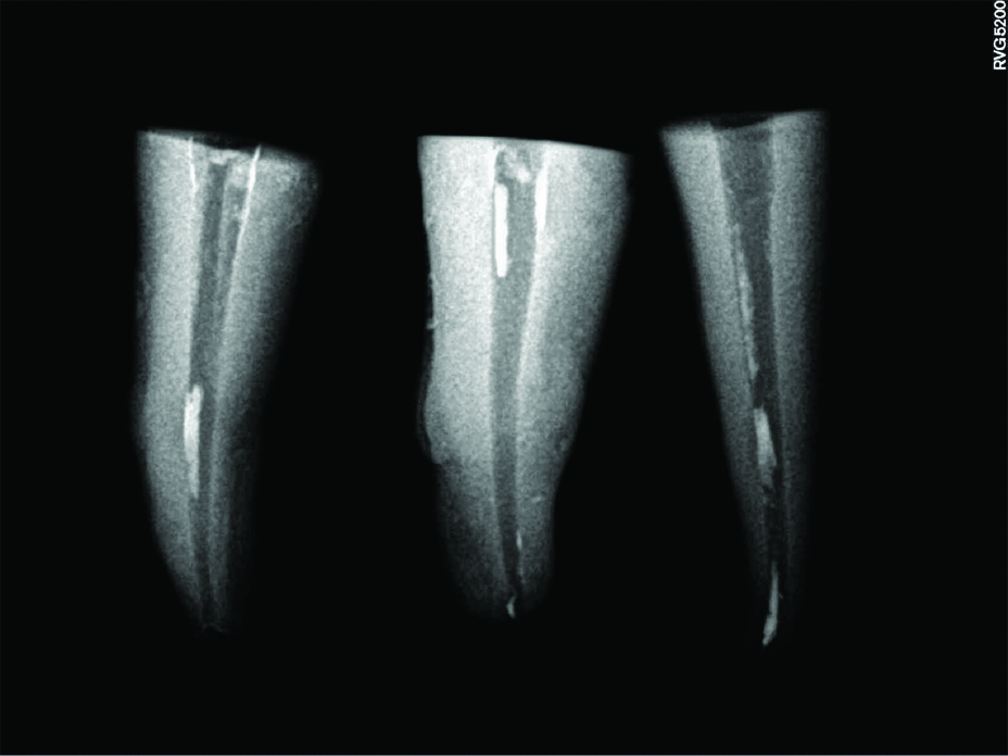
Following retreatment radiographs of all specimens were taken [Table/Fig-2]. However, the radiographs were not analysed for presence of residual filling materials. Instead the specimens were split buccolingually and observed under stereomicroscope for residual RCF. The hand and rotary instruments were discarded after instrumenting five canals. There were no procedural mishaps during the study.
Radiograph showing root canal filling material removed from the canal space.
Assessment of RCF Removal
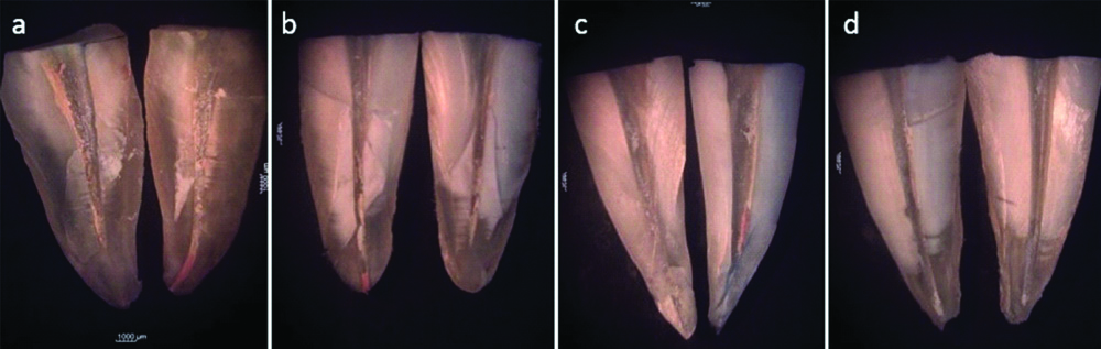
The specimens were further prepared for evaluating the residual filling materials present on the root canal walls stereomicrospically. The specimen were coded before assessment. They were grooved buccolingually using high speed diamond disk and the sections were separated using a chisel. Both the root halves were photographed under a stereomicroscope (Motic DM-39C-N9GO, MoticAsia, Hong Kong) at 10X magnification [Table/Fig-3]. The JPEG images obtained were loaded into Motic image plus 2.0 V software (Motic DM-39C-N9GO, MoticAsia, Hong Kong) for image analysis. No attempt was made to quantify the residual GP and root canal sealer separately. Total percentage of residual filing material was calculated and data was subjected to statistical analysis.
Stereomicroscope images of specimens split buccolingually after retreatment procedures. a) Group 1: Retreatment with H- file; b) Group 2: Retreatment with H-file and PUA; c) Group 3: Retreatment with ProTaper Universal retreatment files; d) Group 4: Retreatment with ProTaper Universal retreatment files and PUA.
The area of the remaining filling material and the total root canal area were traced and quantified in sqmm [Table/Fig-4]. The percentage of the RCF material on the canal walls was calculated using the formula:
An example of the area of the remaining filling material and the total root canal area traced using Motic image plus 2.0 V image analysis software for all groups.


Statistical Analysis
The data obtained was tabulated and subjected to statistical analysis using MedCalc statistical software version 18.2.1 (MedCalc Software bvba, Ostend, Belgium). Normality of data was tested using Shapiro-Wilk test and was normally distributed (p=0.5547). Further analysis was done using one-way ANOVA followed by Tukey-Kramer test for pairwise comparisons. The level of significance was set at 5% and p-values <0.05 was considered significant.
Results
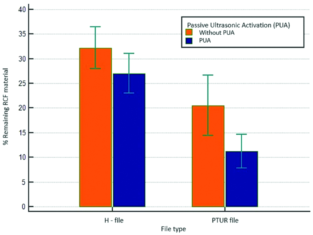
The results of the study showed that PTUR files supplemented with PUA were more efficient in removing RCF material [Table/Fig-5]. [Table/Fig-6] shows the mean and standard deviation of the residual filling materials in various groups. The highest mean value for remaining RCF material was seen for Group 1 where the retreatment was done using H-file followed by Group 2 (retreatment with H-file was supplemented with PUA). The specimens in which rotary retreatment was done with or without PUA showed considerably lesser residual RCF materials [Table/Fig-3]. The ANOVA test showed a statistically significant difference between the groups with p<0.001 [Table/Fig-7]. Intergroup comparison by Tukey Kramer test showed statistically significant difference when Group 4 was compared with Groups 1, 2 and 3 respectively [Table/Fig-8]. Also, statistically significant difference was seen when Group 3, rotary retreatment was compared to Group 1 (p=0.0024). However, a non-significant correlation was observed between Group 3 and Group 2. None of the techniques could remove RCF materials completely [Table/Fig-9]. Most of the specimens were free of RCF remnants in the coronal third of the canals except in those of Group 1 [Table/Fig-9]. All the groups showed residual RCF material on the canal walls predominantly in the apical and middle third [Table/Fig-9]. [Table/Fig-10] shows the type of residual filling materials seen in the different groups. It can be observed that the specimens belonging to the hand retreatment groups showed GP and sealer remnants where as the specimens belonging to the rotary retreatment groups showed lesser or no GP remnants but the sealer residues were present.
Comparison of mean percentage of remaining RCF materials for hand and rotary retreatment with or without PUA of solvent.
Descriptive statistics of percentage area of residual RCF material in all the four groups.
| Group | n | Mean % of remaining RCF | Standard error of mean (SEM) | 95% confidence interval | Minimum value (%) | Maximum value (%) |
|---|
| Group 1(H Files) | 10 | 32.1890 | 2.0268 | 28.0786 to 36.2994 | 21.34 | 41.07 |
| Group 2(H Files+PUA) | 10 | 27.0410 | 2.0268 | 22.9306 to 31.1514 | 18.24 | 37.51 |
| Group 3(Protaper retreatment files) | 10 | 20.5440 | 2.0268 | 16.4336 to 24.6544 | 9.06 | 34.20 |
| Group 4(Protaper retreatment files+PUA) | 10 | 11.2160 | 2.0268 | 7.1056 to 15.3264 | 4.67 | 21.01 |
| Source of variation | Sum of squares | DF | Mean square |
|---|
| Between groups (influence factor) | 2454.0697 | 3 | 818.0232 |
| Within groups (other fluctuations) | 1478.7787 | 36 | 41.0772 |
| Total | 3932.8484 | 39 | |
| F-ratio | 19.914 |
| Significance level | p<0.001 |
*p is value statistically significant; DF: Degree of freedom
Tukey Kramer test for intergroup comparison of percentage area of residual RCF material.
| Group | Group | p-value (<0.005) |
|---|
| Group 1 | Group 2 | 0.0629 |
| Group 3 | 0.0024* |
| Group 4 | <0.001* |
| Group 2 | Group 1 | 0.0629 |
| Group 3 | 0.0610 |
| Group 4 | <0.001* |
| Group 3 | Group 1 | 0.0024* |
| Group 2 | 0.0610 |
| Group 4 | 0.0077* |
| Group 4 | Group 1 | <0.001* |
| Group 2 | <0.001* |
| Group 3 | 0.0077* |
Distribution of residual filling materials observed in the coronal, middle and apical third of the specimens.
| Group | Coronal third (n=10) | Middle third (n=10) | Apical third (n=10) |
|---|
| Group 1 | 6 | 10 | 9 |
| Group 2 | 3 | 7 | 7 |
| Group 3 | 2 | 10 | 10 |
| Group 4 | 2 | 6 | 10 |
Residual filling materials observed on the root canal walls of the specimens.
| Group | GP+sealer | Only sealer |
|---|
| Group 1 (n=10) | 6 | 4 |
| Group 2 (n=10) | 8 | 2 |
| Group 3 (n=10) | 2 | 8 |
| Group 4 (n=10) | 2 | 8 |
Discussion
Success following endodontic retreatment depends on complete removal of RCF material. One of the ideal requirements of an obturating materials stated by Grossman LI is that it should be removed easily from the root canal space when the need arises [14]. Gutta-percha can be removed relatively easily from the canal depending upon the anatomy and quality of obturation.
Single rooted teeth with a single straight root canal were selected for standardisation in the present study. Root canal preparation for all the teeth was accomplished using Protaper Next rotary files (DentsplyMaillefer, Ballaigues, Switzerland) in a crown down technique to an apical size of 30. The canals were obturated by a hybrid technique combining lateral compaction and warm gutta-percha technique. This technique takes the advantage of lateral compaction which provides a tight apical seal through the compaction of several gutta-percha points by a spreader in the apical region and warm gutta-percha that can adapt more effectively to canal irregularities in addition to filling the lateral canals [15]. This technique provides better obturation length control too. It has been reported that warm gutta-percha obturations and resin based sealers pose a challenge during retreatment [16].
The present study evaluated the removal of gutta-percha using a combination of hand/rotary retreatment files, chemical solvent and ultrasonic instruments since a combination technique is recommended for safe, efficient and possibly complete removal of RCF [7,17].
Previous studies have evaluated the RCF removal using radiography, computed tomography, operational microscope and clearing method [18]. In this study, the residual filling material was assessed by longitudinally splitting the roots into two separate halves and photographed under a stereomicroscope. The images were later analysed. This is a well established and effective technique, which the recent studies have used to assess residual filling materials [19]. All the above methods have specific limitations and ideally 3 dimensional visualisation of the root canal system would provide better detection of remaining RCF material [18].
Rotary instruments like PTUR files have been reported to perform better than H-files in retreating teeth obturated with warm gutta-percha obturation techniques as observed in the present study [Table/Fig-3,5] [20,21]. Group 3 and 4 where PTUR with or without PUA showed residual sealer and most of them had no GP residues [Table/Fig-10]. Protaper retreatment files have been shown to leave significantly cleaner walls in lesser time than compared to hand files [Table/Fig-5]. The PTUR files remove large increments of GP in spirals around the instruments resulting in softening of GP and cut it. The active tip of D1 in combination with initial solvent application facilitates further penetration of D2 and D3 which have a non-active tip. The non active tip of D2 and D3 prevents mishaps such as ledging, perforations and instrument separation during filing removal [19]. The tip diameter of the Master Apical File (MAF) used to instrument the canals was a size 30 (PTN X3 file) and the diameter of the retreatment file used at the working length was a size 25 D2 (size 25, .08 taper). It has been shown in a previous study that these retreatment instruments may not remove the RCF materials completely from the root canal as it is of a smaller diameter than compared to the MAF [21]. Supplementary instrumentation performed after the use of PTUR files with a larger apical diameter file than the apical diameter of MAF used in the initial enddontic treatment resulted in more effective RCF removal than the use of only rotary retreatment files in the apical third [21]. However in the present study, root canal refinement after instrumentation with rotary retreatment files was achieved using PTN X3 file at 300 rpm and #30 hand K-files to the working length material. The MAF was used because it has reported that additional instrumentation with a larger size file may result in more apical crack initiation and propagation of existing cracks [22]. The hand and rotary files may mechanically remove the bulk of the gutta-percha and sealer but remnants remaining on the walls will require a solvent for removal [4]. Organic solvents such as chloroform, eucalyptol oil, xylene, halothane, turpentine oil and pandine needle oil have been advocated to soften gutta-percha and RCS to assist in its removal safely and quickly [23].
Chemical solvents used for removal of RCF have been found to form a fine layer of softened gutta-percha and sealer smearing the canal walls [24]. Residual obturation materials, especially sealer present on the root canal walls and in canal ramifications remain largely inaccessible and resist dissolution [25]. Previously, passive ultrasonic irrigation of NaOCl and EDTA for one minute following gutta-percha removal has shown to improve canal and isthmus cleanliness due to acoustic streaming [25,26]. However, the filling materials were not completely removed.
In the present study, PUA of xylene was attempted to evaluate the filling material removal. Xylene (dimethylbenzene), an aromatic compound has been widely investigated and found to be an efficient solvent for root canal obturating materials including epoxy resin based sealers such as AH Plus [23,27] H-files and PTUR files when supplemented with PUA of xylene showed better removal of the RCF which was statistically significant when compared to hand and PTUR files without PUA of xylene. Hence the null hypothesis was rejected. However, in agreement with other studies, none of the techniques in the current study too could remove RCF materials completely from the root canal [18,19,21]. The remnants seen in Group 1 were observed to be present in coronal, middle and apical third [Table/Fig-9]. Moreover in these specimens residues of GP and sealer were noted [Table/Fig-3,10]. In the specimens of Groups 2 and 3, residual filling materials were present mainly in the middle and apical third consisting of both gutta-percha and RCS [Table/Fig-3,9,10]. However, in Group 3 and 4, the residues were mainly of RCS seen in the apical third [Table/Fig-3,10]. It can be appreciated in [Table/Fig-9] that majority of specimens belonging to Groups 2,3 and 4 showed no residues in the coronal third. In the present study, it was observed that both hand and rotary files showed lesser residual debris when instrumentation was supplemented with PUA [Table/Fig-5]. Better results observed in the PUA groups may be due to the increase in temperature of xylene caused by PUA which could have resulted in enhanced removal of guttapercha in addition to acoustic streaming [6,10,26].
The results of this study are in agreement with one study where chloroform and NaOCl were ultrasonically activated, both were found to be efficient in removing gutta-percha and RCS [3]. Though chloroform was the solvent of choice for removal of GP previously, it is no longer in clinical use owing to its cytotoxicity, carcinogenic potential and toxicity to the tissues [6,27,28]. Xylene (dimethylbenzene) is a less toxic alternative to chloroform. Another study reported PUA of an orangewood oil and eucalyptol resulted in better removal of Zinc Oxide Eugenol (ZOE) sealer discs than epoxy resin based sealers [6]. In agreement to the present study, Cavenago BC et al., reported that the use of passive ultrasonic irrigation with 2.5% NaOCl for three activation cycles of 20s each totaling one minute after GP-sealer removal and irrigation with xylene enhanced the removal of filling materials [7]. In contrast to this study Müller GG et al., evaluated the passive ultrasonic irrigation of Endosolv R for the removal of RCF material from the canal walls and observed that ultrasonic activation of Endosolv R was not effective for removal of filling debris [10]. The residual filling material was guttapercha which may not have been susceptible to dissolution by Endosolv R and in addition, the solvent was not replenished in the canal as generally performed to facilitate removal of residual filling material [29]. Similarly, two other studies reported that passive ultrasonic irrigation technique was ineffective in removing residual root canal filling material during endodontic retreatment. However in these studies, passive ultrasonic irrigation with NaOCl was performed for 3 cycles of 20s totaling one minute in one canal followed by 17% EDTA solution [7,30]. In contrast, in the current study, the organic solvent used was xylene and the solvent was replenished after a twenty second PUA in the canal space. Thus, the specimens where PUA was carried out showed cleaner root canal walls. A comparison of the results of the present study and the studies conducted in recent past is shown in [Table/Fig-11].
Comparison of the results of the present study and the studies conducted in recent past.
| Author (s) | Year | Methodology | Conclusions |
|---|
| Wilcox LR et al., [2] | 1987 | Retreatment system: hand instruments like heated pluggers, k files and barbed broachesAfter GP-sealer removal, ultrasonic activation of either NaOCl or chloroform done. | Ultrasonic activation of either NaOCl or chloroform resulted in cleaner canal walls. |
| Müller GG et al., [10] | 2013 | Retreatment system: PTURAfter GP-sealer removal with PTUR.Passive ultrasonic irrigation of Endosolv R(formamide). Solvent was ultrasonically activated for 60 seconds without renewal. | Technique found to be ineffective in removing RCF materials from the root canal. |
| Cavenago BC et al., [7] | 2014 | Retreatment system: Bio Ra Ce;After GP-sealer removal with PTUR. canals were irrigated with xylene.Followed by three activation cycles with 2.5% NaOCl performed for 20s totaling 1 min in each canal. | The use of Passive ultrasonic irrigation after GP-sealer removal enhanced the removal of filling materials of the canals. |
| Latheef AA et al., [8] | 2016 | Retreatment system: PTURPassive ultrasonic irrigation with 1 mL 3% NaOCl for 1 min after GP-sealer removal with PTUR. | The use of Passive ultrasonic irrigation after GP-sealer removal enhanced the cleanliness of canals, but could not remove all the filling debris. |
| Michelona C et al., [30] | 2016 | Retreatment system: PTURHuman mandibular molars having isthmus in the mesial roots were selected. After GP-sealer removal with PTUR, three activation cycles with 2.5% NaOCl was performed for 20s totaling 1 min in one canal, followed by 17% EDTA solution activated ultrasonically for 1 min. | The Passive ultrasonic irrigation technique was ineffective in removing residual root filling material during endodontic retreatment in root canals with a complex anatomy when used after removal of RCF materials. |
| Current study | 2018 | Retreatment system: PTURAfter GP-sealer removal with PTUR passive ultrasonic activation of xylene was done in three cycles of 20s each for a total of 1 minute. The solvent was replenished after each cycle. This was followed by paper wicking. | Passive ultrasonic activation of xylene enhanced the removal of RCF though the walls were not completely free of debris. |
Limitation(s)
In-vitro study design and inclusion of teeth with straight canals, hence the complex intraoral conditions, canal curvatures, isthmi and ramifications could not be accounted for. Also, AH plus being an epoxy resin based sealer, the efficacy of removal using other resin sealer solvents has to be assessed. In addition, ultrasonic files can be used only in the straight part of the canal hence the efficacy of PUA in the presence of curved and narrow canals have to be evaluated. Further studies need to be carried out to investigate the canal cleanliness at different intensities of PUA, different solvents and apical extrusion of gutta-percha and solvent.
Conclusion(s)
Within the limitations of this in-vitro study, it was observed that hand and rotary instrumentation when supplemented with PUA of xylene is more efficient in removing RCF materials from the root canal space.
*p is value statistically significant; DF: Degree of freedom