Bilateral Fusion of Primary Mandibular Lateral Incisors and Canines: A Case Report
Sahar Fahad Alotaibi1, Raya Almufleh2, Hamed Alshamrani3, Fouad Salama4
1 Dental Intern, College of Dentistry, King Saud University, Riyadh, Saudi Arabia.
2 Dental Intern, Dental Intern, Riyadh, Saudi Arabia.
3 Consultant, Department of Paediatric Dentistry and Orthodontics, College of Dentistry, King Saud University, Riyadh, Saudi Arabia.
4 Professor, Department of Paediatric Dentistry and Orthodontics, College of Dentistry, King Saud University, Riyadh, Saudi Arabia.
NAME, ADDRESS, E-MAIL ID OF THE CORRESPONDING AUTHOR: Sahar Fahad Alotaibi, 102050, Riyadh, Riyadh, Saudi Arabia.
E-mail: dr.sahar1994@hotmail.com
Fusion is known as a developmental anomaly described by the union of two neighbouring teeth. The worldwide prevalence of bilaterally fused teeth in the primary dentition is about 0.02% which is considered very rare. Only few cases of bilateral fusion have been reported in Indian population. This paper presents a rare case of three-year-old boy with bilateral fusion of primary mandibular lateral incisors and canine teeth. Treatment consisted of pit and fissure sealants and topical fluoride application. To conclude, fused primary teeth are considered as harmless asymptomatic abnormality.
Absent teeth, Bifid crown, Developmental anomaly, Double tooth, Gemination
Case Report
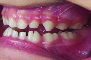
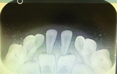
A three-year-old boy was referred to the primary care dental clinic for a routine dental examination. The medical history was non-relevant. There was no past dental history. The family history was insignificant, and the parents reported no consanguinity. Intraoral examination showed primary dentition with good oral hygiene and no carious lesion. In addition, examination revealed the presence of two large teeth bilaterally in the mandibular canine and lateral incisor area. Teeth counting revealed two missing teeth when the anomalous teeth were counted as one. The fused tooth on the right side had a groove on the labial surface that starts from the incisal edge to the gingival third marking the outline of each individual tooth [Table/Fig-1a]. While the fused tooth on the left side had a similar groove on the labial surface but terminates at the middle third making the gingival third completely fuse [Table/Fig-1b]. The provisional diagnosis was of fused mandibular lateral incisor and canine bilaterally. For further diagnosis, informed consent was obtained from the patient’s parent. Radiographs were taken to confirm fusion of primary teeth and check the development of permanent successors [Table/Fig-2]. A mandibular anterior radiograph showed bilateral single-rooted mandibular canine and lateral incisor and double pulp chamber [Table/Fig-2]. After thorough clinical and radiographic examination, it was diagnosed as bilateral fusion involving the mandibular primary lateral incisors and canines.
Intraoral lateral view of the fused teeth in the mandibular right side.
Intraoral lateral view of the fused teeth in the mandibular left side.
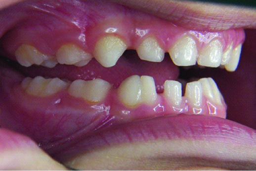
Intraoral frontal view of the fused teeth in the mandibular anterior area.
As no caries was detected between the fused teeth, pit and fissure sealants were applied to the groove and topical fluoride application was performed as well. Moreover, the parents were informed about the need for regular six months follow-up but the patient lost to follow-up.
Discussion
Double tooth is the term frequently used to describe the anomaly of conjoined teeth, which include fusion, gemination and concrescence [1]. Fusion is defined as a developmental anomaly characterised by the union of two adjacent teeth. The union may be complete or incomplete depending on the developmental stage at the time the union occurs. Fused teeth are joined by the dentin, but their pulp chambers and canals may be separated or joined [2]. On the other hand, gemination is known as an attempt by a single tooth bud to divide which result in formation of either a large tooth with a bifid crown or two divided teeth completely in both the crown and root [3]. Fusion may be distinguished from gemination by counting the teeth, which reveals a missing tooth when the malformed tooth is counted as one [3]. In contrast to fusion and gemination, concrescence is a condition involving fusion of the roots area only of adjacent teeth by cementum [4] and was not included in this differential diagnosis as there was fusion in the coronal part of the teeth in the present case.
Fusion occurs more often in the primary teeth with an estimated worldwide prevalence of 0.5% to 2.5% [5] compared to permanent teeth with an estimated prevalence of 0.1% [6]. In Indian population, the overall prevalence of fusion in primary teeth is 0.27% [7]. The most common cases of fusion involved unilateral mandibular canines and lateral incisors [8]. The bilateral presentation of fusion in the deciduous teeth is about 0.02%, which is considered very rare [5]. To the best of our knowledge, only 17 cases of bilateral fusion of mandibular primary lateral incisor and canine have been reported in the English literature since 1940 [Table/Fig-3] [2,3,5,6,9-19].
Cases of bilateral fusion of mandibular primary lateral incisor and canine reported in the literature since1940.
| Sl. No. | Author | Age | Gender | Year |
|---|
| 1 | Tinn CA [9] | Not reported | Not reported | 1940 |
| 2 | Menczer LF [10] | Not reported | Not reported | 1955 |
| 3 | Pogrel H [11] | 8 | Female | 1956 |
| 4 | Munro D [12] | Not reported | Not reported | 1958 |
| 5,6 | Brook AH, Winter GB [13] | Not reported | Female | 1970 |
| 7 | Nik-Hussein NN [14] | 11 | Female | 1989 |
| 8 | Alpoz AR et al., [5] | 9 | Male | 2003 |
| 9 | Chalakkal P, Thomas AM [16] | 8 | Male | 2009 |
| 10 | Rajashekhara B et al., [6] | 8 | Female | 2010 |
| 11 | Tewari N, Panday R [17] | 1 | Male | 2011 |
| 12 | Şekerci AE et al., [2] | 8 | Female | 2012 |
| 13 | Barbério GS et al., [18] | 5 | Female | 2013 |
| 14 | Prabhu RV et al., [3] | 4 | Male | 2013 |
| 15 | Reddy VR et al., [15] | 7 | Male | 2017 |
| 16,17 | Bhavna Dave SS [19] | 9,12 | Male | 2018 |
| 18 | AlOtaibi (Present Case) | 3 | Male | 2019 |
Fused teeth are usually associated with multiple concerns. The major concern is the risk of caries due to the presence of vertical labial and/or lingual grooves on the crown surface [5]. Fissure sealants or composite restorations are highly recommended to be placed in these grooves [20]. In addition to caries, periodontal problems may occur if the grooves extend to the root surface [5]. However, no caries or periodontal problems were present in this case even though the teeth were fused completely. Fissure sealants were placed as a preventive measure.
Furthermore, due to the increased area of the fused root surface and the greater root mass, delayed resorption of the root may result, leading to a delayed or ectopic eruption of their permanent successor [2,6]. In fact, studies have found that half of the fused teeth were followed by permanent dentition anomaly. Moreover, the incidence of hypodontia or supernumerary teeth in family histories was found in some cases [13]. Reviewing the family history of this case, we found that fused teeth have not been detected among the parents and siblings of the patient.
Another concern usually presents with fusion cases is compromised aesthetic, as the fused teeth appear to be large and solitary. Diastema may be present because of excess dental space [2]. In the present case, visible diastema was seen between the fused teeth and the central incisors bilaterally, but aesthetic was not a problem and the family was unaware of the fused teeth until the patient visited the dental clinic. The aesthetic might even be worse in the permanent dentition when there is a congenital absence of lateral incisor. Hagman FT has reported that 75% of the fusion cases in primary teeth are presented with congenitally missing permanent lateral incisors [21]. Congenital absence of permanent teeth will affect the occlusion which will require both cosmetic and orthodontic consideration [5]. Bilateral mandibular fusion cases reported by Şekerci AE et al., Alpoz AR et al., and Bhavna Dave SS et al., exhibited missing succedaneous lateral incisors [2,5,19]. However, lower occlusal radiograph confirmed the presence of permanent lateral incisors in this case. Orthopantamograph was needed to confirm presence of permanent canines but cannot be provided because the child is three-year-old and Orthopantamograph is only recommended by the American Academy of Pediatric Dentistry around six-year-old when first permanent molar erupt [22].
The proper management of fused teeth depends on several factors, which include the level of fusion, the morphologic result and the teeth that included [15]. If the fused teeth are primary, teeth should be retained as they are. However, if extraction is indicated, before starting the procedure, it is important to determine the presence of their succedaneous teeth. If the fused teeth are caries-free, preventive advice is to be given to the child and parent. Also, dietary changes, periodic follow-ups, topical fluoride application, fissure sealant, as preventive approach should be implemented. The restorative procedure should be done in case of the presence of caries while pulpal involvement mandates endodontic treatment. Functional occlusion and aesthetics improvement should be insured by considering orthodontic and prosthodontic management [3]. If needed it is critical to have a multidisciplinary approach with multiple practitioners in different specialties of dentistry to achieve the best functional and aesthetic success to treat these rare cases [6]. Long-term and regular follow-ups have been emphasised in the fusion cases [3].
Conclusion(s)
Fused primary teeth are considered harmless asymptomatic abnormality. However, their presence could lead to several dental consequences including caries, periodontal disease and congenital absence of their permanent successors affecting both aesthetic and occlusion. Consequently, it’s considerably important to provide early diagnosis, preventive approach, regular follow-up and multidisciplinary treatment to achieve functional and aesthetic success in management of such rare cases.
Author Declaration:
Financial or Other Competing Interests: No
Was informed consent obtained from the subjects involved in the study? Yes
For any images presented appropriate consent has been obtained from the subjects. Yes
Plagiarism Checking Methods: [Jain H et al.]
Plagiarism X-checker: Sep 28, 2019
Manual Googling: Dec 10, 2019
iThenticate Software: Dec 30, 2019 (13%)
[1]. Yuen SW, Chan JC, Wei SH, Double primary teeth and their relationship with the permanent successors: A radiographic study of 376 casesPediatr Dent 1987 9(1):42-48. [Google Scholar]
[2]. Şekerci AE, Şişman Y, Ertaş ET, Gümüş H, Ertaş H, Clinical and radiographic evaluation and comparison of six cases of fusion involving the primary dentitionJ Dent Child (Chic) 2012 79(1):34-39. [Google Scholar]
[3]. Prabhu RV, Chatra L, Shenai P, Prabhu V, Bilateral fusion in primary mandibular teethIndian J Dent Res 2013 24(2):27710.4103/0970-9290.11668023965464 [Google Scholar] [CrossRef] [PubMed]
[4]. Tapadiya DV, Ramanojam DS, Gelada DK, Sethi DS, Oswal DN, Concrescence of erupted second molar and impacted third molar: A rare case reportIOSR J Dent Med Sci 2017 16(4):74-76.10.9790/0853-1604067476 [Google Scholar] [CrossRef]
[5]. Alpöz AR, Munanoğlu D, Oncag O, Mandibular bilateral fusion in primary dentition: Case reportJ Dent Child (Chic) 2003 70(1):74-76. [Google Scholar]
[6]. Rajashekhara B, Dave B, Manjunatha B, Poonacha K, Sujan SG, Bilateral fusion of primary mandibular lateral incisors and canines: A report of a rare caseRev Odonto Ciência 2010 25(4):427-29.10.1590/S1980-65232010000400020 [Google Scholar] [CrossRef]
[7]. Gupta SK, Saxena P, Jain S, Jain D, Prevalence and distribution of selected developmental dental anomalies in an Indian populationJ Oral Sci 2011 53(2):231-38.10.2334/josnusd.53.23121712629 [Google Scholar] [CrossRef] [PubMed]
[8]. Chen YH, Cheng NC, Wang YB, Yang CY, Prevalence of congenital dental anomalies in the primary dentition in TaiwanPediatr Dent 2009 32(7):525-29. [Google Scholar]
[9]. Tinn CA, Excess, deficiency and gemination in the deciduous and permanent dentition of school childrenBr Dent J 1940 68:236-38. [Google Scholar]
[10]. Menczer LF, Anomalies of the primary dentitionAnomalies Prim dentition 1955 22:57-62. [Google Scholar]
[11]. Pogrel H, Case of bilateral gemination of deciduous incisors with congenital absence of permanent successorsDent Pr 1956 7:13-14. [Google Scholar]
[12]. Munro D, Gemination in the deciduous dentitionBr Dent J 1958 104:238-40. [Google Scholar]
[13]. Brook AH, Winter GB, Double teeth. A retrospective study of ‘geminated’ and ‘fused’ teeth in childrenBr Dent J 1970 129(3):123-30.10.1038/sj.bdj.48025335272471 [Google Scholar] [CrossRef] [PubMed]
[14]. Nik-Hussein NN, Bilateral symmetrical fusion of primary and permanent mandibular lateral incisors and caninesJ Pedod 1989 13(4):378-83. [Google Scholar]
[15]. Reddy Vr, Chowdhary N, Kumar G, Ambareen Z, Bilateral fusion of mandibular primary teeth with partial anodontia of permanent teeth: A report of a rare caseSaudi J Heal Sci 2017 6(3):18210.4103/sjhs.sjhs_43_17 [Google Scholar] [CrossRef]
[16]. Chalakkal P, Thomas AM, Bilateral fusion of mandibular primary teethJ Indian Soc Pedod Prev. Dent 2009 27(2):108-10.10.4103/0970-4388.5533619736504 [Google Scholar] [CrossRef] [PubMed]
[17]. Tewari N, Pandey R, Bilateral fusion in primary mandibular teeth: A report of two casesJ Indian Soc Pedod Prev Dent 2011 29(1):5010.4103/0970-4388.7993621521919 [Google Scholar] [CrossRef] [PubMed]
[18]. Barbério GS, da Costa SV, Rios D, de Oliveira TM, de Andrade Moreira Machado MA, Rare case of bilateral gemination in deciduous teethInt J Morphol 2013 31(2):575-77.10.4067/S0717-95022013000200035 [Google Scholar] [CrossRef]
[19]. Bhavna Dave SS, Bargale S, Patel R, Bilateral fusion of mandibular primary lateral incisor and canine with missing lateral succedaneous teeth: A rare dental anomalyClinical Dentistry 2018 12(6):10-14. [Google Scholar]
[20]. Mochizuki K, Yonezu T, Yakushiji M, Machida Y, The fusion of three primary incisors: Report of caseASDC J Dent Child 1999 66(6):421-25.:367 [Google Scholar]
[21]. Hagman FT, Anomalies of form and number, fused primary teeth, a correlation of the dentitionsASDC J Dent Child 1988 55(5):359-61. [Google Scholar]
[22]. American Academy of Pediatric Dentistry, “Prescribing Dental Radiographs for Infants, Children, Adolescents, and Individuals with Special Health Care Needs,” 2017 [Google Scholar]