Leg Ulcers Secondary to Antiphospholipid Syndrome in a Young Female
Aditya Dhanawat1, Partisha Gupta2, Prasanta Padhan3, Lalatendu Mohanty4
1 PG Resident, Department of Internal Medicine, Kalinga Institute of Medical Sciences, Bhubaneswar, Odisha, India.
2 PG Resident, Department of Internal Medicine, Kalinga Institute of Medical Sciences, Bhubaneswar, Odisha, India.
3 Associate Professor, Department of Rheumatology, Kalinga Institute of Medical Sciences, Bhubaneswar, Odisha, India.
4 Professor, Department of Internal Medicine, Kalinga Institute of Medical Sciences, Bhubaneswar, Odisha, India.
NAME, ADDRESS, E-MAIL ID OF THE CORRESPONDING AUTHOR: Dr. Lalatendu Mohanty, Professor, Department of Internal Medicine, Kalinga Institute of Medical Sciences, Bhubaneswar, Odisha, India.
E-mail: lalatendumohanty3@gmail.com
Eighty percent of leg ulcers have a vascular aetiology. These ulcers are often debilitating for the patients and difficult to treat owing to lack of clinical suspicion leading to significant morbidity and mortality. Here we report a case of bilateral leg ulcers in a young female with Anti Phospholipid Syndrome (APS). She also had associated Evans’ syndrome in the absence of lupus. She responded well with oral anticoagulant and immunosuppressives on follow-up after 6 weeks and 12 weeks. Thus, APS should be kept in mind while evaluating a case with leg ulcers in a young patient as in the present case.
Arterial ulcers, Autoimmune, Deep vein thrombosis, Evans’ syndrome, Lower limb ulcers
Case Report
A 27-year-old married female was admitted with complaints of ulcers on right leg and dorsum of left foot for three months. They were gradually progressive, painful, non-healing, non foul-smelling and there was no history of discharge from the ulcers. She was a known case of recurrent deep vein thrombosis of the left lower limb for the last eight years and she was not on any anti-coagulant. She was put on Inferior Vena Cava (IVC) filter six years back. There was no history of rashes, photosensitivity, arthralgia, oral ulcers, alopecia or abortions. She is a non-diabetic and non-hypertensive. Family history was not suggestive of any chronic illness. She was vegetarian, literate, married since four years, nulligravid, normal menstrual cycle and normal bowel and bladder habit.
Treatment history revealed that she had been applying topical antibiotics for the last one month over the ulcers and reconstructive surgery was done for similar ulcer over left leg one year back.
On examination, she was conscious, oriented to time, place and person. Her Body Mass Index (BMI) was 23 kg/m2. There was no evidence of pallor, icterus, cyanosis, clubbing, lymphadenopathy or oedema. Her vitals were stable and systemic examination was within normal limits.
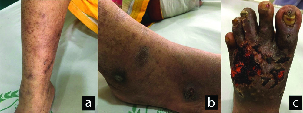
Local examination revealed presence of multiple ulcers on the medial and lateral aspect of right leg [Table/Fig-1a] and foot [Table/Fig-1b], largest measuring 3×3 cm. They were well-defined, rounded, painful and with a necrotic base. There was also a 6×5 cm ulcer over the dorsum of the left foot which was tender, necrotic base, well-defined edges with punched out appearance and erythematous periphery [Table/Fig-1c]. With these findings, a clinical diagnosis of arterial ulcers secondary to peripheral arterial disease was made. Differential diagnoses of diabetic foot ulcer, infectious ulcers, pyoderma gangrenosum and Antineutrophil cytoplasmic antibody (ANCA)-associated vasculitic ulcers were also considered.
Right leg (a), right foot (b) and left foot (c) ulcer at the time of diagnosis.
On investigations, her leucocyte count was 6500/mm3 and platelet count was 1,40,000/mm3. Her haemoglobin was 8.2 g/dL, Erythrocyte Sedimentation Rate (ESR) was 96 mm in 1st hour. Her serum iron was 22 mcg/dL and transferrin saturation was 8%. Peripheral smear revealed normocytic and normochromic anaemia.
Her serum Lactate Dehydrogenase (LDH) was raised (432 U/L) and Direct Coomb’s Test was positive. Prothrombin time was 16.7 seconds and INR was 1.62. Screening Antinuclear antibody (ANA) and Anti-double stranded DNA antibody (Anti-dsDNA) were negative. Test for lupus anticoagulant by Dilute Russel Viper Venom Test (DRVVT) method was negative. β2-Glycoprotein-1 IgM was positive by EIA (>150 SMU, Normal range- <20 SMU). Her Anti-Cardiolipin IgM antibody was also positive (9.74 U/mL; Normal range <7 U/mL). Subsequently, Anti-Cardiolipin IgM antibody was still elevated when tested after 12 weeks (9.39 U/mL; Normal range < 7 U/mL).
A 2D Echocardiography was normal. Venous doppler of both lower limbs were normal. Contrast CT Thorax did not reveal any evidence of pulmonary embolus. Her liver and kidney function tests were within normal limits. Thus, with the history of recurrent venous thrombosis, examination finding of arterial ulcers and subsequent laboratory findings, a diagnosis of Primary Antiphospholipid Syndrome (APS) was made.
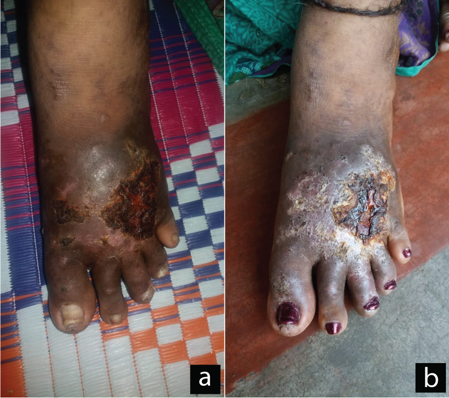
She was started on oral warfarin 2 mg once daily with a target INR of 2.5-3.5 and hydroxychloroquine 200 mg once daily as an anti-thrombotic. For Evan’s syndrome (autoimmune haemolytic anaemia with thrombocytopenia), she was given oral prednisolone 30 mg once daily with azathioprine 50 mg once daily. She responded to treatment and her ulcers regressed significantly when reviewed at 6 weeks and 12 weeks of medication [Table/Fig-2]. Repeat complete blood count at 12 weeks was within normal limits.
Left foot ulcer after 6 weeks of treatment (a) and 12 weeks of treatment (b).
Discussion
Eighty percent of leg ulcers have a vascular aetiology (48% venous, 15% arterial and 17% mixed) according to a large epidemiological study [1]. About 20-23% of patients have ulcers due to complex etiologies such as vasculitis, pyoderma gangrenosum and other autoimmune diseases [2].
Antiphospholipid Syndrome (APS) is an autoimmune disease entity diagnosed by two criteria-clinical and laboratory. The clinical criteria encompass either of three presentations such as arterial, venous thrombosis or pregnancy morbidity. The laboratory criteria constitute presence of either of three autoantibodies such as anti-cardiolipin, anti-β2-glycoprotein or lupus anticoagulant on two separate occasions 12 weeks apart [3].
APS could either be primary or secondary to Systemic Lupus Erythematosus (SLE), malignancies, infections or drugs. Thirty nine percent patients present with deep vein thrombosis followed by early foetal loss (less than 10 weeks) in 35% and stroke in 20%. Leg ulcers or digital gangrene is seen in 9% patients [4]. The prevalence of APS is estimated to be about 40-50 cases per 1,00,000 population [5].
The present case had recurrent deep vein thrombosis and arterial thrombosis as evidenced by the bilateral leg and foot ulcers coupled with laboratory findings of anti-β2-glycoprotein IgM and anti-cardiolipin IgM positive. She also had autoimmune haemolytic anaemia and thrombocytopenia, known as Evans’ syndrome which is present in 10% patients of APS [6].
In a retrospective study of 450 patients suspected of APS in southeast Asia by Jatuworapruk K et al., stroke was found to be the most common finding followed by deep vein thrombosis and pulmonary embolism and the majority of patients (79%) tested positive for lupus anticoagulant whereas the present case had history of recurrent deep vein thrombosis and examination finding suggestive of leg ulcers due to arterial thrombosis [7]. Also, lupus anticoagulant was negative in the present case.
Apart from leg ulcers, other cutaneous manifestations of primary APS include Raynaud’s phenomenon, acral necrosis, livedo reticularis and subcutaneous nodules [8]. In a study, 60 patients with clinical findings suggestive of APS were screened. Thirty-nine were found to be positive out of which 25 patients were primary APS. Forty percent of the APS patients had cutaneous manifestations of which four patients had ulcers [9]. In a case report by Tishler M et al., a 51-year-old woman with unilateral foot ulcers as the presenting symptom was found to have primary APS and treatment with anticoagulants resulted in complete disappearance of the ulcers [10]. Leg ulcers are usually severe in APS and respond to treatment with immunosuppressives and anticoagulants such as warfarin [11].
Conclusion
The present case had recurrent history of deep vein thrombosis for eight years and reconstructive surgery was done one year back for the left leg ulcer but the cause was never ascertained which led us to believe that awareness about primary APS and its clinical suspicion is warranted in a woman of reproductive age-group presenting with venous, arterial thrombosis or recurrent pregnancy morbidity. Thus, APS should be kept in mind in a female presenting with leg ulcers.
Author Declaration:
Financial or Other Competing Interests: No
Was informed consent obtained from the subjects involved in the study? Yes
For any images presented appropriate consent has been obtained from the subjects. Yes
Plagiarism Checking Methods: [Jain H et al.]
Plagiarism X-checker: Oct 08, 2019
Manual Googling: Nov 08, 2019
iThenticate Software: Nov 20, 2019 (4%)
[1]. Körber A, Klode J, Al-Benna S, Wax C, Schadendorf D, Steinstraesser L, Etiology of chronic leg ulcers in 31,619 patients in Germany analyzed by an expert surveyJDDG: Journal der DeutschenDermatologischen Gesellschaft 2011 9(2):116-21.10.1111/j.1610-0387.2010.07535.x20946240 [Google Scholar] [CrossRef] [PubMed]
[2]. Shanmugam VK, Schilling A, Germinario A, Mete M, Kim P, Steinberg J, Prevalence of immune disease in patients with wounds presenting to a tertiary wound healing centreInternational Wound Journal 2012 9(4):403-11.10.1111/j.1742-481X.2011.00899.x22168783 [Google Scholar] [CrossRef] [PubMed]
[3]. Miyakis S, Lockshin MD, Atsumi T, Branch DW, Brey RL, Cervera R, International consensus statement on an update of the classification criteria for definite antiphospholipid syndrome (APS)J ThrombHaemost 2006 4(2):295-306.10.1111/j.1538-7836.2006.01753.x16420554 [Google Scholar] [CrossRef] [PubMed]
[4]. Cervera R, Piette JC, Font J, Khamashta MA, Shoenfeld Y, Camps MT, Antiphospholipid syndrome: Clinical and immunologic manifestations and patterns of disease expression in a cohort of 1,000 patientsArthritis Rheum 2002 46(4):1019-27.10.1002/art.1018711953980 [Google Scholar] [CrossRef] [PubMed]
[5]. Litvinova E, Darnige L, Kirilovsky A, Burnel Y, de Luna G, Dragon-Durey MA, Prevalence and significance of non-conventional antiphospholipid antibodies in patients with clinical APS criteriaFront Immunol 2018 9:297110.3389/fimmu.2018.0297130619328 [Google Scholar] [CrossRef] [PubMed]
[6]. Moutsopoulos HM, Antiphospholipid Syndrome. In: Kasper Dennis L, Fauci Anthony S, Longo Dan L, Hauser Stephen L, Jameson J Larry, Loscalzo JHarrison’s Principles of Internal Medicine 2018 20edU.S.A.McGraw-Hill Publishing:2526-27. [Google Scholar]
[7]. Jatuworapruk K, Bhoopat L, Hanvivadhanakul P, Clinical and immunological characteristics of antiphospholipid syndrome in an Asian population: a retrospective studyAsian Pac J Allergy Immunol 2019 37(3):171-78. [Google Scholar]
[8]. Van Beek N, Schumacher N, Haase O, Zillikens D, Kahle B, Schmidt E, Primary antiphospholipid syndrome: Newly developed leg ulcer and history of strokeHautarzt 2013 64(9):666-70.10.1007/s00105-013-2601-623744031 [Google Scholar] [CrossRef] [PubMed]
[9]. Diógenes MJ, Diógenes PC, de Morais Carneiro RM, Neto CC, Duarte FB, Holanda RR, Cutaneous manifestations associated with antiphospholipid antibodiesInt J Dermatol 2004 43(9):632-37.10.1111/j.1365-4632.2004.01939.x15357740 [Google Scholar] [CrossRef] [PubMed]
[10]. Tishler M, Papo J, Yaron M, Skin ulcer as the presenting symptom of primary antiphospholipid syndrome-resolution with anticoagulant therapyClin Rheumatol 1995 14(1):112-14.10.1007/BF022080967743736 [Google Scholar] [CrossRef] [PubMed]
[11]. Cañas CA, Durán CE, Bravo JC, Castaño DE, Tobón GJ, Leg ulcers in the antiphospholipid syndrome may be considered as a form of pyoderma gangrenosum and they respond favorably to treatment with immunosuppression and anticoagulationRheumatol Int 2010 30(9):1253-57.10.1007/s00296-010-1418-120349240 [Google Scholar] [CrossRef] [PubMed]