Multiple Dental Anomalies in a Patient with Down Syndrome
Kriss Mélani Gárate1, Darlle Santos Araújo2, Fernanda Da Silva3, Gloria Fernanda Castro4
1 MSD, Department of Paediatric Dentistry and Orthodontics, Universidade Federal do Rio de Janeiro-Faculdade de Odontologia, Rio de Janeiro, Brazil.
2 PhD, Department of Paediatric Dentistry, Dental School, Universidade de Campinas (UNICAMP), Campinas, São Paulo, Brazil.
3 PhD Student, Department of Paediatric Dentistry and Orthodontics, Universidade Federal do Rio de Janeiro-Faculdade de Odontologia, Rio de Janeiro, Brazil.
4 Professor, Department of Paediatric Dentistry and Orthodontics, Universidade Federal do Rio de Janeiro-Faculdade de Odontologia, Rio de Janeiro, Brazil.
NAME, ADDRESS, E-MAIL ID OF THE CORRESPONDING AUTHOR: Gloria Fernanda Castro, Rua Rodolpho Paulo Rocco 325, Cidade Universitária, Rio de Janeiro Faculdade de Odontologia, UFRJ CEP: 21941-971 Rio de Janeiro-RJ-Brazil, Rio de Janeiro, Brazil.
E-mail: gfbacastro@yahoo.com.br
Dear Sir,
Down’s Syndrome (DS) is a genetic alteration described in 1866, caused by a trisomy of chromosome 21 and the prevalence is 1 in 1,000 births worldwide. Around 90% of the population with DS has some type of dental anomalies and in this case we reported a rare case of several concomitant dental anomalies in a patient with DS.
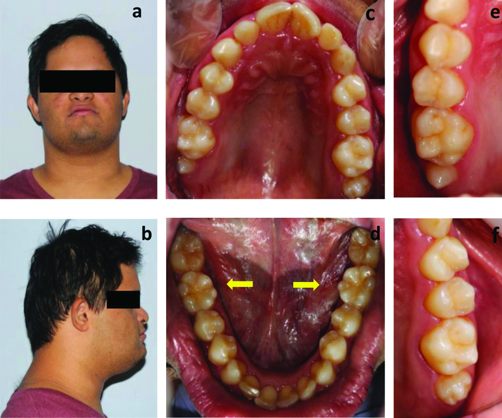
An 18-year-old male was presented to the Special Patients Clinic for a routine dental evaluation. In regard to his medical history, the patient presented a previous diagnosis of DS, hypothyroidism, a history of heart alteration treated at birth, and a mild intellectual disability. Family histories were non-contributory. An extra oral examination revealed an underdeveloped or hypoplastic mid-face, slant-eyes, hypertelorism, a protruding tongue, short neck, and the fingers of the hands were short and stubby. A detailed intraoral examination revealed healthy soft tissues, macroglossia, good oral hygiene, absence of caries, gingival inflammation, agenesis in relation to 22, 18, 28, 38, 48, tooth conic in relation to 17, microdontia in relation to 27, supernumerary cusp in relation to 36 and 46 and tooth impaction and translocation in relation to 23 [Table/Fig-1]. Prolonged retention of teeth 62 and 63, absence of teeth 22 with suspected agenesis, alterations in size and shape affecting teeth 17 and 27 (tooth 17 showed small size and peg shape, and tooth 27 was small in size with cusp definition), suggestive of teeth conic and microdontia respectively. Teeth 36 and 46 showed supernumerary cusps located on the lingual surface [Table/Fig-2].
Intraoral findings of the patient.
| Tooth |
|---|
| Agenesis | 22, 18, 28, 38, 48 |
| Tooth conic | 17 |
| Microdontia | 27 |
| Supernumerary cusp | 36, 46 |
| Tooth impaction | 23 |
| Tooth translocation | 23 |
a,b) Extra oral images of the patient; (c) Upper arch; (d) Lower arch 36, 46 molars with supranumerary cusp; (e) Teeth conic in tooth 17; (f) Microdontia in tooth 27.
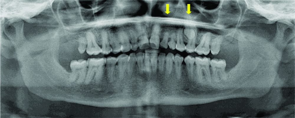
Orthopantograph examination confirmed the absence of the tooth 22, as well as the absence of the all third molars. In addition, another two anomalies were observed; the translocation and impaction of tooth 23, located between the roots of the premolars [Table/Fig-1,3].
Orthopantomograph showing absence the tooth 22, all third molars, and impaction and translocation of tooth 23.
After evaluation with the orthodontic team, the treatment plan included extraction of teeth 23, 62, and 63, prior to orthodontic treatment. Although caregivers have been informed about treatment and possible sequels, at the present time the parents’ choice was monitoring the impacted tooth. Periodic consultations are made at every 6 months.
A study has reported that patients with DS presented at least one type of dental anomaly, with an incidence of 95.92% [1]. Most of these patients presented one or two associated anomalies, and only 4% manifested four concomitant anomalies [1-3]. The literature did not report any previous cases of six dental anomalies associated in the same DS patient; which highlights the novelty of this case report.
The prevalence of agenesis in DS individuals reported in the literature is 38.6%, the upper lateral incisor being the most commonly affected (63%) [2]. Also, the occurrence of third molar agenesis was reported to be around four times greater in DS individuals than the normal population, more often in the maxilla [2] which is in accordance with the present case.
On the other hand, microdontia of the left upper molar and shape change (conoid) of the contralateral molar was also observed. Lateral incisor microdontia is reported previously, as well as its association with the agenesis of the contralateral tooth [2-4]. However, microdontia or molar conoids in patients with DS and their possible association has not been observed in the literature.
Dental transposition is defined as the exchange of position between two adjacent teeth within the same dental arch or hemiarch, most commonly in the maxillary arch. Its prevalence in syndromic patients is higher, with a rate of 14.29% in individuals with SD [4]. Its aetiology is unknown; however, it is normally associated with other anomalies, such as agenesis and peg-shaped incisors, or with a genetic factor, retention of primary teeth, trauma, and abnormality of the eruptions [4]. As in present case, the most frequent transposition occurs between the canine and first premolar superior, commonly known as, Mx.C.P1 [4].
Additionally, this report was most complicated because the transposed canine was impacted. This would be associated with the agenesis of permanent lateral incisor and retention of deciduous maxillary lateral incisor and canine. When the lateral is absent or diminished, the rush guide is lost and the canine can impact [4].
In relation to the extra cusp formation in the mandibular molars, it has two forms, namely, tuberculum sextum and tuberculum intermedium [5,6]. In this case, it is tuberculum intermedium because a third lingual cusp was developing on the mandibular molars on the lingual surface. The several clinical alterations of DS manifesting in the orofacial region should not be seen as separate but associated with the Syndrome. The reports of cases with various patterns of dental abnormalities are informative and important for the dissemination of information, as well as to highlight the fundamental role it plays in detailed oral clinical examination, to contribute to better dental care for these patients. Thus, this is the first case reported in the literature with six types of concomitant dental anomalies in a Down’s syndrome patient.
Author Declaration:
Financial or Other Competing Interests: No
Was informed consent obtained from the subjects involved in the study? Yes
For any images presented appropriate consent has been obtained from the subjects. Yes
Plagiarism Checking Methods: [Jain H et al.]
Plagiarism X-checker: Jul 09, 2019
Manual Googling: Sep 14, 2019
iThenticate Software: Nov 01, 2019 (7%)
[1]. de Moraes ME, de Moraes LC, Dotto GN, Dotto PP, dos Santos LR, Dental anomalies in patients with Down syndromeBrazilian Dental Journal 2007 18(4):346-50.10.1590/S0103-6440200700040001418278307 [Google Scholar] [CrossRef] [PubMed]
[2]. Sekerci AE, Cantekin K, Aydinbelge M, Ucar Fİ, Prevalence of dental anomalies in the permanent dentition of children with Down syndromeJournal of Dentistry for Children 2014 81(2):78-83. [Google Scholar]
[3]. Nieminen P, Arte S, Pirinen S, Peltonen L, Thesleff I, Gene defect in hypodontia: Exclusion of MSX1 and MSX2 as candidate genesHuman Genetics 1995 96(3):305-08.10.1007/BF002104127649547 [Google Scholar] [CrossRef] [PubMed]
[4]. Watted N, Abu-Hussein M, Hussein E, Proff P, Watted A, A dental transposition: Literature review and clinical managementJournal of Dental and Medical Sciences 2015 14:80-85. [Google Scholar]
[5]. Scheid RC, Woelfel’s dental anatomy 2012 Lippincott Williams & Wilkins [Google Scholar]
[6]. Sedano HO, Ocampo-Acosta F, Naranjo-Corona RI, Torres-Arellano ME, Multiple dens invaginatus, mulberry molar and conical teeth. Case report and genetic considerationsMed Oral Patol Oral Cir Bucal 2009 14(2):69-72. [Google Scholar]