Among the lymphoid organs, spleen is the largest one with rich blood supply. It serves to filter the blood, shows immune responses to antigens and in fetal life acts as a haematopoietic organ to produce erythrocytes and granulocytes [1].
As of known, the human lymphoid organs show variations, spleen lacks the definite histogenetic changes [5]. The present study tried to fill the lacunaes in the histogenesis of spleen that is known in the literature. The present study aims to find out the histogenesis of spleen in different gestational ages.
Hypertension is the most common medical disorder of pregnancy which leads to complications, the prevalence in worldwide ranges from 3% to 8% of all pregnancies [6]. The maternal deaths due to hypertension in pregnancy is common throughout the world. Hypertension in pregnancy also results in still births and neonatal morbidity and mortality [7].
High blood pressure in pregnancy is classified into following categories: gestational hypertension, chronic hypertension, preeclampsia, and pre-eclampsia superimposed on pre-existing hypertension by the the national high blood pressure education program of the national heart lung and blood institute. Hypertension is known to cause intrauterine growth restriction [8]. An interesting outcome by a study done by Singh MV et al., revealed the fact that spleen a source of T lymphocytes, itself can cause hypertension which is said to be immune mediated hypertension [9].
Keeping in mind the effect of hypertension on fetal growth, an attempt was made to compare the histogenesis of spleen in normal pregnancy and hypertension in pregnancy in the study. The patients included all the above said category of hypertensive cases.
Materials and Methods
Source of Data
The present observational study was conducted in the Department of Anatomy, BLDEDU’s Shri B M Patil Medical College, Vijayapura from May 2016 to April 2018. The study was approved by the Ethical Committee of the Institution IEC no:169/2016-17. Written consent was obtained from the parents.
Null hypothesis: There is no difference in Histological Features of fetus between normal and hypertensive pregnancies.
Sample size calculation- If there is truly no difference between the normal and hypertensive pregnancy, then 100 patients (50 per group) are required to be 90% sure that the limits of a two-sided 98% confidence interval will exclude a difference in means of more than 0.4 with SD ±0.5.
Calculation based on the formula:
n=f(α,β/2)×2×σ2/d2where, σ is the standard deviation, and d is equivalence limit.
All the foetuses which apparently looked normal grossly were included in the study. Hundred human aborted and still born fetuses were procured from Department of Pathology and Department of Obstetrics and Gynaecology, Bldedu’s shri B M Patil medical college, Vijayapura. Out of 100 fetuses, 50 belonged to normal pregnancy and 50 belonged to hypertension in pregnancy. Keeping in mind the effect of hypertension on fetal growth, an attempt was made to compare the histogenesis of spleen in normal pregnancy and hypertension in pregnancy in the study. The patients included all the above said category of hypertensive cases.
The samples belonging to Hypertension in pregnancy, Pre-eclampsia and eclampsia were included in this study.
Study Plan
Dissection of the fetus was done following the standard protocol as described by Romanes. Midline incision starting from Xyphisternum to pubic symphysis was taken. Second incision was taken along the costal margin starting from Xyphisternum extending up to the left midaxillary line. Third incision was taken starting from the pubic symphysis to Anterior superior iliac spine. The anterior abdominal wall flap was reflected laterally. Peritoneum was cut and reflected. After cutting the gastro-splenic and lieno-renal ligaments the spleen was removed en mass [10]. The entire spleen was dissected into two parts. From one part bits of entire spleen starting from the hilum were taken, processed, blocks prepared and sections as thin as 5 mm were taken. Slides were prepared and stained with Haematoxylin and Eosin.
The slides were studied using 4x, 10x, 40x and 100x objectives and interpreted.
The different gestational ages were categorised and fetuses of each age group were studied. Distribution of fetuses in gestational group: in each group 10 fetuses were studied.
Group Age
Group I 12-18 weeks
Group II 18.1-24 weeks
Group III 24.1-30 weeks
Group IV 30.1-36 weeks
Group V 36.1-40 weeks
Results
The following microscopic features at different gestational ages in normal pregnancy were observed.
In Group I: 12-18 weeks, as seen in [Table/Fig-1,2], At 12 weeks spleen was lined by thin capsule, parenchyma with few blood vessels, reticular cells and haematopoietic cells were seen, at 17 weeks central arteriole started appearing, Venous sinuses were detected, with hematopoietic cells scattered, the lymphocytes were seen around the central arteriole. The vascularity was increased.
Group I, Microscopic view of spleen at 12 weeks of gestation stained with haematoxylin and eosin as observed under 4x, 10x, 40x and 100x objectives.
1-Capsule; 2- The Interstitium containing blood vessels; 3- red blood cells; 4-reticular cells cells; 5-hematopoietic cells
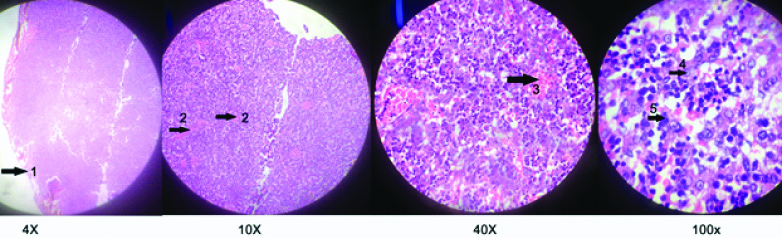
Group I, Microscopic view of spleen at 17 weeks of gestation stained with haematoxylin and eosin as observed under 4x,10x,40x and 100x objectives.
1-capsule, 2-lymphocytes, 3-venous sinus, 4-central arteriole, 5-reticular cell, 6-Red blood cell
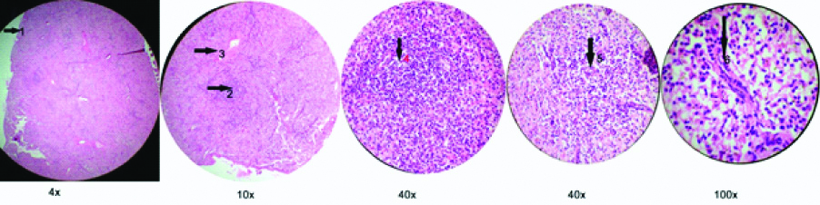
In Group II: 18-24 weeks, as seen in [Table/Fig-3,4], at 20 weeks, the capsule was more thicker, trabeculae were seen, venous sinuses increased in number, central arteriole surrounded by few lymphocytes could be seen. At 20 weeks red and white pulp marked their appearance, Peri arteriolar lymphatic sheath was seen. At 24 weeks reticular fibres were seen around the white pulp.
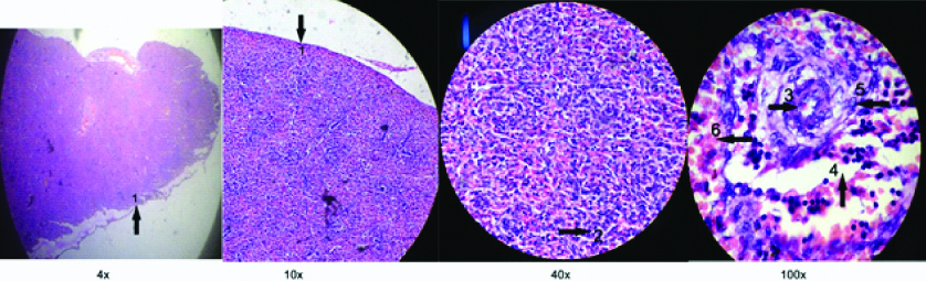
Group II, Microscopic view of spleen at 20 weeks of gestation stained with haematoxylin and eosin as observed under 4x, 10x, 40x and 100x objectives.
1-capsule, 2-white pulp, 3-red pulp, 4-central arteriole, 5-lymphocytes, 6-venous sinus, 7-thick capsule
Group II, Microscopic view of spleen at 24 weeks of gestation stained with haematoxylin and eosin as observed under 4x, 10x, 40x and 100x objectives.
1-capsule; 2-trabeculae; 3-reticular fibres around white pulp; 4- white pulp; 5 and 6-red pulp; 7-central arteriole
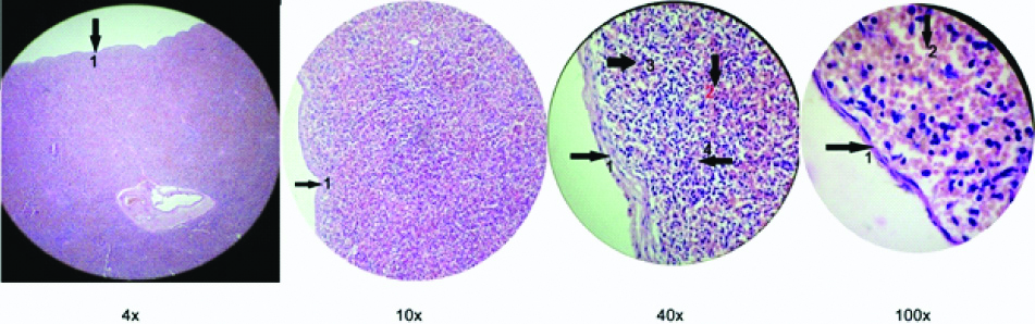
In group III: 24-30 week, as seen in [Table/Fig-5], At 28 weeks the germinal centre was seen. Eccentrically placed central arteriole was seen, along with the ring fibres. The haematopoietic cells had diminished in number.
Group III, Microscopic view of spleen at 28 weeks of gestation as observed under 4x,10x, 40x and 100x objectives.
1-white pulp; 2-red pulp; 3-germinal centre; 4- eccentric arteriole
In group IV: 30-36 weeks, as seen in [Table/Fig-6], At 31weeks, the capsule was thick, white pulp was well-defined with germinal center, marginal zone and mantle zones seen.
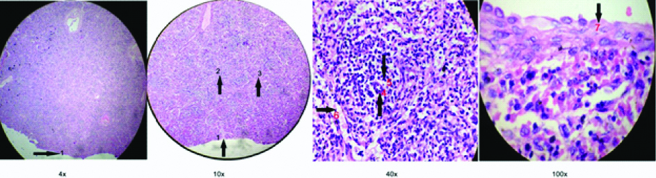
Group IV, Microscopic view of spleen at 31weeks of gestation stained with haematoxylin and eosin as observed under 4x, 10x, 40x and 100x objectives.
1-Thick capsule; 2-Red blood cells; 3-lymphocytes; 4- Reticular fibres
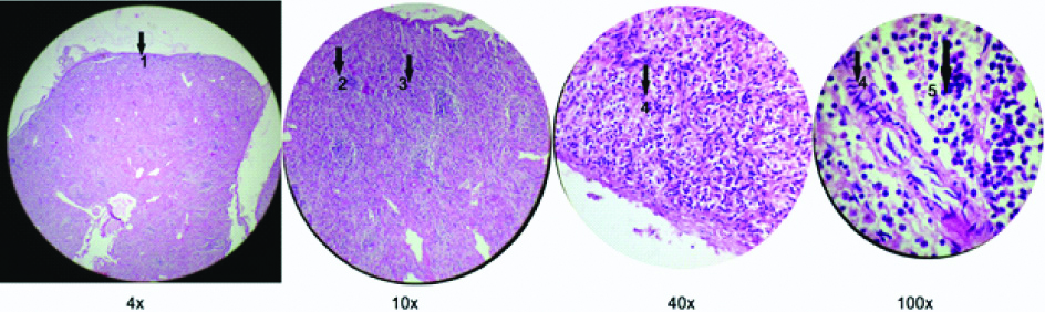
In Group V: 36-40 weeks, as seen in [Table/Fig-7], at 37 weeks, the capsule showed increased thickness. The red and white pulp were well-defined. the hematopoietic activity seems to be completely absent. At 38-40 weeks the fetal spleen resembled that of an adult spleen.
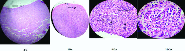
Group V, Microscopic view of spleen at 37weeks of gestation stained with haematoxylin and eosin as observed under 4x, 10x, 40x and 100x objectives.
1-Thick capsule; 2-white pulp; 3-Red pulp; 4- ring fibres; 5-lymphocytes
The present study tried studying the histogenesis of fetal spleen in hypertension in pregnancy. The microscopic features of spleen did not show differences between normal pregnancy and that in hypertension in pregnancy.
Discussion
Gray’s has described the dual origin of development of spleen as early as 6th week of IUL as a localised thickening of the coelomic epithelium of the dorsal mesogastrium near its cranial end. The proliferating cells invade the underlying angiogenetic mesenchyme, which become condensed and vascularised [11]. In 13th century, Gray’s has opined about the relative scarcity in the study of histogenesis of spleen [12]. During the 20th century splenic histogenesis gained attention. Van Furth R et al., started the study as early as 1965. He has contributed by studying the immunoglobulins produced by the fetal spleens in different gestational ages [13]. Contributions from Weiss L in 1973 served a major component in studying the lacunaes in the histogenesis. In his ultramicroscopic study of spleens from 5 human fetuses, he described development of the primary vascular reticulum and of the capsule [14]. In 1983 Wolf BC et al., observed haematopoiesis in spleen to begin from early second trimester till fifth month of gestation when the intramedullary haematopoiesis begins and splenic blood formation reportedly declines. At birth only occasional hematopoietic cells are seen [15].
In 1985, Vellguth S et al., described the histogenesis in three stages. The primary vascular reticulum upto 14 weeks, transformation stage from 15 to 17 weeks, lymphoid colonisation at 18 weeks and at 23 weeks had observed B cell primary follicles [16]. In the 21st century, several studies have been going on to understand the histogenesis. The observations done by different authors are shown in the [Table/Fig-8] [5,17-21].
Shows different observations done by various authors on histogenesis of spleen [5,17-21].
| Author | Year | No. of spleens | Capsule | Lymphocyte aggregation | Lymphoid follicles | Central arteriole | Resemblance to adult spleen |
|---|
| Radhika D et al., [17] | 2012 | 50 | | 11 wk | 20 wk | 32 wks | 36 wk |
| Pal M et al., [18] | 2013 | 74 | | | 17 wk | 31 wk | |
| Dsouza A et al., [19] | 2015 | 15 | 1st trim | 2nd trim | 20 wks, 23rd wk Red Pulp, White Pulp | 30 wk | 36 wk |
| Alex L et al., [20] | 2015 | 70 | | | 6th month | Full term | |
| Mukhia R et al., [5] | 2016 | 50 | 12-14 wk | | 18 wk | 22 wk | 38 wk |
| Holkunde A et al., [21] | 2018 | 30 | 13-14 wk | | 20 wk | 22 wk | 38 wk |
| Present study | 2019 | 50 | 12 wks | 17 wks | 20 wk | 24 wk | 38 wk |
In the present study, as observed in the [Table /Fig-8], the following findings can be compared with other studies done. The thin capsule covering the spleen could be seen at 12 weeks in the present study with similar findings by Mukhia R et al., D Souza A et al., and Holkunde A et al., [5,19,21]. The lymphocyte aggregation was seen at 17 weeks in the present study with a similar results by D Souza A et al., [19]. The Lymphoid follicles were observed around 20 weeks in the present study, with differential observation done by remaining authors with range of 17 to 23 weeks. The central arteriole was seen at 24 weeks in the present study with near similar findings of Mukhia R et al., and Holkunde A et al., [5,21]. The resemblance to adult spleen was seen at 38 weeks in the study with almost similar findings of other authors.
Hypertension in pregnancy is known to cause growth retardation in the fetus resulting in Intrauterine growth restriction and low birth weight [22]. When the histogenesis of spleen was observed there were no changes observed in the present study. So this study could not establish any comparison to the normal pregnancy fetus. There are no studies done on hypertension and histogenesis of spleen in the literature.
Singh MV et al., studied the relationship between hypertension and the immune system. The conclusion drawn by his study was that dysregulated immune system leads to enhanced sympathetic activity. This sympathetic over activity leads to mobilisation of the haematopoietic stem cells, monocytes and lymphocytes from bone marrow and spleen to the vasculature, heart, kidneys and the CNS where these cells significantly contribute to end organ damage seen in hypertension [9].
A study conducted on mice by Carnevale D et al., demonstrate that Placental growth factor mediates the neuroimmune interaction in the spleen, leading to a surprising and non superfluous response that allows the onset of hypertension [23].
Limitation
Limitation of the present study is that it did not focus on the gross features of spleen. The study could not establish any comparable features in the two situations.
Conclusion
The present study served to study the splenic histogenesis to some extent. The findings in individual author variations are still existing. The study could not conclude difference in the histogenesis of spleen in normal pregnancy and hypertension in pregnancy. This study may serve the Anatomists and the Pathologists for studying the individual variations in histogenesis of spleen.
Future Recommendation
Future recommendations of the study are using special stains and Immunohistochemistry can be done to detect markers such as placental growth factor in spleen which may be the factor for onset of immune mediated hypertension.