Introduction
The Major and Minor blood vessel and the optic disc are the important factors for finding the presence of retinal disease like diabetic retinopathy.
Aim
To segment the blood vessel using Major vessel extraction and Minor vessel classification methods.
Materials and Methods
The retinal blood vessels are analysed from the images, for determining the changes in the blood vessels depending on the vessel branching pattern, width and density. High pass filter is used for easily segmenting the major vessels and also to make the retinal nerves lighter and the eye background dark to avoid fovea. A morphological operation like the top hat is performed for zooming and shrinking the nerves to adjust the outer structure of the eye. The Minor vessels are obtained by using the derivative of Gaussian filter without any crack or without any breaks in the structure of the retinal nerves which is used to determine the presence of blood clot in the eye. Gabor Filter is used to strengthening the pixels of the image.
Conclusion
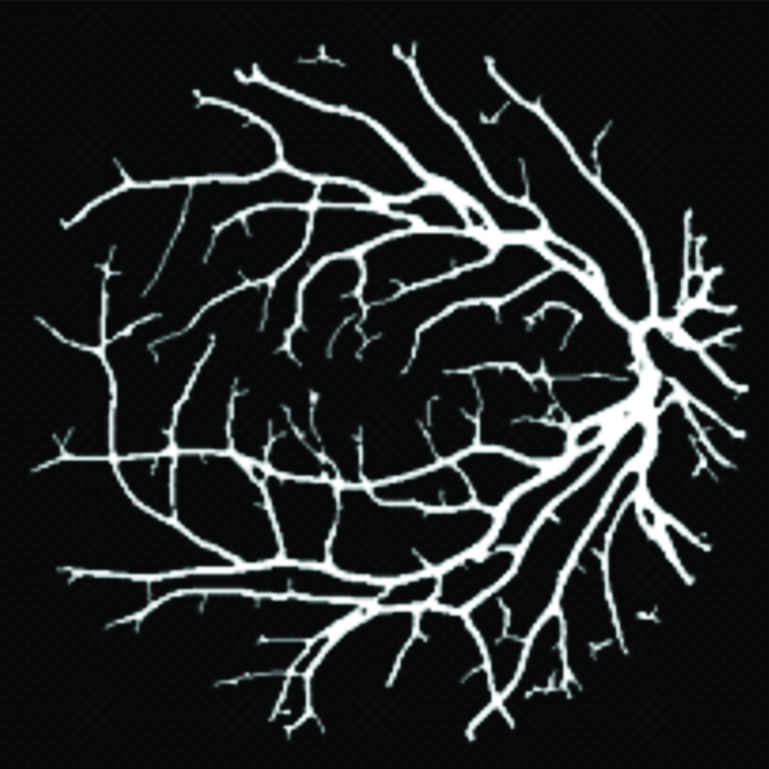
The segmented outputs of the Major vessel and Minor vessels are together added to get the resultant image of the eyes. Thus extracted and classified vessels can be sent to an intelligent artificial neural network for comparison with threshold values for diagnosing the problems due to high glucose level, cardiac disorders, etc.
Diabetic retinopathy, Extraction and classification, Morphological operation Prediction of eye health, Retinal disease
Introduction
The blood vessel segmentation system is used to determine the changes in the blood vessels depending on the factors which are vessel branching pattern, width and density. Major vessels are easily segmented using filters and the Minor vessels are studied with the derivative of Gaussian and Gabor filter [1]. The goal is to facilitate the early detection of certain pathologies such as ischemia, necrosis etc., and to avoid specialist intervention. Principal Component Analysis (PCA) is the mathematical method that is proposed for optic disc extraction. This PCA is used to reduce the dimensionality of a dataset comprising of numerous variables correlated with each other either heavily or lightly. It makes use of different operations such as a variant of the watershed transformation. PCA is also used to obtain the input of the segmentation method. The other purpose of using PCA is to achieve the grey-scale image that better represents the original Red-Green-Blue (RGB) image.
Existing System
In this existing model, the threshold is used and connecting the broken line components is done to obtain the blood vessel images. This method focuses on vessel extraction from bright images and it also does not perform very effectively on retinal images with red lesions. Only Major vessels can be determined. This method provides an incomplete structure of veins due to blood clots (these are the disadvantages of the existing system) [2]. The green channel of the RGB image is chosen for vessel enhancement. The major blood vessels are obtained by applying Low-Power Field (LPF) and High-Power Field (HPF) (edge detection). The advantages of the proposed system are Minor vessels, Major vessels, partial retinal nerve structure can be obtained and the presence of clots or any black dots cannot be determined.
Major Vessel Extraction and Sub-image Classification
The most important feature of the proposed segmentation algorithm is to separate segmentation of Major vessel and fine vessel regions. The intersecting regions between threshold versions of two pre-processed images, i.e., the high-pass filtered image and top-hat transformed image are identified as Major vessels regions [3]. The pixel threshold “p” which is used to increase or decrease the number of pixels identified as Major vessels can be varied across images. The number of pixels subjected to classification in the vessel sub-image increases if the number of Major vessel pixels is decreased by varying the pixel thresholds for the two pre-processed images. This process will support the automated segmentation of very fine vessel branches. These vessel branches are necessary for detecting retinal abnormalities such as Intra-Retinal Micro Vascular Abnormalities (IRMA) or vessel beading.
The Detection and Quantification of Retinopathy
The present article presented a scheme based on image segmentation. Although the fields are identified mainly by comparing the key points cited in the image, the present authors cannot simply classify the proposed scheme as one key point-based. This can also be looked like a mixture of existing schemes because both key points and pixel features are employed in the two stages of the matching process [4].
Segmentation into Semantically Independent Patches
Our main contribution can be concluded in the following two aspects. We propose to segment the image into semantically independent patches by considering that the regions usually have a certain meaning. By segmenting in that manner, the problem can be solved by partial matching among these segmented patches. There are two stages in the matching process between segmented patches. An accurate estimation of the transform matrix can be obtained by an EM-based algorithm in the second stage [5]. If using some efficient methods, we can segment an image in several seconds. The number of pixels subjected to classification in the vessel sub-image increases if the number of Major vessel pixels is decreased by varying the pixel thresholds for the two pre-processed images. One may concern the computational complexity of the proposed scheme. The image segmentation and the transform estimation refinement are the two more steps that are mainly needed in the proposed scheme compared with the key point-based schemes.
Proposed System
The green channel of the RGB image is chosen for vessel enhancement. The major blood vessels are obtained by applying LPF and HPF (edge detection). Minor vessels are obtained by using the Gaussian filter and Gabor is used for strengthening. Both Major and Minor vessels are obtained [6,7]. Complete retinal nerve structure is obtained. Presence of Blood clots or any black dots can also be determined. This Gaussian is used for removing the noise present in between the minor blood vessels. Gabor filter is used for strengthening the image and obtained from the Gaussian filter.
Pre-processing
The first pre-processing stage necessitates the green plane of the fundus image and a fundus mask which is superimposed on image, this is followed by contrast adjustment and vessel enhancement, leading to vessel enhanced image. Two different pre-processing strategies are implemented to extract the dark blood vessel regions from the image. A smoothened Low-Pass Filtered version of image {LPF (i.e.,)} is subtracted from enhancement image at first to obtain a high-pass filtered image. The Input Image can be a colour image or a gray scale image. The input chosen here is a colour image. The Major and Minor nerves of this colour image are obtained as the output. The colour image consists of three planes which are Red, Green and Blue. These planes are separately called as gray scale image [8]. The Green plane is chosen to determine the Major and Minor nerves as the domination of nerves are greater in the green plane. These are some of the pre-processing process done to find the nerves.
Major Blood Vessel Extraction
From the negative of enhancement image, the Red regions corresponding to the dark pixels are extracted and use morphological Top-Hat operations. To fit, the length of pixels for the linear structuring element is selected to roughly fit the diameter of the biggest vessels in the images and select the threshold to extract the Major vessels for the retinal images. The Green plane image is extracted from the colour image [7,9]. The Green plane is selected as the domination of the nerves are greater in this plane. This Green plane is a single gray scale image. These planes are separately called as gray scale image. These are some of the pre-processing process done to find the nerves. The Input Image can be a colour image or a gray scale image. The input chosen here is a colour image. The Major and Minor nerves of this colour image are obtained as the output. The contrast of the image is adjusted by making the white pixel more whiter and darker pixels darker [8,10]. This is done to display the nerves more brightly and distinctly for the edge of the nerves to be displayed clearly. This is done to distinguish the difference between the gray and white shades clearly. Since the contrast is adjusted the whiter pixel become more whiter and the darker pixels become more darker.
Sub-Image Extraction Process
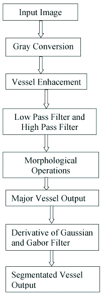
This approach enhances the robustness of vessel segmentation on normal and abnormal retinal images in two stages[9,11]. In the first stage, the Major vessel regions comprising of 50-70% of the total blood vessel, pixels are segmented. In the second stage, we are extracting the sub-image pixel using both Gabor filter and derivative of Gaussian filter. Top Hat is a combination of erosion and dilation. Ada-tive Histogram equalisation is used to redistribute the pixels. This is to redistribute the lighter pixels evenly in all the directions. Block representation of process flow are shown in [Table/Fig-1].
Block representationof process flow.
The green plane is chosen to determine the major and minor nerves as the domination of nerves are greater in the green plane. These are some of the pre-processing process done to find the nerves. The Input Image can be a colour image or a gray scale image. The input chosen here is a colour image. The Major and Minor nerves of this colour image are obtained as the output [10,12]. The contrast of the image is adjusted by making the white pixel whiter and darker pixels darker. This is done to display the nerves more brightly and distinctly for the edge of the nerves to be displayed clearly. This distinguishes the difference between the gray and white shades clearly. Since the contrast is adjusted the whiter pixel become whiter and the darker pixels become darker. This Adaptive Histogram equalisation is the process of determining the pixel values, calculating the occurrence of those pixels based on their values and arranging them in order based on the pixel values. This is the process of adaptive histogram equalisation which is a morphological process.
The blood vessel segmentation system is used in determining the changes in the blood vessels depending on the vessel branching pattern, width and density. The filter is used for easily segmenting the Major vessels and the Minor vessels are examined by the derivative of Gaussian and Gabor filter. In this existing model, the threshold is used and connecting the broken line components is done to obtain the blood vessel images [11,13]. This method focuses on vessel extraction from bright images and it also does not perform very effectively on retinal images with red lesions. Only Major vessels can be determined, incomplete structure of veins due to blood clots, these are the two disadvantages of this existing system [12,14]. The green channel of the RGB image is chosen for vessel enhancement. The major blood vessels are obtained by applying LPF and HPF (Edge Detection). Minor vessels are obtained by using the Gaussian filter and Gabor is used for strengthening. Both Major and Minor vessels are obtained. Complete retinal nerve structure is obtained. Presence of Blood clots or any black dots can also be determined. This method is more useful in finding and curing vision problems due to diabetes by comparing the extracted features of blood vessels with the threshold values of healthy blood vessels using an intelligent ANN back propagation network [13,15].
Output and Explanation
The structure of major and minor nerves is obtained either from the colour image or also from the Grey scale image. The input image that is given is a colour image. The fundus mask is superimposed on the image. It is followed by contrast adjustment and vessel enhancement, resulting in a vessel enhanced image. Two different pre-processing strategies are implemented to extract the dark blood vessel regions from the image. A smoothened low-pass filtered version of image {lpf (i.e.,)} is subtracted from enhancement image at first to obtain a high-pass filtered image [14,16]. The colour image consists of three planes they are Red, Green and Blue. These planes are separately called as gray scale image. The Green plane is extracted for this purpose of extracting the nerves. The Green plane is chosen since the domination of nerves is greater in the Green plane [17]. The Input Image can be a colour image or a gray scale image. The input chosen here is a colour image. The Major and Minor nerves of this colour image are obtained as the output [18]. Pre-processing is done before the main process [19]. The Pre-processing is the process of both Enhancement and Masking. Masking is the process of obtaining the area of interest. This is done by setting a threshold value considering the area of interest. The value greater than the threshold is given as one and the value lesser are given as zero [15,17]. Thus, the area of interest is obtained by this masking. Enhancement is the process done for redistribution of the lighter pixels. This is done in order to avoid the lighter pixels in the same place. The pixel value of all the pixels is noted and their number of occurrences is also noted. Filters are used in order to determine the edge of the nerves and also for smoothing the image. Both High pass filter and Low pass filter are used.
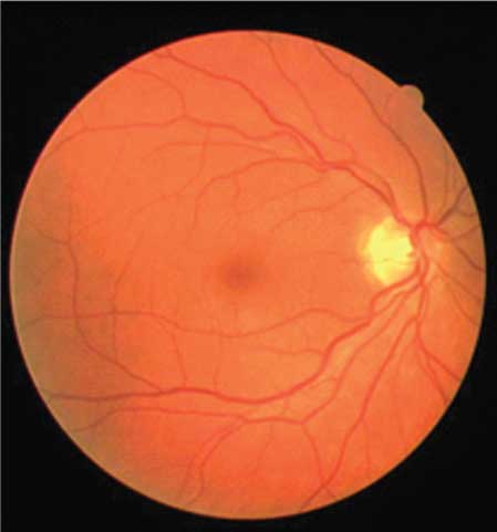
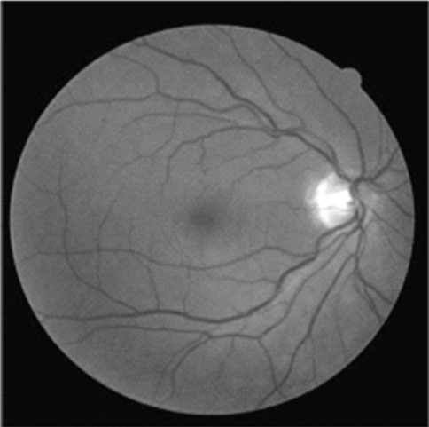
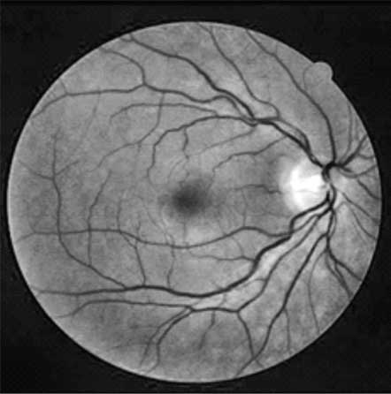
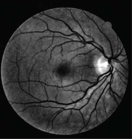
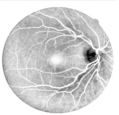
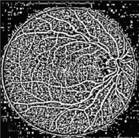
The fundus mask is superimposed on the image. It is followed by contrast adjustment and vessel enhancement, resulting in a vessel enhanced image. Two different pre-processing strategies are implemented to extract the dark blood vessel regions from the image. This is done by setting a threshold value considering the area of interest. The green plane image is extracted from the colour image. The colour image consists of three planes Red, Green, Blue. The green plane is selected as the domination of the nerves is greater in this plane. This green plane is a single gray scale image. The colour image consists of three planes they are Red, Green and Blue. These planes are separately called as gray scale image. The green plane is chosen in order to determine the major and minor nerves as the domination of nerves are greater in the green plane. These are some of the pre-processing process done in order to find the nerves [8,18]. The Input Image can be a colour image or a gray scale image. The input chosen here is a colour image. The Major and Minor nerves of this colour image are obtained as the output. The contrast of the image is adjusted by making the white pixel much whiter and darker pixels much darker. The Output images of Major vessel and Minor Vessel are shown in [Table/Fig-2,3,4,5,6,7 and 8].
Conclusion
Two threshold binary images are obtained in the first stage. One image is obtained by high pass filtering and another image is obtained by top hat reconstruction of the red regions in the green plane image. A vessel sub-image is created by combining the remaining pixels in both the binary images which are extracted as the Major vessels. The pixels in the vessel sub-image are subjected to a two-class in the second stage using Gabor Filter and Derivative of Gaussian. In the third post-processing stage, the classifier classifies all the pixels in the sub-image as vessels which are combined with the Major vessels to obtain the segmented vasculature. The segmented vessels are further enhanced to ensure higher vessel segmentation accuracy depending on the resolution of the fundus images. More oftenly medical community uses fundus images method to diagnose the complications due to cardiovascular disease, stroke, arteriosclerosis, glaucoma, diabetic retinopathy (DR) and hypertension. Variations in the blood vessels can be determined by using automated blood vessel segmentation systems. These are based on the vessel branching patterns, vessel width, tortuosity and vessel density as the pathology progress. This can be used for retinal health observation for a diabetic patient and the prediction of any complains related to vision. When this, retinal diagnosis result is added as one of the essential observation parameters in dynamic wireless sensor network-based remote patient monitoring [20], the loss of vision due to high glucose level, blood pressure, heart problems, etc., can be prevented effectively by the assistance of expert physicians through telemedicine technology.
Author Declaration:
Financial or Other Competing Interests: No
Was Ethics Committee Approval obtained for this study? Yes
Was informed consent obtained from the subjects involved in the study? Yes
For any images presented appropriate consent has been obtained from the subjects. Yes
PLAGIARISM CHECKING METHODS: [Jain H et al.]
Plagiarism X-checker: Feb 27, 2019
Manual Googling: Jul 17, 2019
iThenticate Software: Sep 13, 2019 (15%)
[1]. Chaudhuri S, Chatterjee S, Katz N, Nelson M, Goldbaum M, Detection of blood vessels in retinal images using two-dimensional matched filtersIEEE Transactions on Medical Imaging 1989 8(3):263-69.10.1109/42.3471518230524 [Google Scholar] [CrossRef] [PubMed]
[2]. Eswaran C, Reza A, Hati S, Extraction of the contours of optic disc and exudates based on marker-controlled watershed segmentationIn Proc Int Conf Comput Sci Inf Technol 2008 :719-23.10.1109/ICCSIT.2008.13 [Google Scholar] [CrossRef]
[3]. Kauppi T, Kälviäinen H, Simple and Robust Optic Disc Localisation Using Colour Decorrelated Templates. In: Blanc-Talon J, Bourennane S, Philips W, Popescu D, Scheunders P. (eds)Advanced Concepts for Intelligent Vision Systems 2008 Berlin, HeidelbergSpringer:525910.1007/978-3-540-88458-3_65 [Google Scholar] [CrossRef]
[4]. Lalonde M, Beaulieu M, Gagnon L, Fast and robust optic disc detection using pyramidal decomposition and Hausdorff-based template matchingIEEE Trans Med Imag 2001 20(11):1193-200.10.1109/42.96382311700746 [Google Scholar] [CrossRef] [PubMed]
[5]. Li H, Chutatape O, Automated feature extraction in colour retinal images by a model based approachIEEE Trans Biomed Eng. vol 2004 51(2):246-54.10.1109/TBME.2003.82040014765697 [Google Scholar] [CrossRef] [PubMed]
[6]. World Health Org. Action plan for the prevention of blindness and visual impairment 2009-2013, 2010 [Google Scholar]
[7]. Lowell J, Hunter A, Steel D, Basu A, Ryder A, Fletcher E, Kennedy L, Optic nerve head segmentationIEEE Trans Med Imag 2004 23(2):256-64.10.1109/TMI.2003.82326114964569 [Google Scholar] [CrossRef] [PubMed]
[8]. Zhou L, Rzeszotarski MS, Singerman LJ, Chokreff JM, The detection and quantification of retinopathy using digital angiogramsMedical Imaging IEEE Transactions on 1994 13(4):619-26.10.1109/42.36310618218540 [Google Scholar] [CrossRef] [PubMed]
[9]. Martinez-Perez ME, Hughes AD, Thom SA, Bharath AA, Parker KH, Segmentation of blood vessels from red-free and fluorescein retinal imagesMedical Image Analysis 2007 11(1):47-61.10.1016/j.media.2006.11.00417204445 [Google Scholar] [CrossRef] [PubMed]
[10]. Osareh A, Mirmehdi M, Thomas B, Markham R, Comparison of colour spaces for optic disc localisation in retinal imagesin Proc. 16th Int. Conf. Pattern Recognit 2002 1:743-46. [Google Scholar]
[11]. Park M, Jin JS, Luo S, Locating the Optic Disc in a Retinal ImagesProceedings of the International Conference on Computer Graphics, Imaging and Visualization 2006 10.1109/CGIV.2006.63 [Google Scholar] [CrossRef]
[12]. Pascolini D, Mariotti SP, Global estimates of visual impairment: 2010Br J Ophthalmol 2011 :614-21.10.1136/bjophthalmol-2011-30053922133988 [Google Scholar] [CrossRef] [PubMed]
[13]. Soares J, Leandro J, Cesar R, Jelinek H, Cree M, Retinal vessel segmentation using the 2-d gabor wavelet and supervised classificationIEEE Transactions on Medical Imaging 2006 25(9):1214-22.10.1109/TMI.2006.87996716967806 [Google Scholar] [CrossRef] [PubMed]
[14]. Staal J, Abramoff MD, Niemeijer M, Viergever MA, Van Ginneken B, Ridge-based vessel segmentation in colour images of the retinaIEEE Transactions on Medical Imaging 2004 23(4):501-09.10.1109/TMI.2004.82562715084075 [Google Scholar] [CrossRef] [PubMed]
[15]. Priya CL, Gowthami D, Poonguzhali S, Lung pattern classification for interstitial lung diseases using an ANN-back propagation network, Proceedings of the 2017IEEE International Conference on Communication and Signal Processing, ICCSP 2017 2018 10.1109/ICCSP.2017.8286732 [Google Scholar] [CrossRef]
[16]. Welfer D, Scharcanski J, Kitamura CM, Pizzol MMD, Ludwig LW, Marinho DR, Segmentation of the optic disk in colour eye fundus images using an adaptive morphological approachComput Biol Med 2010 40(2):124-37.10.1016/j.compbiomed.2009.11.00920045104 [Google Scholar] [CrossRef] [PubMed]
[17]. Xu L, Luo S, A novel method for blood vessel detection from retinal imagesBiomedical Engineering Online 2010 9(1):1410.1186/1475-925X-9-1420187975 [Google Scholar] [CrossRef] [PubMed]
[18]. Xu J, Sung CE, Zheng C, Kuan PCT, Optic disk feature extraction via modified deformable model technique for glaucoma analysisPattern Recognit 2007 40:2063-76.10.1016/j.patcog.2006.10.015 [Google Scholar] [CrossRef]
[19]. Zana F, Klein JC, Segmentation of vessel-like patterns using mathematical morphology and curvature evaluationImage Processing, IEEE Transactions on. vol 2001 10(7):1010-19.10.1109/83.93109518249674 [Google Scholar] [CrossRef] [PubMed]
[20]. Poonguzhali S, Advanced fetal essential parameters observation using wsnInternational Journal of Pharmacy and Technology (IJPT) 2016 8(4):21629-34. [Google Scholar]