Introduction
The association of adhesive systems and desensitising agents could influence the values of the bond strength. That is because these desensitising agents cause the obliteration of the tubular holes and minimise the movement of the dentin fluids, promoted by environmental stimulus that tends to cause hydrolysis at the base of the hybrid layer over time.
Aim
To evaluate the microtensile bond strength of total-etch (Single Bond II, SB, 3M ESPE, USA) and self-etch (Clearfil SE Bond, SE, Kuraray, Japan) adhesive systems in dentin conditioned after the application of oxalate-based and fluoride- based desensitising agents.
Materials and Methods
Discs obtained from human molars were used, which had its occlusal enamel removed to expose flat surfaces of dentin. Six groups were formed, according to the materials used: G1 (control)- hybridization with SB; G2- desensitizer based on oxalate (BisBlock, BB) and SB; G3- desensitizer based on fluoride (Aqua Prep F, AF) and SB; G4 (control)- hybridization with SE; G5- BB and SE; G6- AF and SE. On these surfaces, blocks of composite resin Filtek Z350 of 5 mm high were prepared to obtain sticks of (1×1×10 mm). The samples were stored in deionized water at 37°C for 24h and submitted to microtensile mechanical trial at 1mm/min speed till the fracture. The results were statistically analysed by one-way ANOVA followed by Tukey’s post hoc test (p<0.05).
Results
The mean values of bond strength of the studied groups in MPa (SD) were as follows: G2: 46.48 (4.02), G3: 44.87 (4.68), G5: 39.02 (4.72), G4: 36.95 (3.57), G1: 36.66 (3.42), G6: 32.71 (3.41) which did not show statistically significant differences (p>0.05). Regarding the fractures analysis, the mixed type prevailed. The fracture was on the base region of the hybrid layer for the total-etch adhesive system, and at the top of the hybrid layer for the self-etch system.
Conclusion
There is no significant difference in bond strength between the combined use of desensitizers with total-etch and self-etch adhesive systems on human dentine.
Introduction
Bonding procedure is very critical for both total-etch and self-etch adhesive systems and can result in areas with collagen fibrils not wrapped by the monomers, at the base of the hybrid layer [1-4] despite the creation of the hybrid layer with different morphological standards by using these distinct bonding techniques [5]. In the total-etch adhesive systems, the excessive humidity of the dentine can hamper the bonding procedure. The areas that were not ideally hybridised are not able to completely seal the base of the hybrid layer and thus become more vulnerable to the enzymes action that damages the exposed collagen, called metalloproteinases, as much as the hydrolytic degradation by intratubular fluids [6,7].
The hybridised region resists better to hydrolysis than the exposed collagen fibrils. As there can be failures in the bonding techniques, with areas likely to be degraded, some materials have been used as alternatives to be associated to adhesive systems, with the main objective of optimising the bonding procedure and helping in the longevity of restorations [6,8]. Now-a-days, materials such as chlorhexidine have been suggested to inhibit the proteolytic enzymes action (metalloproteinases), responsible for the collagen degradation. The non-infiltrated fibrils tend to be more vulnerable to the metalloproteinases action [7,9]. Many ways have been recently used to improve resin-dentin bond strength such as, the use of collagen crosslinking agents; the use of antioxidants, the use of protease inhibitors, the bonding procedure modification using the ethanol wet-bonding technique or by applying an additional adhesive (hydrophobic) coating, the laser treatment of the substrate that cause specific topographic changes in the surface of dental substrates, and the reinforcement of the resin matrix with inorganic fillers [10].
The material that can be associated during the bonding procedures is the dentin desensitizers. This material can cause the obliteration of the tubular holes and minimise the movement of the dentin fluids promoted by environmental stimulus that tends to cause hydrolysis at the base of the hybrid layer over time [11-14].
Perdigão J recommended the utilisation of desensitizer as an alternative to minimise the increase of the dentine permeability postconditioning, mostly in deeper dentin areas [6]. This factor tends to deteriorate the bond interface over time. The dentin desensitizers are rated with regard to the mechanism of action and the chemical composition. According to Yiu CKY et al., the oxalate based desensitizers promote the tubular occlusion by forming soluble crystals of calcium oxalate, by the chemical reaction between the oxalate and the calcium of the tooth structure [15]. Components like fluorides have an effective action on the dentin surface by chemical reaction with the calcium [13].
Pinto SCS et al., and Dall’Orologio GD et al., compared the effectiveness of the desensitizers in reducing the dentin permeability [16,17]. They observed through in vitro and in vivo studies that the desensitizers were able to partly occlude the dentin tubules, which allowed the reduction of the fluids passage.
The association of adhesive systems and desensitizers, from a morphologic point of view, can result in reduction of the fluids passage and help the hydrophobic materials bonding [12]. However, some studies about the mechanical resistance of the materials demonstrated that this association can influence the bond resistance values [18-22].
Sadek FT et al., evaluated the compatibility of the desensitizers based on oxalate with total-etch adhesive systems of two and three clinical steps [12]. The treatment with desensitizers on the conditioned dentin surface reduced the permeability and improved the bond resistance, increasing its values. This was justified by the authors as an optimization process of the bonding with the hydrophobic resin, mostly in areas of deeper dentin. Dündar M et al., evaluated the interaction of total-etch systems of two and three steps and desensitizers based on fluoride/HEMA and triclosan/PENTA, during resinous luting, and they observed that the desensitising agents did not interfere negatively in the bond strength [23]. The desensitizer based on fluoride and HEMA had the best performance, probably due to the capacity of the flour to react with the dentine calcium, and the capacity of the monomer HEMA to seep into the conditioned fibrils net.
The association of desensitizers and self-etch adhesive systems of one clinical step, demonstrated by Akca T et al., revealed that the products based on fluoride, laser and potassium nitrate interfere negatively in bond resistance [24]. But when the adhesive system itself was used as a desensitizer, satisfactory results were obtained in terms of mechanical resistance.
The current study aimed to evaluate the bond strength of the total-etch and self-etch adhesive systems after the application of desensitising agents in human dentine. The null hypothesis was that, there was no difference in the bond strength after the application of the desensitising agents.
Materials and Methods
The present study was an in vitro research done at Federal University of Juiz de Fora which was conducted for a duration of two years six months from February 2009 to July 2011. To accomplish this study, 30 healthy extracted human third molars were selected after the approval of Ethics Committee of Human, in the Federal University of Juiz de Fora, MG, Brazil-CEP/UFJF under the protocol number (0009.0.180.000-09). These teeth were extracted for orthodontic reason. Healthy complete third molars were included in the study. Teeth with dental caries or which were fractured were excluded.
The teeth were cleaned and frozen in physiological saline until they were used. The occlusal enamel was removed with diamond disc (Isomet, Buehler Ltd., Lake Bluff, IL) under water cooling to expose the dentin surface (the dentin located in the middle portion of this substrate). Then, a second cut was made to remove the root region below the cementoenamel junction of the teeth. These cuts led to the attainment of 5 mm high discs. The space left by the pulp chamber was restored with the adhesive system Single Bond II (3M/ESPE, St Paul, MN, USA) and the composite resin Filtek Z350 (Color A3; 3M/ESPE, St Paul, MN, USA) following the manufacturer’s indications. To standardise, the dentin surfaces were manually worn with silicon carbide sandpapers of 400 and 600 granulation, under water cooling for one minute.
Six groups were formed, two control and four trial groups, in each group there were 5 dentin discs. The groups G1 and G4, were both control groups – with the application of the adhesive systems Single Bond (SB) and Clearfil SE Bond (SE) over the dentin surface; G2 and G5: with the application of the desensitising agent Bis Block after acid conditioning and acid primer; G3 and G6: with the application of the desensitising agent Aqua-Prep F after acid conditioning and acid primer [Table/Fig-1]. On these hybridised dentin, blocks were built with composite resin Filtek Z350 (Color A3; 3M/ESPE) which were 5 mm high, by incremental technique. This was important to prepare the blocks for the microtensile test, where the dentin and resin parts must be at the same height. Each 2 mm of increment was photopolymerized with halogen light (Demetron LC, Kerr, EUA) for 40 seconds. Afterwards, all the groups were stored in deionized water at 37°C for 24 hours.
Adhesives used in this study, trade names (lot), composition, techniques and application manufacturer of the adhesives used in this study.
| Interaction methods | Trade name (Lot) | Composition | Application technique | Manufactures |
|---|
| Etch and rinse system (G1) “two steps” | Single Bond (8RY) | Etching: phosphoric acid 35%Bond: water ethanol, Bis-GMA, HEMA, copolymer of acid polialquenóico, dimethacrylate, camphoroquinone – pH: 3.6 | A(15s), b(15s), c, d, e, h(20s) | 3M ESPE, St Paul, MN, USA |
| Self-etch system (G4) “two steps” | Clearfil SE Bond (012DA) | Primer: 10 MDP; HEMA; hydrophilic dimethacrylates; camphoroquinone; N,N diethanol-p-toluidine; watterBond: 10 MDP; Bis-GMA; HEMA; hydrophobic dimethacrylates; camphoroquinone; N,N diethanol-p-toluidine; silanized colloidal silica. – pH: 2.0 | f(20s), e, g, h (10s) | Kuraray, Osaka, Japão |
| Etch and rinse system and desensitizer (G3)Self-etch system and desensitizer (G6) | Aqua-Prep F(1000001065) | 30 a 40%HEMA, 1 a 2% sodium fluoride and water– pH:5-7 | a(15s), b (15s), c, I (30s), h (10s), d, e, h (20s)f(20s), e, i (30s), h (10s), g, h (10s) | Bisco, Schaumburg, USA |
| Etch and rinse system and desensitizer (G2)Self-etch system and desensitizer (G5) | BisBlock(1000004959) | Calcium oxalate anda water-pH: 1.8 | a(15s), b (15s), c, i (30s), d, e, h (20s)f(20s), e, g, h(10s) | Bisco, Schaumburg, USA |
Bis-GMA; bisphenol- A diglicidil eter dimetacrylate; HEMA, 2-hidroxietil metacrylate; 10-MDP, 10-metacriloxietil dihidrogeniun phosphate
*Technical bonding: a) phosphoric acid etching; b) rinse; c) drying the surface with absorbent paper; d) primer application; e) bond application; f) applying the primer-bond; g) drying the solvent for volatilization; h) application of the acid primer on the dry dentin surface doing slight pressure; i) bond application; j) photoactivation
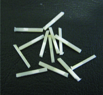
At the end of the storage period, the samples were taken to the digital cutter (Isomet, Buehler Ltd., Lake Bluff, IL) to get the “sticks” (1×1×10 mm) (n=20, the number of the sticks that were obtained for each group) [Table/Fig-2]. These samples were submitted to the microtensile test in the universal testing machine Emic DL 2000, at 1 mm/min speed till the fracture.
The sticks used in the study that have 10 mm in length; 5 mm in dentin and 5 mm in composite resin.
The fractured sticks were mounted in aluminum stubs, covered with gold and analysed by scanning electron microscopy (JEOL, model JSM-5800 LV, Tokio, Japan), operating in 20 Kv, in order to verify the fracture areas and the surface morphology. The types of fractures were classified as cohesive in dentin; cohesive in resin, adhesive, when it occurred at the bond interface; and mixed, when it involved more than one type of fracture [14,22].
Statistical Analysis
The difference in the mean values of bond strength between the studied groups was statistically analysed by one-way ANOVA followed by Tukey’s post-hoc test at the significance level of α=0.05 using the program IBM® SPSS® Statistics 20.0.
Results
The mean (standard deviation) values of the groups in MPa (SD) were: G2: 46.48 (4.02), G3: 44.87 (4.68), G5: 39.02 (4.72), G4: 36.95 (3.57), G1: 36.66 (3.42), and G6: 32.71 (3.41) [Table/Fig-3]. It was observed that the bond strength values of Single Bond and Clearfil SE Bond were not modified by the application of the desensitizers p=0.069 and p=0.246, respectively. The bond strength values did not change significantly after Bisblock or Aqua Prep F application. Tukey’s post-hoc test did not show any differences between the groups.
Values of bond strength in MPa.
| Groups (n=20) | Values of bond strength in MPa and standard deviation |
|---|
| Adhesive systemsDesensitizer | Single Bond | Clearfil SE Bond |
| Control | 36.66±3.42 | 36.95±3.57 |
| BisBlock | 46.48±4.02 | 39.02±4.72 |
| Aqua Prep F | 44.87±4.68 | 32.71±3.41 |
| p-value (ANOVA) | p=0.069 | p=0.246 |
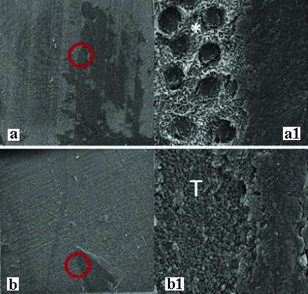
According to the morphological analyses of the fractured surfaces [Table/Fig-4], predominantly mixed fractures were found in all the groups, followed by adhesive fractures (occurred at the bond interface) [Table/Fig-5]. It was possible to note fractures at the base region of the hybrid layer in the groups formed by the total-etch adhesive system (Single Bond), with areas of conditioned dentin not infiltrated by the adhesive. In the groups in which the self-etch adhesive system was used, it was noticed that the fracture was at the top region of the hybrid layer.
Distribution of the modes of fracture.
| Groups | Fracture modes | |
|---|
| Cohesive in resin | Cohesive in dentin | Adhesive | Mixed |
|---|
| Single bond-control | 0 | 0 | 4 | 16 |
| Single bond+Bis block | 0 | 0 | 3 | 17 |
| Single bond+Aqua Prep F | 0 | 0 | 4 | 16 |
| Clearfil SE bond-control | 0 | 0 | 5 | 15 |
| Clearfil SE Bond+Bis block | 0 | 0 | 4 | 16 |
| Clearfil SE bond+Aqua Prep F | 0 | 0 | 5 | 15 |
Representative SEM images of the dentin of fractured specimens bonded with the adhesives. (a-a1) Specimens bonded with Adper Single Bond (SB); (b – b1) Specimens bonded with Clearfil SE Bond (SE); (a-b) Low-power magnification of whole area of the fractured specimens (130x). (a1-b1) Higher magnification of the circled area (5000x). (*) Bottom of the hybrid layer; (T) Top of the hybrid layer.
Discussion
The bond strength of total-etch and self-etch adhesive systems in human dentin conditioned and treated with desensitizers was evaluated. No statistically differences were observed in the bond strength between these materials when compared to the control groups, therefore the initial null hypothesis was accepted.
The association of the desensitizers did not modify the performance of the adhesive systems in face of the mechanical tests. According to Silva SMA et al., Sadek FT et al., Yiu CKY et al., and Acka T et al., the tubules capacity to seal/obliterate, caused by desensitizers, can reduce the dentin permeability and help the bonding [11,12,15,24].
As suggested by Perdigão J and Shafiei F et al., these materials association can reduce the degradation process of the hybrid layer and benefit the longevity of the bonding interface [6,25]. However, Silva SMA et al., affirmed that studies combining desensitizers and adhesive systems are difficult to reproduce, and this fact can prejudice the achievement of the results [8]. They noticed that, although this association reduced the bond resistance of the studied adhesives, the desensitizers lessened the degradation of the hybrid layer over time.
It is important to observe that the desensitizers selected in the current study were compatible with the adhesive systems Single Bond and Clearfil SE Bond, widely used in bonding procedures. According to the results of our study, the application of desensitizers based on oxalate on dentin conditioned with 37% phosphoric acid did not change the bond resistance of the Single Bond adhesive system (total-etch technique). This fact is in agreement with the studies of Sadek FT et al., Tay FR et al., and Baseggio W et al., [12,18,26]. When they used a desensitising treatment on conditioned dentin they did not notice changes in the bond resistance of the total-etch adhesive systems and considered the good performance of the desensitizers as a beneficial factor that can optimise the bonding procedure. The possible explanation to this result is the acidic performance of the oxalates, which will penetrate and react with the calcium of the dental structure through chelation, in a sub superficial portion, that would not interfere in the hybridization process.
According to the morphological analysis by SEM in this study, it was possible to observe that the oxalate crystals formation did not prevent the resinous tag shaping, neither the hybridization process with the total-etch adhesive system. Vachiramon V et al., noticed a reduction of the bond resistance when they used oxalate and the adhesive system, and suggested that the associated use of oxalate may have caused a negative interference in the hybrid layer formation, which is not in accordance with the present study [27].
Regarding the oxalates interaction with the total-etch adhesive system, it is suggested that compatibility between them is responsible for the good performance in the mechanical tests when they are associated. As noticed byYiu CKY et al., the oxalates have high compatibility with the Single Bond adhesive, because it has less acidic pH and reduced concentration of fluorides in its chemical composition when compared to other total-etch adhesives, which do not interfere in the bond resistance [15]. According to Tay FR et al., the use of desensitizers based on oxalates with no monomers in its composition was not capable of modifying the resistance of the total-etch adhesive surveyed [18].
The combined use of the desensitizer based on fluoride and HEMA (Aqua Prep F) did not alter the bond resistance of Single Bond. However, these results are not in agreement with the study by Dündar M et al., who observed an increase in bond resistance of the total-etch adhesive systems of two and three steps when associated with desensitizers [23]. The performance of the agent based on fluoride and HEMA was attributed to the infiltration capacity of the monomer HEMA in conditioned dentin and the reaction capacity between the fluoride and the calcium of the dental structure. Nevertheless, some differences between the adhesive systems can be found in the studies. Regarding the morphological evaluation in SEM, areas that suggest the existence of crystals inside the dentin tubules were observed, which can only be confirmed by analysis of the chemical elements (EDS). But these findings do not negatively affect the hybridization of the dentin surface, indicating compatibility among the materials.
The association of the desensitizers based on oxalate or fluoride and HEMA with the self-etch adhesive system Clearfil SE Bond do not significantly alter the bond resistance of the adhesive system. In agreement to the results, the Clearfil SE Bond performance was satisfactory, even when associated with desensitizers. These results suggest that, the oxalates can remove the smear layer and penetrate the inner part of the tubules with no prejudice to the hybrid layer formation. Huh JB et al., verified that the association between self-etch and oxalate free from monomers in its composition and concluded similar bond resistance values when compared to the control group (only with the adhesive appliance) [19].
The one containing monomers affected the formation of the hybrid layer because they formed copolymer mass that interfere in the reaction between the self-etch primer and the dental subtrate, consequently, weakening the adhesive bond resistance. In regard to the fluorides use, Acka T et al., concluded that using desensitizers with fluoride reduced the bond resistance values when compared to the control group (which only used self-etch adhesive), because of the precipitation of calcium fluoride crystals in the dentin tubules that interfere in the self-etch adhesive infiltration [24]. According to Acar O et al., [22], the use of desensitizer agents Bisblock and Aqua Prep-F adversely affected bond strength self-etch cements. The morphology of the surfaces treated with desensitizers and the self-etch system, observed by SEM, suggests that neither oxalates nor fluorides interfere in the hybridization, presenting a fracture predominantly at the top of the hybrid layer.
Limitation and Future Recommendations
The present study was an in-vitro study and the sample size was small. The effect of oral environment was also neglected. Therefore, in-vivo studies are required to investigate the effect of application of desensitising agents on the bond strength of adhesive systems. Also, more adhesive systems with desensitizers should be worthy investigated. Furthermore, this study focuses on the importance of the application of adhesive systems in the clinial procedures.
Conclusion
The use of the total-etch and the self-etch adhesive systems with the oxalates and fluorides desensitizers, surveyed in this study, was able to produce satisfactory results that motivate more studies about the advantages of this combination. The compatibility of these materials can represent a favourable factor to the durability of the bond and consequently to the longevity of the restorations. Though the results were not significant, Single Bond did not interfere in bond resistance when dentine desensitizers were applied, while the Clearfil SE Bond showed the lowest values when associated with the desensitizer Aqua Prep F. Predominantly mixed fractures were found in all groups followed by adhesive fractures (occurred at the bond interface).
Bis-GMA; bisphenol- A diglicidil eter dimetacrylate; HEMA, 2-hidroxietil metacrylate; 10-MDP, 10-metacriloxietil dihidrogeniun phosphate
*Technical bonding: a) phosphoric acid etching; b) rinse; c) drying the surface with absorbent paper; d) primer application; e) bond application; f) applying the primer-bond; g) drying the solvent for volatilization; h) application of the acid primer on the dry dentin surface doing slight pressure; i) bond application; j) photoactivation
[1]. Van Meerbeek B, De Munck J, Yoshida Y, Inoue S, Vargas M, Vijay P, Adhesion to enamel and dentin: current status and future challengesOper Dent 2003 28:215-35. [Google Scholar]
[2]. Cortiano FM, Rached RN, Mazur RF, Vieira S, Freire A, De Souza EM, Effect of desensitizing agents on the microtensile bond strength of two-step etch-and-rinse adhesives to dentinEur J Oral Sci 2016 124:309-15.10.1111/eos.1226327038226 [Google Scholar] [CrossRef] [PubMed]
[3]. Silva e Souza MH Jr, Carneiro KG, Lobato MF, Silva e Souza Pde A, de Góes MF, Adhesive systems: important aspects related to their composition and clinical useJ Appl Oral Sci 2010 18:207-14.10.1590/S1678-7757201000030000220856995 [Google Scholar] [CrossRef] [PubMed]
[4]. Pashley DH, Smear layer: physiological considerationsOper Dent Suppl 1984 3:13-29. [Google Scholar]
[5]. Van Meerbeek B, Inokoshi S, Braem M, Lambrechts P, Vanherle G, Morphological aspects of the resin-dentin interdiffusion zone with different dentin adhesive systemsJ Dent Res 1992 71:1530-40.10.1177/002203459207100813011506519 [Google Scholar] [CrossRef] [PubMed]
[6]. Perdigão J, Dentin bonding-variables related to the clinical situation and the substrate treatmentDent Mater 2010 26:e24-37.10.1016/j.dental.2009.11.14920005565 [Google Scholar] [CrossRef] [PubMed]
[7]. Carrilho MR, Carvalho RM, de Goes MF, di Hipólito V, Geraldeli S, Tay FR, Chlorhexidine preserves dentin bond in vitroJ Dent Res 2007 86:90-94.10.1177/15440591070860011517189470 [Google Scholar] [CrossRef] [PubMed]
[8]. Silva SMA, Malcarne-Zanon J, Carvalho RM, Alves MC, De Goes MF, Anido-Anido A, Effect of oxalate desensitizer on the durability of resin-bonded interfacesOper Dent 2010 35:610-17.10.2341/09-202-L21179999 [Google Scholar] [CrossRef] [PubMed]
[9]. Carrilho MR, Carvalho RM, Sousa EM, Nicolau J, Breschi L, Mazzoni A, Substantivity of chlorhexidine to human dentinDent Mater 2010 26:779-85.10.1016/j.dental.2010.04.00220472282 [Google Scholar] [CrossRef] [PubMed]
[10]. Münchow EA, Bottino MC, Recent advances in adhesive bonding-The role of biomolecules, nanocompounds, and bonding strategies in enhancing resin bonding to dental substratesCurr Oral Health Rep 2017 4:215-27.10.1007/s40496-017-0146-y29177123 [Google Scholar] [CrossRef] [PubMed]
[11]. Silva SMA, Marquezini L, Manso AP, Garcia FP, Carrilho MRO, Pashley DH, Effects of a combined application of potassium oxalate gel/adhesive agent on dentin permeability in vitroJ Adhes Dent 2007 9:505-12. [Google Scholar]
[12]. Sadek FT, Pashley DH, Ferrari M, Tay FR, Tubular occlusion optimizes bonding of hydrophobic resins to dentinJ Dent Res 2007 86:524-28.10.1177/15440591070860060717525351 [Google Scholar] [CrossRef] [PubMed]
[13]. Paes Leme AF, Santos JCRG, Giannini M, Wada RS, Occlusion of dentin tubules by desensitizing agentsAm J Dent 2004 17:368-72. [Google Scholar]
[14]. Pei D, Liu S, Huang C, Du X, Yang H, Wang Y, Effect of pretreatment with calcium-containing desensitizer on the dentine bonding of mild self-etch adhesivesEur J Oral Sci 2013 121:204-10.10.1111/eos.1204723659244 [Google Scholar] [CrossRef] [PubMed]
[15]. Yiu CKY, King NM, Suh BI, Sharp LJ, Carvalho RM, Pashley DH, Incompatibility of oxalate desensitizers with acidic, fluoride-containing total-etch adhesivesJ Dent Res 2005 84:730-35.10.1177/15440591050840080916040731 [Google Scholar] [CrossRef] [PubMed]
[16]. Pinto SCS, Pochapski MT, Wambier DS, Pilatti GL, Santos FA, In vitro and in vivo analyses of the effects of desensitizing agents on dentin permeability and dentinal tubule occlusionJ Oral Sci 2010 25:23-32.10.2334/josnusd.52.23 [Google Scholar] [CrossRef]
[17]. Dall’Orologio GD, Ishihata H, Finger WJ, Sasaki K, In vitro and in vivo evaluation of the effectiveness of three dentin desensitizing treatment regimensAm J Dent 2014 27:139-44. [Google Scholar]
[18]. Tay FR, Pashley DH, Mak YF, Carvalho RM, Lai SCN, Suh BI, Integrating oxalate desensitizers with total-etch two-step adhesiveJ Dent Res 2003 82:703-07.10.1177/15440591030820090912939354 [Google Scholar] [CrossRef] [PubMed]
[19]. Huh JB, Kim JH, Chung MK, Lee HY, Choi YG, Shim JS, The effect of several dentin desensitizers on shear bond strength of adhesive resin luting cement using self-etching primerJ Dent 2008 36:1025-32.10.1016/j.jdent.2008.08.01218986747 [Google Scholar] [CrossRef] [PubMed]
[20]. Bhatia S, Krishnaswamy MM, Effect of two different dentin desensitizers on shear bond strength of two different bonding agents to dentin: an in vitro studyIndian J Dent Res 2012 23:703-08.10.4103/0970-9290.11124223649049 [Google Scholar] [CrossRef] [PubMed]
[21]. Shafiei F, Doozandeh M, Impact of oxalate desensitizer combined with ethylene-diamine tetra acetic acid-conditioning on dentin bond strength of one-bottle adhesives during dry bondingJ Conserv Dent 2013 16:252-56.10.4103/0972-0707.11132723833461 [Google Scholar] [CrossRef] [PubMed]
[22]. Acar O, Tuncer D, Yuzugullu B, Celik C, The effect of dentin desensitizers and Nd:YAG laser pre-treatment on microtensile bond strength of self-adhesive resin cement to dentinJ Adv Prosthodont 2014 6:88-95.10.4047/jap.2014.6.2.8824843392 [Google Scholar] [CrossRef] [PubMed]
[23]. Dündar M, Çal E, Gökçe B, Türkün M, Özcan M, Influence of fluoride-or triclosan- based desensiting agents on adhesion of resin cements to dentin. Clinical Oral InvestigationClin Oral Investig 2010 14:579-86.10.1007/s00784-009-0328-719690902 [Google Scholar] [CrossRef] [PubMed]
[24]. Akca T, Yazici AR, Çelik Ç, Özgünaltay G, Dayangaç B, The effect of desensitizing treatments on the bond strength of resin composite to dentin mediated by a self-etching primerOper Dent 2007 32:451-56.10.2341/06-13017910221 [Google Scholar] [CrossRef] [PubMed]
[25]. Shafiei F, Memarpour M, Doozandeh M, Effect of oxalate desensitizer on the bonding durabilkity of adhesive resin cements to dentinJ Prosthodont Res 2012 56:187-93.10.1016/j.jpor.2011.11.00322264675 [Google Scholar] [CrossRef] [PubMed]
[26]. Baseggio W, Conslomagno EC, Carvalho FLN, Ueda JK, Schimitt VL, Formighieri LA, Effect of deproteinization and tubular occlusion on microtensile bond strength and marginal microleakage of resin composite restorationsJ Appl Oral Sci 2009 17:462-66.10.1590/S1678-7757200900050002119936527 [Google Scholar] [CrossRef] [PubMed]
[27]. Vachiramon V, Vargas MA, Pashley DH, Tay FR, Geraldeli S, Qian F, Effects of oxalate on dentin bond after 3-month simulated pulpal pressureJ Dent 2008 36:178-85.10.1016/j.jdent.2007.11.01118241968 [Google Scholar] [CrossRef] [PubMed]