Abnormal Twin Foramina on Fifth Lumbar Vertebra-Case Report
Rajani Singh1, Karishma Sharma2
1 Additional Professor, Department of Anatomy, AIIMS, Rishikesh, Uttarakhand, India.
2 PhD Student, Department of Anatomy, AIIMS, Rishikesh, Uttarakhand, India.
NAME, ADDRESS, E-MAIL ID OF THE CORRESPONDING AUTHOR: Dr. Rajani Singh, Additional Professor, Department of Anatomy, All India Institute of Medical Sciences, Virbhadra Marg, Pashulok, Rishikesh, Dehradun, Uttarakhand, India.
E-mail: nani_sahayal@rediffmail.com
Normally lumbar vertebrae in general and L-5 vertebra in particular are devoid of such foramen as explored in this case. Two mutually communicating foramina were detected in L-5 vertebra during demonstration classes of 1st year MBBS students in the Department of Anatomy, AIIMS Rishikesh, Uttarakhand, India. This is a unique and rare anomaly having enormous clinical significance in very prevalent lower back pain therefore, the case is worth reporting. One foramen was located on lateral aspect near the inferior margin of right transverse process and the other between the superior articular process and right transverse process equivalent to retro-transverse foramen. First foramen was oval in shape with transverse diameter of 1 cm and vertical diameter 0.6 cm and may provide pathways to vessels irrigating surrounding structures. Second foramen was circular in shape having diameter of 0.8 cm and acts as a conduit for medial branch of dorsal ramus. The knowledge of these foramina is clinically significant to orthopaedic and neurosurgeons, anaesthesists and anatomists.
Dorsal ramus, Lumbar vertebra, Retro-transverse foramen, Superior articular process, Transverse process
Case Report
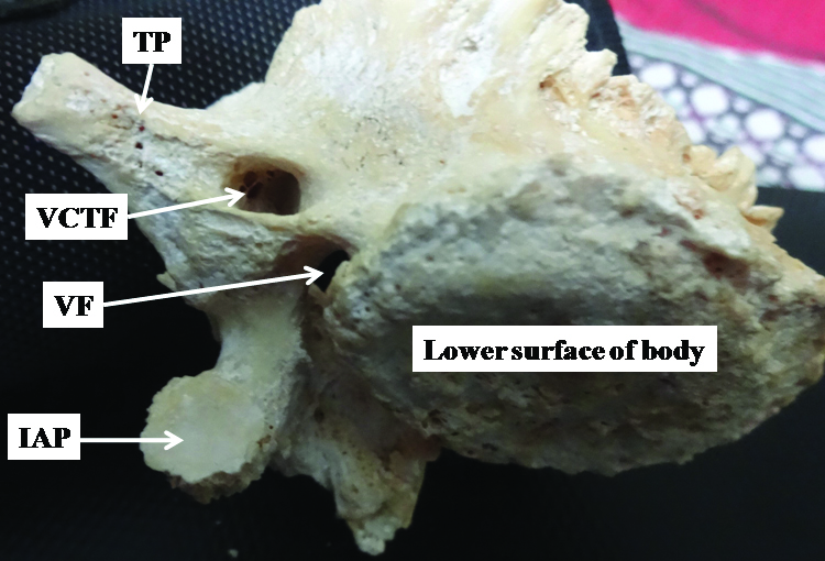
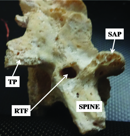
During osteology, demonstration classes of 1st year MBBS students, twin foramina anomalously communicating with each other were detected on a fifth lumbar vertebra. The age and sex of the vertebra was not known. One foramen called as costo-transverse foramen was located on the lateral aspect near the inferior margin at the base of right transverse process of fifth lumbar vertebra [Table/Fig-1]. This was oval in shape with transverse diameter of 1 cm and vertical diameter 0.6 cm. This foramen was oblique to axial plane and continued into a canal of 2 cm long and opened into vertebral foramen. In addition to this, another foramen described as retro-transverse foramen in literature was found located between the right superior articular and transverse processes [Table/Fig-2]. It was circular in shape. Diameter of this foramen was 0.8 cm. It continued into a canal of one cm in length and opened into canal of first foramen. This foramen was perpendicular to axial plane. The two foramina were communicating with each other.
Shows first foramen on the lateral aspect of right transverse process.
TP: Transverse process; VCTF: Variant costo-transverse foramen; VF: Vertebral foramen; IAP: Inferior articular process
Shows second foramen between superior articular process and transverse process.
TP: Transverse process; SAP: Superior articular process; RTF: Retro-transverse foramen
Discussion
Normally, lumbar vertebra is characterised by absence of foramen on the transverse process. Dwight T and Szawlowski J described foramen on the transverse process which according to Manners-Smith T was costo-transverse foramen [1-3]. Manners-Smith T observed two foramina, one each on right and left transverse process of fifth lumbar vertebra [3]. These foramina were present on the superior surface of transverse process and lateral to vertebral foramen classified as ‘costo-transverse foramen’ but it was vertical. Retro-transverse foramen also called Mammilo-Accessory Foramen (MAF) situated between articular process and transverse process. Manners-Smith T classified the foramina on transverse process as costo-transverse foramina to differentiate them from retro-transverse foramina occurring between the mamillary and accessory processes [3]. While carrying out routine computed tomography of the lower lumbar spine in patients with backache and sciatica, Beers GJ et al., observed four foramina including one on sacral vertebra; one similar to retro-transverse foramen and two vertical foramina through the medial aspect of transverse process of fifth lumbar vertebra [4].
The foramen observed by above investigators was present on the superior aspect of transverse process lateral to vertebral foramen whereas one of the twin foramen detected by present authors though present on the lateral aspect of transverse process near its inferior margin is location and orientation wise different so it may be taken as another variant of costo-transverse foramen. The structures passing through costo-transverse foramen are not known. But authors of present study consider that the foramen is vascular in nature and might transmit spinal branch of lumbar artery to irrigate the structures in the lumbar spinal canal. These structures may be compressed in the foramen leading to ischemia of the structures irrigated or if any nerve passes through it, there are chances that it may get compressed giving rise to neurological complications.
Large part of the lumbar transverse process described in literature to be homologous to the ribs and develop embryology wise from the “costal element” but medial and posterior part including the accessory process is homologous to the transverse process of the thoracic vertebrae and so might have developed from the “transverse element” [5].
Therefore, lumbar costo-transverse foramina like the foramina transversaria in the cervical region, caused due to incomplete fusion of transverse and costal elements [3]. In the lumbosacral region in the embryo anastomotic vessels passed between the costal and transverse elements similar to thoracic region. If a lumbar inter-vertebral vessel became atrophied during development, a vertical foramen might persist to transmit an enlarged anastomotic vessel [2]. Authors of present study support this view of SzawIowski.
The Mammillo-Accessory Ligament (MAL), a characteristic of lumbar vertebra extends between mammilary and accessory processes. The MAL spread over the gap between the mammilary and accessory processes forming the groove of variable depth to form a tunnel about 6 mm long in lumbar vertebra [6]. The foramen formed by ossification of MAL was named as MAF [7] and was observed in 10% of cases. Maigne JY et al., reported the incidence of MAF 26% on the left side and in 13.5% on the right [8]. This tunnel/foramen acts as a conduit for the passage of vessels to supply the dorsal paraspinal muscles [3] and the medial branch of the dorsal ramus formed of spinal nerves exiting from the inter-vertebral foramen. MAL is ossified very frequently in elderly age or congenitally controlled by expression of genes to form Mammilo-accessory foramen. This was termed as ‘retro transverse foramen’ by Manners-Smith [3]. Second foramen/tunnel of present study is equivalent to retro-transverse foramen described in literature. It is a manifestation of osteoarthritic changes and these may irritate or compress the dorsal ramus along its passage [8]. Mammilo-accessory foramen forms not merely due to osteoarthritic ossification of the MAL but could also be related to facet dimensions or degree of MP-facet fusions that lie close to the mamillo-accessory junctions [9].
Conclusion
MAF or retro-transverse transmits medial branch of dorsal ramus as advocated by various investigators. This foramen may compress or causes entrapment of medial branch leading to back pain and sciatica. In addition to this various percutaneous techniques to stimulate, anaesthetize or destroy the medial branch are used. Ossification of MAL may create difficulties during some percutaneous denervation techniques. MAF may be the site of entrapment of medial branch and may cause back pain. Costo-transverse foramen might transmit spinal branch of lumbar artery to irrigate the structures in the lumbar spinal canal. These structures may be compressed in the foramen leading to ischemia of the structures irrigated or if any nerve passes through it, there are chances that it may get compressed giving rise to neurological complications.
[1]. Dwight T, A transverse foramen in the last lumbar vertebraAnat Anz 1901 20:571-72. [Google Scholar]
[2]. Szawlowski J, Ueber einige seltene Variationen an der Wirbel saeule beim MenschenAnat Anz 1901 20:305-20. [Google Scholar]
[3]. Manners-Smith T, The variability of the last lumbar vertebraJ Anat 1908 43:146-60. [Google Scholar]
[4]. Beers GJ, Carter AP, McNary WF, Vertical foramina in the lumbosacral region: CT appearanceAJR 1984 143:1027-29.10.2214/ajr.143.5.10276333144 [Google Scholar] [CrossRef] [PubMed]
[5]. Gardner E, Gray DJ, O’Rahilly A, Anatomy: a regional study of human structure 1975 4th edPhiladelphiaSaunders [Google Scholar]
[6]. Bradley KC, The anatomy of backacheAust NZ J Surg 1974 44:227-32.10.1111/j.1445-2197.1974.tb04409.x4282245 [Google Scholar] [CrossRef] [PubMed]
[7]. Bogduk N, The lumbar mamillo-accessory ligament: Its anatomical and neurosurgical significance 1981 6:162-67.Spine10.1097/00007632-198103000-000106456553 [Google Scholar] [CrossRef] [PubMed]
[8]. Maigne JY, Maigne R, Guerin-Surville H, The lumbar mamillo-accessory foramen: a study of 203 lumbosacral spinesSurg Radiol Anat 1991 13:29-32.10.1007/BF016231371905063 [Google Scholar] [CrossRef] [PubMed]
[9]. Mahato NK, Mamillo-accessory notch and foramen: Distribution patterns and correlation with superior lumbar facet structureMorphologie 2014 98:176-81.10.1016/j.morpho.2014.03.00224889272 [Google Scholar] [CrossRef] [PubMed]