Letter to Editor- Oligodontia in Non-syndromic Children: Series of three Cases
Fernanda Felix Da Silva1, Christiane Cruz2, Flavia Martinez De Carvalho3, Marcelo Castro Costa4, Andrea Fonseca Gonçalves5
1 Phd Student, Department of Paediatric Dentistry and Orthodontics, School of Dentistry, Universidade Federal do Rio de Janeiro, Rio de Janeiro, Brazil.
2 PhD, Department of Paediatric Dentistry and Orthodontics, School of Dentistry, Universidade Federal do Rio de Janeiro, Rio de Janeiro, Brazil.
3 PhD, Laboratory of Congenital Malformation Epidemiology, Oswaldo Cruz Institute-FIOCRUZ, Rio de Janeiro, Brazil.
4 Associate Professor, Department of Paediatric Dentistry and Orthodontics, School of Dentistry, Universidade Federal do Rio de Janeiro, Rio de Janeiro, Brazil.
5 Adjunct Professor, Department of Paediatric Dentistry and Orthodontics, School of Dentistry, Universidade Federal do Rio de Janeiro, Rio de Janeiro, Brazil.
NAME, ADDRESS, E-MAIL ID OF THE CORRESPONDING AUTHOR: Dr. Andrea Fonseca Gonçalves, Rua Rodolpho Paulo Rocco, 325- Ilha do Fundão, Rio de Janeiro, RJ, Brazil.
E-mail: andrea.goncalves@odonto.ufrj.br
Dear Editor,
Tooth agenesis is an anomaly that can occur in both dentitions. The aetiology may be related to several genetic and environmental factors. Usually, it can cause aesthetic and functional damage to the patient. We highlight three cases of non-syndromic children with tooth agenesis to better understand the aetiological factors of this anomaly, even when genetic analyses are not performed.
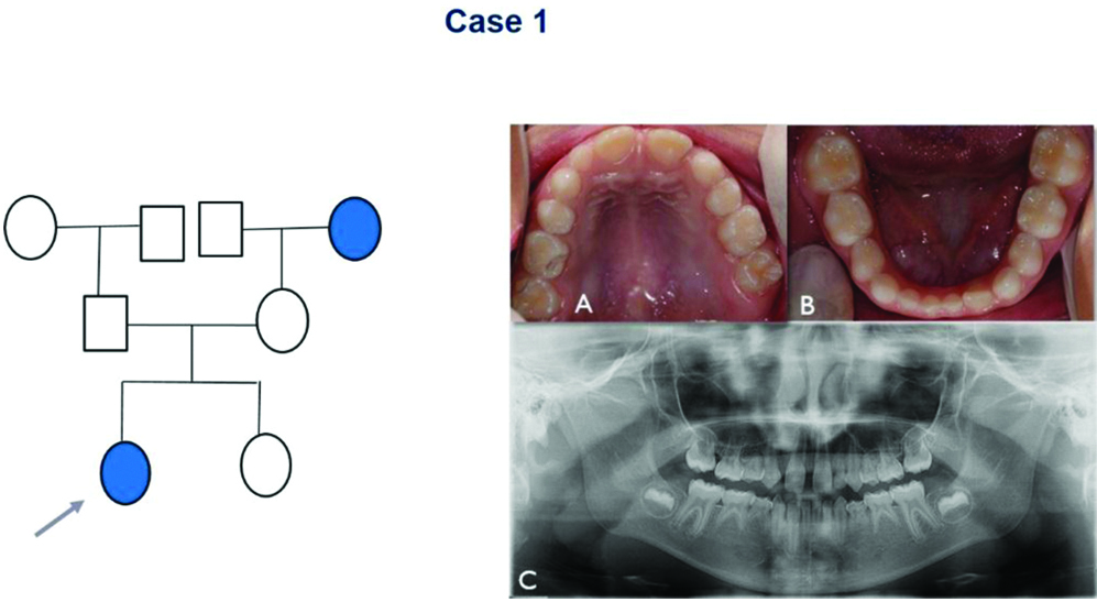
The first case was a seven-year-old girl who reported with delay in eruption of permanent lower front teeth. The anamnesis revealed no systemic involvement or syndromes and the parents reported tooth absence in family members [Table/Fig-1] and no complications during pregnancy. On examination, the patient was in the mixed dentition stage, with no carious lesions [Table/Fig-1a,b]. The radiographic examination showed the absence of the maxillary premolars, maxillary permanent canines and maxillary permanent lateral incisors [Table/Fig-1c]. In addition, it was observed absence of all mandibular premolars, mandidular permanent canines, mandibular permanent central incisors, mandibular left permanent lateral incisor (#32) and all permanent second molars [Table/Fig-1c]. Also, a delay in the root formation of the maxillary permanent first molars (#16, #26) was detected [Table/Fig-1c]. Patient was informed about the findings and the treatment plan was the maintenance of the deciduous teeth in the arch and to keep a regular follow-up every 6 months. Once the deciduous teeth are physiologically reabsorbed, an orthodontic and prosthetic rehabilitation treatment was planned.
Non-syndromic patient with agenesis (gray arrow) and the family pedigree. (a) Maxillary intraoral photographs that shows the permanent central incisors and the absence of the left primary lateral incisor. (b) Mandibular intraoral photograph shows the primary teeth and the left permanent central incisor. (c) Panoramic radiography that shows absence of maxillary permanent lateral incisors, maxillary premolars, maxillary and mandibular canines and a root formation delay in the maxillary permanent first molars.
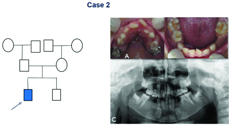
The second case was a 10-year-old boy with absence of permanent tooth attended for his first dental appointment, with caries in multiple teeth as the chief complaint. The patient has already had hepatitis A, anemia and severe convulsions, for which he is on medication since the age of 6-years. Furthermore, his mother reported no complications during pregnancy and no syndrome. After the intraoral examination, it was observed that the patient was in mixed dentition stage with carious lesions in teeth #54, #64 and #75. Palate atresia and a vestibular inclination of the maxillary permanent left central incisor (#21) [Table/Fig-2a,b] was also observed. The panoramic radiograph revealed absence of all maxillary and mandibular premolars [Table/Fig-2c]. The extraction of teeth #54 and #64 and restorative treatment for tooth #75 [Table/Fig-2a,b] were conducted. Furthermore, the patient was referred for orthodontic treatment to correct the bone and tooth discrepancies.
Non-syndromic patient with agenesis (gray arrow) and the family pedigree. (a) Maxillary photographs shows a palate atresia and a vestibular inclination of the maxillary permanent left central incisor, besides the absence of the right second primary molar and left first primary molar. (b) Mandibular photographs shows absence of the left first primary molar. (c) Panoramic radiography that exhibit absence of maxillary and mandibular premolars.
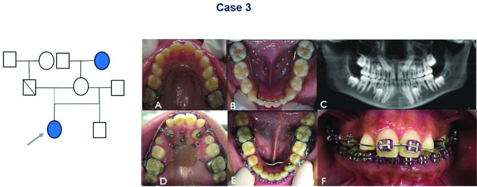
The last case was a 12-year-old girl with no systemic or syndrome involvement and her mother reported no complication at her pregnancy period. The complaint was absence of permanent tooth, with history of tooth agenesis of her paternal grandmother. The intraoral examination showed that patient was in mixed dentition with absence of dental caries [Table/Fig-3a,b]. on radiographic examination, we observed absence of the maxillary second premolar (#15,#25), maxillary permanent lateral incisor (#12,#22), mandibular permanent central and lateral incisors (#41,#31,#32, #42) and mandibular second premolars (#35,#45) [Table/Fig-3c]. The treatment plan was the orthodontics preventive treatment [Table/Fig-3a,b], following by corrective orthodontics devices [Table/Fig-3d-f] for correction of tooth discrepancies and better aesthetics.
Non-syndromic patient with agenesis (gray arrow) and the family pedigree. (a) Maxillary photograph shows the permanents central incisor. (b) Mandibular photograph shows all the primary teeth present in the arch. (c) Panoramic radiography that shows absence of the maxillary second premolar, maxillary permanent lateral incisor, mandibular permanent central and lateral incisors and mandibular first and second premolars. (d), (e) and (f) Intraoral Images that exhibit a mixed dentition with absence of dental caries and 1-year follow-up of the patient in orthodontic treatment.
Tooth agenesis is the most common congenital anomaly that is seen in humans. The prevalence of which varies from 3.2% to 13.3% [1]. Some studies proposed that tooth agenesis may result from genetic factors [2,3]. Among the candidates are the polymorphisms (MMPs), responsible for the craniofacial development and the BMP (Bone morphogenetic protein) genes that participate directly in the early stages of odontogenesis [3]. However, environmental factors cannot be ignored. Case 1 and 3 present history of agenesis in a family member and the child of Case 2 showed systemic changes during his early childhood, which probably may have influenced the aetiology of the agenesis. In the first case, no reports of damages, during tooth formation, was observed. Thus, reporting the case is very important for better understanding of the aetiological factors of this anomaly.
Usually, the most commonly affected teeth are the third molars, followed by mandibular second premolars, maxillary lateral incisors, maxillary second premolars and mandibular incisors [1]. The absence of permanent canines, first and second molars are extremely rare, that can be seen in association with oligodontia, mainly in syndromic cases [4]. The children did not have syndromes, but two showed agenesis of canines and one presents absence of second molars that lead us to believe they are rare cases.
The consequences of agenesis include malocclusions, deficiency of the alveolar processes, infraocclusion of deciduous teeth, inadequate angulations of adjacent teeth and psychological and social impact to patients [5]. There are various treatment options ranging from orthodontic to prosthetic rehabilitation [5]. The choice of treatment will depend on the patient’s age, physical and social conditions. For all the present cases, the options for future treatments were discussed with the parents, such as adhesive prosthesis or implant placement. Children from Case 2 and 3 were immediately referred for orthodontic treatment.
Absence of various teeth can generate serious implications for the patient. Thus, an early diagnosis and working with a multidisciplinary team are the main factors for a better planning of treatment to achieve success. The present authors suggest more detailed studies of oligodontia, with the investigation of the children’s quality of life.
[1]. Albashaireh ZS, Khader YS, The prevalence and pattern of hypodontia of the permanent teeth and crown size and shape deformity affecting upper lateral incisors in a sample of Jordanian dental patientsCommunity Dental Health 2006 23(4):239-43. [Google Scholar]
[2]. Coelho Neto OL, Reis MF, de Sabóia TM, Tannure PN, Antunes LS, Antonio AG, Clinical and genetic analysis of a nonsyndromic oligodontia in a childCase Rep Dent 2014 2014:13762110.1155/2014/13762125215247 [Google Scholar] [CrossRef] [PubMed]
[3]. Küchler EC, Menezes R, Callahan N, Costa MC, Modesto A, Meira R, MMP1 and MMP20 contribute to tooth agenesis in humansArch Oral Biol 2011 56(5):506-11.10.1016/j.archoralbio.2010.11.00721144496 [Google Scholar] [CrossRef] [PubMed]
[4]. Kambalimath HV, Jain S, Patil RU, Asokan A, Kambalimath D, Permanent maxillary canine agenesis: a rare case reportInt J Clin Pediatr Dent 2015 8(3):242-46.10.5005/jp-journals-10005-132226604546 [Google Scholar] [CrossRef] [PubMed]
[5]. McNamara C, Foley T, McNamara CM, Multidisplinary management of hypodontia in adolescents: case reportJ Can Dent Assoc 2006 72(8):740-46. [Google Scholar]