Osteoblasts and osteoclasts are continuously renovating alveolar bone under a process called remodeling. The regulation of osteoclasts is done mainly by osteoblasts through the Receptor Activator of Nuclear factor-Kappa B Ligand (RANKL) and Osteoprotegerin (OPG) [1,2].
Factors such as medications can interfere with orthodontic treatment through alveolar remodeling and, consequently, dental movements [3]. Many of the analgesic medications are prostaglandin inhibitors and these reduce Orthodontic Tooth Movements (OTM) [4,5].
Exogenous opioids that are used to control pain also interfere with bone metabolism and cause changes in OTM through a different mechanism [6,7]. Opioids can reduce or enhance OTM. For example, internal opioids accelerate OTM by reacting with Nitric Oxide (NO), but external opioids reduce OTM by affecting a receptor of the osteoblast cells [8].
Opioids, depending on the duration of the effect and stability of the drug, are divided into two groups; Morphine and codeine are Short-Acting Opioids (SAO’s), and methadone is Long-Acting Opioids (LAO’s) [9,10].
Opioid receptors are present in osteoblast-like cells (MG-63) that are related to bone modeling and remodeling [7]. Recently, the results of a study showed that self-medication for pain control in high school and university students was common and the most common analgesic drug used was acetaminophen codeine [11]. In the United States, the National Drug Early-Warning System (NDEWS) reported that incorrect and personal use of drugs had reached an explosion, and among prescription drugs, almost half of them are over-the-counter drugs [3].
Orthodontic tooth movements are associated with pain and patients have a potential for self-medication with opioids. Opioids can affect OTM and relieves the pain very well. Also, information about opioids in OTM is not consistent [8,12].
This animal study decided to evaluate and compare the effects of three different types of opioids (regarding the duration of their effects) on OTM.
Materials and Methods
This animal study protocol was approved by the Ethics Committee of Shahid Sadoughi University of Medical Sciences (No: IR.SSU.REC.1395.244), Yazd, Iran.
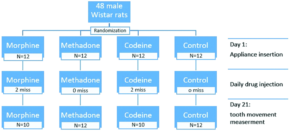
Forty-eight male Wistar rats weighing between 250-300 grams and the same age of six weeks were obtained from the animal room of the School of Medicine of Yazd University of Medical Sciences. The experimental sample size of each group was calculated to be 12, considering standard deviation 0.02 and mean difference 0.03 between groups for OTM, respecting confidence level 95% and power test 80% [8].
The rats were housed in plastic cages with a 12-hour light/dark cycle at 21±2 °C and 55% humidity. They were fed by a standard laboratory diet (consisted of calcium, phosphorus and vitamin D) with access to the sufficient amount of water for one week before the study, for accommodation with the environment and these conditions were maintained during the study until day twenty-one, in July 2018.
All of the processes were conducted following the U.S. National Institutes of Health’s (NIH’s) Guide for the Care and Use of Laboratory Animals.
The rats were randomly divided into four groups according to the random table of numbers. The rats were weighted once before the placement of the orthodontic appliance and once at the day of sacrification to evaluate the effect of the drug on the weight of the rats [Table/Fig-1].
Orthodontic Appliance
In order to place the orthodontic appliances, the rats were anesthetized with an intraperitoneal injection of 44 mg/kg ketamine hydrochloride (Gedeon Richter Ltd., Budapest, Hungary) and 2 mg/kg xylazine (Rompoun, Bayer, Leverkusen, Germany). The applied orthodontic appliance consisted of a 3 mm NiTi closed coil spring 0.006*0.022 inch (GAC International, Inc., USA) that was anchored between the upper left incisor and first molar with a 0.010-inch stainless steel ligature wire (Dentaurum, Ispringen, Germany) [Table/Fig-2].
a) Actual view and b) Schematic view of installed appliance.
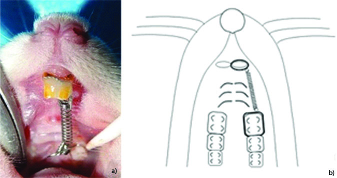
In the posterior region, the ligature wire crossed under the contact area of the first and second molars and was wrapped around the first molar at the gingival margin and then tied to the close coil spring. In the anterior region, the Ligature wire crossed under the contact area of the incisors, was wrapped around the left incisor, placed in a shallow groove that was carved with a diamond bur at the bucco-gingival and disto-gingival surfaces of the left incisor (to secure ligature from sliding out of cone shape incisor), and tied to a closed coil spring. Then, the groove area was etched with 37% phosphoric acid and covered with a small amount of light-cured composite resin to protect the Ligature wire from damage. Activation of the spring by 5-7 mm delivered about 25 grams of force to the first molar and incisor. After the placement of the appliance, the crown of the lower incisors was reduced 1.5 mm to prevent cutting of ligature wire.
Measurement of Tooth Movement
We measured the movement of the first molar at the beginning and twenty-one days after the placement of the orthodontic appliance, this timeline was set in order to have enough time to generate a mesial movement of the first molar [13].
The amount of mesial movements of the molar was measured by a feeler gauge (Mitutoyo Co, Kawasaki-Shi, Japan) by measuring the distance between the first and second molars. At the beginning of the study, there was no space between the molars. At the end of the study, the distances between the molars were measured before removing the appliance by the feeler gauge to prevent immediate relapse.
The measurements were performed twice by the same operator who was blinded to the intervention, and the mean of that was allocated for the OTM.
Drug Prescription
Medications were prepared by an operator with a digital coding for each group daily, and the injections were performed peritoneally by a second operator who was blinded to the intervention.
The first group received morphine at a dose of 6 mg/kg/day, the second group received methadone at a dose of 3 mg/kg/day, and the third group received codeine phosphate 30 mg/kg/day mixed in normal saline, and the fourth group received a normal saline injection. All injections were administered intra-peritoneal at 24 hours’ interval for twenty-one days and doses selected from similar studies in pain control [8,14].
Histological Analysis
After sacrificing the rats by an overdose of ether for 15 minutes in a plastic container, then the animals were decapitated with a scalpel, their maxilla was dissected and fixed by 10% formalin for ten days and decalcified with 10% hydrochloric acid for four days. The specimens were then embedded in paraffin blocks and sectioned along the sagittal plane at the site of the mesio-buccal root of the first molar with a thickness of 4-6 μm, and the samples were stained with Haematoxylin and Eosin (H&E) staining.
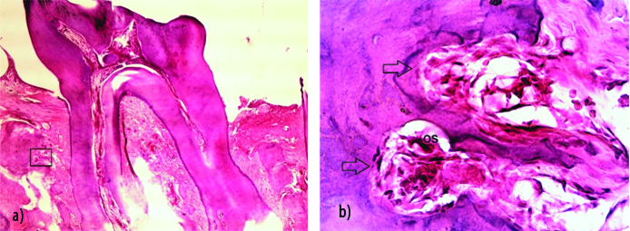
The slides were read by an operator who was blinded to the intervention with optical MB-41 Olympus microscope with 40x magnification in five random fields, and the average number of osteoclasts in five regions were considered as the number of osteoclasts. Multi-nucleotide large cells with foamy cytoplasm associated with a bone resorption region were considered as osteoclasts [Table/Fig-3].
a) Sagittal section of rat molar with H&E staining (x40). b) Arrows show two Howship lacunae on the resorptive layer of the adjacent tooth. OS shows an osteoclast (x400).
According to the resorptive lacuna on the left side of the alveolar bone, force delivered to the left side. There was no undermining resorption, and all was direct resorption through the bone. The box in figure a) is magnified to create figure b).
Statistical Analysis
According to the result of Kolmogorov-Smirnov’s test for examining the normality of data, Kruskal-Wallis’s test was used for the comparison of OTM between four groups and for multiple comparisons analysis Mann-Whitney test was implemented. In this study, the mean of OTM and osteoclasts was measured. The significance level (p<0.05) was considered statistically significant, and all statistical analyses were performed by SPSS version 11.
Results
A total of forty-eight rats including twelve in each group were included in this study, of which forty-four rats reached the end of the study and were analysed. In this study, four rats, two in the morphine group and two in the codeine group were excluded because of detachment of orthodontic appliance or weight loss (less than 250 mg).
All groups showed evidence of weight loss with no statistical significance. (p>0.05) There was no statistically significant difference between the average weights of the groups (p>0.05).
Mesial movement of the first molar was observed in all groups [Table/Fig-4].
Descriptive statistics of orthodontic tooth movement after 21 days.
| Groups | N | Mean (mm) | Median (mm) | Std. deviation | Minimum (mm) | Maximum (mm) |
|---|
| Morphine | 10 | 0.160 | 0.150 | 0.06 | 0.05 | 0.25 |
| Methadone | 12 | 0.162 | 0.125 | 0.08 | 0.05 | 0.35 |
| Codeine | 10 | 0.164 | 0.150 | 0.06 | 0.05 | 0.20 |
| Control | 12 | 0.279 | 0.250 | 0.12 | 0.15 | 0.55 |
Among the groups, the highest rate of OTM was observed in the control group (mean=0.27 mm) and the lowest rate was related to the morphine group (mean=0.16 mm). The difference between the three groups of morphine, methadone, and codeine was not statistically significant (p>0.05). However, there was a statistically significant difference between the three intervention groups and the control group in reducing OTM (p<0.05).
Osteoclasts were seen at the mesial alveolar crest of the first molar in all groups [Table/Fig-5].
Descriptive statistics of number of osteoclasts after 21 days.
| Groups | N | Mean | Median | Std. deviation | Minimum | Maximum |
|---|
| Morphine | 10 | 2.22 | 2.00 | 1.09 | 0.8 | 4 |
| Methadone | 12 | 2.00 | 2.00 | 1.36 | 1 | 5 |
| Codeine | 10 | 2.08 | 1.20 | 1.42 | 1 | 5 |
| Control | 12 | 2.81 | 2.70 | 1.36 | 0.8 | 5 |
Although the mean osteoclast count was higher in the control group than the other groups, there was no statistically significant difference between the groups. (p>0.05)
Discussion
We studied the magnitude of OTM in rats following the use of three exogenous opioids, two short acting with different affinity to opioid receptors (morphine and codeine) and one long-acting (methadone) through measuring the separation between the first and second molars after mesial movements of the first molar using a simple orthodontic appliance (Pull Coil).
Although we expected a difference in OTM between experimental groups because of the difference in duration of effects and affinity of the drugs to the receptors, this three opioid drugs in therapeutic doses [8,13] reduced OTM to the same degree without significant statistical differences.
Previous studies have shown that opioids play a role in osteoblastic activity and bone remodeling [8,12,15,16]. The results of the present study can be compared with Nilforoushan D et al., which stated that internal opioid (morphine) in the plasma of cholestatic rats are increased and can affect the rate of OTM through bone remodeling [12]. Increased OTM was attributed to the effect of internal opioids on NO which is a factor in tooth movement acceleration. In that study, internal opioids accelerated the dental movement which was not neutralised by Naltrexone (opioid antagonist). However, in the present study, exogenous opioids reduced OTM in healthy rats. This different behaviour of morphine may be because of intermediate compounds in the production path of opioids that can interfere in nitric oxide path and also the negligible dose of morphine in the plasma of cholestatic rat that was not neutralised by Naltrexone. It has been suggested in the previous studies that morphine stimulates the mu receptor which inhibits adenylate cyclase and reduces CAMP levels and cause a reduction in bone remodeling and tooth movement that agreed with the present study [16].
Limitation
This study was conducted on rats, therefore, the results may not be completely applicable to human. Further studies should evaluate the clinical and cytological changes in humans.
Conclusion
Morphine, Methadone, and Codeine reduced orthodontic tooth movement in rats. There is no difference between morphine, methadone, and codeine as a matter of reduction of OTM. This study was unable to show the relation between osteoclasts and OTM; although opioids had a small insignificant effect on the number of osteoclasts. Further studies are required to assess the effect of opioids on orthodontic tooth movement and bone resorption in human.