The smile is a key component to the self-esteem of an individual. At times, from a clinical perspective, it seems that concepts of denture aesthetics are being masked by mechanistic concerns for denture stability and function. The role of dental professionals is to promote oral health and dental esthetics. The anterior teeth are primarily related to the esthetics as they play an important role in the functions of lip support and phonetics. Size, form, and colour of anterior teeth must be in harmony with the surrounding facial environment for a completely edentulous patient. All these objectives are difficult to achieve when pre-extraction records are not available [2].
Several efforts have been made to precisely quantify the selection of the anterior teeth. Some of the more conversant extraoral factors are bizygomatic width, interpupillary distance, intercanthal distance, interalar width, intercommissural width and some novel extraoral anatomical measurements such as philtral width and circumference of skull. Some conversant intraoral factors like maxillary arch length, maxillary arch width, pterygomaxillary notch, palatal width, length, depth have also been considered. But, there are no studies, which proves single esthetic factor that can be used reliably for selection of artificial teeth [2].
Studies on anthropometric facial characteristics and their inter-relation with the natural teeth have provided data on their common individual agreement. Numerous studies on human face demonstrate the presence of significant disparities in parameters amid diverse races, nations, populations and individuals as well. They have recommended a ratio among the facial size and tooth size that could be used as a guide in selecting artificial denture teeth. But the chief limitation is that the soft tissue measurements are subjective to variation. This can also be troublesome to one, who has no natural teeth left and no pre-extraction records are existing [3-5].
The resolution to this problem is the practice of using stable facial references that are not subjective to change. One of such landmarks is the interpupillary distance. Gomes VL et al., found that the extraoral factor interpupillary distance could help reliably for the selection of maxillary anterior teeth [6]. Cesario VA et al., reported that interpupillary distance could be used reliably in selecting maxillary anterior teeth width, because their measurements showed consistent relationship for sexual and racial differences [7]. The distance between the right and left pupils was found to be 6.6 times the width of the maxillary central incisor. {X (width of max central incisor)=IPD (Interpupillary Distance)/6.6} [6,7].
Selection of teeth varies in each facial form. We have different set of commercially available teeth set, which also depends on facial form. The width of maxillary anterior teeth varies in each form and is not the same for all. Therefore, it is necessary to distinguish the participants in different facial forms so that teeth selection could be more reliable.
There is no single anthropometric measurement that can be used to quantify the width of maxillary anterior teeth. The anthropometric measurement used depends on the population group. Therefore, this research was carried out as an attempt to better understand and analyse biometric parameters of our study population. Until now there have been no similar studies conducted representing the whole of Indian population including different facial and tooth forms. Therefore, the aim of the study is to determine and correlate the width of maxillary anterior teeth using extraoral factor Interpupillary width in different facial and tooth forms among Indian population.
Materials and Methods
The study design was cross-sectional survey. The study was conducted in Department of Prosthodontics, Saveetha Dental College, Saveetha University from May 2017 till June 2018 in duration of 1 year and 2 months. A total of one thousand and two hundred (n=1200) dentulous individuals who visited to our department for general dental check-up were included in this study with age ranged from 18 to 55 years and those who had no significant medical problems were selected. All subjects had no history of smoking, alcohol abuse or use of specific drugs. Subjects were selected from each state based on Multistage Sampling (To select the study respondents two stage sampling design is used that is cluster sampling methodology is applied. In the first stage, each state is considered as a cluster, which are selected randomly and in second stage from the selected states the respondents are selected randomly using simple random sampling procedure).
Inclusion Criteria
Subjects who met the following criteria were included in this study:
Natives of India.
Angle’s Class I molar and canine relationships
Permanent teeth with no history of orthodontic treatment or extraction
All the teeth were morphologically normal with no defects in enamel
Above 18 years of age
Having full complement of teeth
Exclusion Criteria
Following subjects were excluded in this study:
Artificial crowns, fillings, attrition on anterior teeth
Crowding or spacing in the anterior teeth
Gingival inflammation or hypertrophy
Below 18 years of age
Facial asymmetry.
Congenitally missing anterior tooth or teeth
All the subjects selected for the study fulfilled the above criteria.
The study protocol was duly approved by the Institutional Ethics Committee (Saveetha Medical College and Hospital, Chennai) (Ethics Committee no: 002/04/2017/IEC/SU; dated 27/04/2017) and written informed consent was obtained from those who agreed to participate voluntarily in the research. Confidentiality of the information was maintained.
Impression making and preparation of cast models: (Preparation of Subjects)
The subjects were made to sit comfortably on the dental chair in a relaxed state and Alginate impression (Tulip Alginate Impression Material, Cavex, Holland Bv, Haarlem Holland) was made for maxillary arch and cast was poured immediately with hard setting dental stone (Type III-Ultrarock, Kalabhai Karson Pvt., Ltd., Mumbai, India). All intraoral measurements (CMA, MCIWR, MCIWL, Tooth form) were carried out on the artificial stone casts of maxillary arches using dental floss, flexible ruler and digital vernier caliper (with 0.01 mm accuracy). Extraoral facial measurements (IPD, Facial form) were recorded upto two decimals using precise digital Vernier caliper (Mitutoyo, UK Ltd.,) [Table/Fig-1]. Each parameter was measured three times and the average value were computed and recorded in a Proforma.

Determination of Inter-Pupillary Distance (IPD) in mm: The interpupillary width was measured from mid-pupil of one eye to mid-pupil of the other [Table/Fig-2]. The distance between pupil to pupil was measured using a digital vernier caliper without the application of pressure.
Measurement of interpupillary distance using digital caliper.
Patient's consent was obtained for publication of the image.

Facial Form (Based on Leon Williams) [8].
The face form was classified based on William’s method as follows [Table/Fig-3]:
Classification of facial forms based on Leon Williams method [8]: a) Square face form; b) Tapering face form; c) Oval face form.
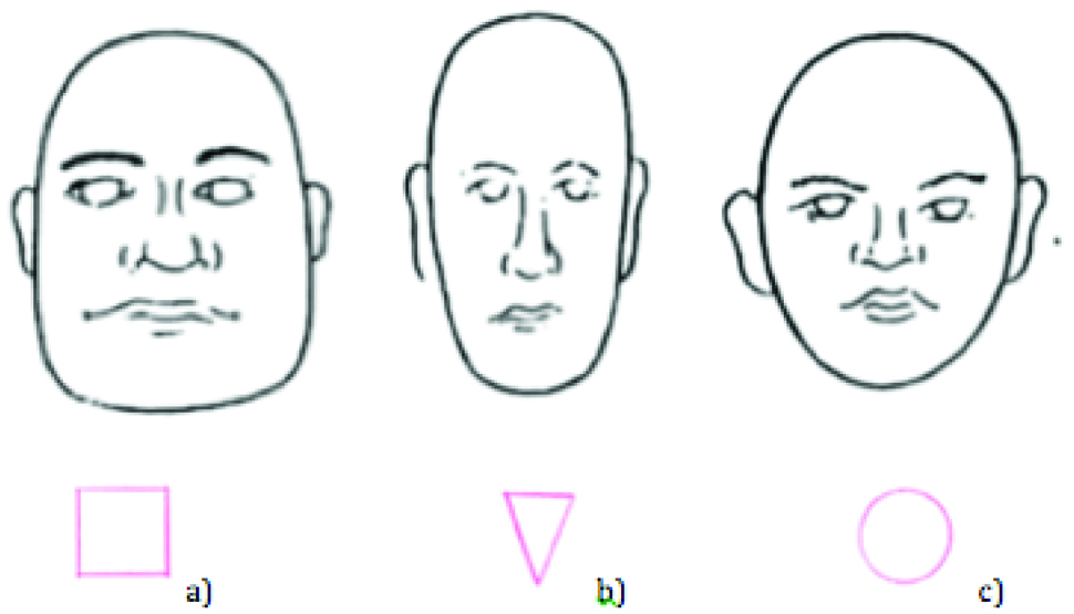
a) Square face-the outline of the face between Temporal, Zygomatic, Gonial were parallel vertically
b) Tapering face-the outline of face from temporal bone to the gonion was inwards vertically
c) Ovoid face-the outline of face from temporal bone to the gonion was outwards vertically
Measurements from Models: (Intraoral Measurements)
Teeth Dimensions
1. CMA- (Combined Width of Maxillary Anterior Teeth): The circumferential arc distance between the distal surface of the left and right canines was measured with a dental floss placed at the greatest facial curvature superio-inferiorly. (Disto-proximal Contacts of upper canines) [Table/Fig-4]. It was then sectioned and measured between the marks with the help of a digital caliper.
Combined width of maxillary anterior teeth measured using dental floss.

2. Maxillary Central Incisor Width (MCIW): The width dimension was obtained by measuring the maximum distance between the mesial and distal contact points of the tooth using digital vernier caliper that could be fixed in position with finely pointed ends that fit interdentally [Table/Fig-5]. The mesiodistal width of each maxillary Central Incisor (CIW) was recorded on stone cast. The widths were summed and divided by 2 to yield the width of a single tooth. The measurements were made in a straight line, with the pointed members of the caliper held parallel to the incisal edges and vertical (on a line perpendicular to long axis) to the facial surface of the tooth.
Measurement of mesiodistal width of central incisor using digital caliper.

3. Tooth form: (Based on Leon Williams) [8].
The tooth form was classified by William’s method as follows [Table/Fig-6].
Classification of tooth forms based on Leon Williams method [8]: a) Square tooth form; b) Oval tooth form; c) Tapering tooth form.
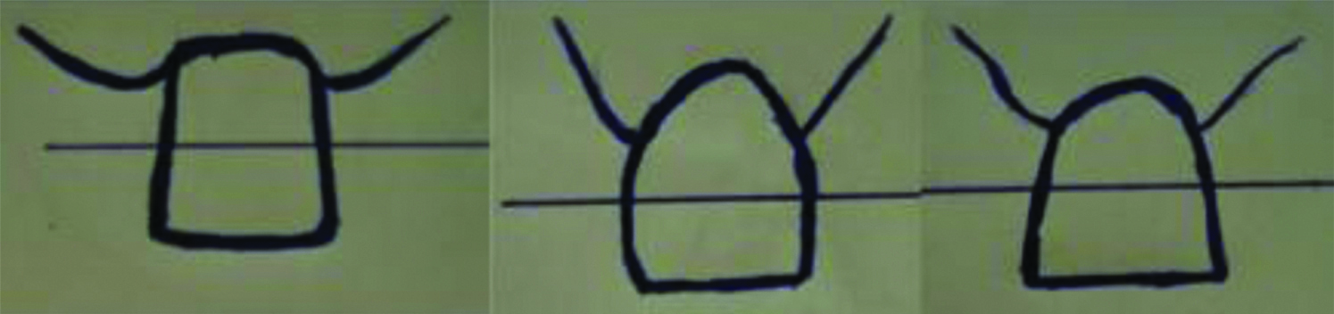
a) Square incisor tooth-Parallel mesial and distal proximal surfaces when viewed from front for atleast half the length of tooth; mesial and distal proximal surfaces are parallel for at least half of the cervico-incisal length of the crown.
b) Ovoid incisor tooth-Mesial and distal proximal surfaces moves outwards from incisal to cervical end.
c) Tapering incisor tooth-Mesial and distal proximal surfaces moves inwards from incisal to cervical end.
Statistical Analysis
The recorded data were compiled and entered in a spreadsheet (Microsoft Excel 2015) computer program and then exported to data editor page of SPSS (IBM SPSS Statistics for Windows, Version 22.0, Armonk, NY: IBM Corp. Released 2013). Descriptive Statistics were carried out for all 1200 participants and then for male and female subjects and in different zones of Indian population with level of significance at 5% (0.05) and power of the study at 95%. Independent t-test was used to compare two variables and Pearsons Correlation was used to know interconnection between IPD and CMA, MCIWR, MCIWL by linear Correlation analysis and summarised numerically with the linear correlation coefficient (r). Simple and multiple regression analysis was carried out to predict the width of maxillary anterior teeth and One-way ANOVA to compare more than two means between different facial forms.
Results
To analyse cluster sampling surveys, weights were applied to the dataset. By the end of the survey, out of 1200 subjects, only 1189 subjects were included in the data analysis since 11 subjects were removed from the survey during analysis as it had extreme values (Extreme values were Interpupillary width more than 75 mm for these 11 subjects only, out of 1200 subjects others were in average of 55-65 mm) which would influence study results. A total of 364 subjects were from South zone, 212 subjects were from North Zone, 312 subjects were from East zone, 301 subjects were from West zone. Out of 1189 subjects, 717 subjects were males and 472 subjects were females.
The correlation of 86.2% was observed between tooth form and face form. The dominant type of facial form in the studied population was oval with an incidence of 800 subjects of which 624 subjects were males. About 176 subjects were females, followed by Square with an incidence of 235 subjects of which 89 subjects were male and 146 subjects were females and Tapering with a frequency of 154 subjects of which 4 subjects were male and 150 subjects were females. The distribution of tooth form in the studied population was oval with an incidence of 640 subjects of which 497 subjects were males 143 subjects were females followed by Square with an incidence of 385 subjects of which 205 subjects were males and 180 subjects were females and Tapering with a frequency of 164 subjects of which 15 subjects were male and 149 subjects were females.
The mean Interpupillary distance, Mean CMA, Mean MCIWR and MCIWL were statistically significant among males and females and different facial and tooth forms mentioned in [Table/Fig-7]. Interpupillary distance was strongly positively correlated with CMA (r=0.983, p<0.001), MCIWR (r=0.959, p<0.001), MCIWL (r=0.953, p<0.001) mentioned in [Table/Fig-7]. Prediction of width of CMA, MCIWR, MCIWL using IPD as Predictor is shown in [Table/Fig-7]. Results of linear regression and derived equations are shown in [Table/Fig-8] to predict maxillary central incisor width at right and left side and combined width of maxillary anterior teeth taking IPD as predictors.
Analysis of variables among various facial and tooth forms.
| Overall | Males | Females | FF-Oval | FF-Square | FF-Tapering | TF-Oval | TF-Square | TF-Tapering |
|---|
| Mean (IPD) p<0.001 | 59.07 | 61.10 | 55.99 | 60.12 | 58.20 | 54.89 | 60.09 | 58.95 | 55.33 |
| Mean (CMA) p<0.001 | 41.73 | 43.14 | 39.58 | 42.46 | 41.11 | 38.80 | 42.44 | 41.65 | 39.11 |
| Mean (MCIWR) p<0.001 | 8.90 | 9.20 | 8.45 | 9.06 | 8.76 | 8.23 | 9.06 | 8.88 | 8.30 |
| Mean (MCIWL) p<0.001 | 8.92 | 9.22 | 8.46 | 9.08 | 8.78 | 8.25 | 9.07 | 8.89 | 8.33 |
| r (CMA) p<0.001 | 0.983 | 0.954 | 0.964 | 0.971 | 0.984 | 0.973 | 0.974 | 0.981 | 0.980 |
| r (MCIWR) p<0.001 | 0.959 | 0.867 | 0.905 | 0.945 | 0.937 | 0.919 | 0.945 | 0.945 | 0.943 |
| r (MCIWL) p<0.001 | 0.953 | 0.850 | 0.885 | 0.934 | 0.925 | 0.915 | 0.935 | 0.933 | 0.941 |
| R2 (CMA) | 0.967 (96%) | 0.910 (91%) | 0.929 (92%) | 0.943 (94%) | 0.969 (96%) | 0.948 (94%) | 0.948 (94%) | 0.962 (96%) | 0.969 (96%) |
| R2 (MCIWR) | 0.920 (92%) | 0.751 (75%) | 0.820 (82%) | 0.893 (89%) | 0.878 (87%) | 0.845 (84%) | 0.893 (89%) | 0.894 (89%) | 0.890 (89%) |
| R2 (MCIWL) | 0.907 (90%) | 0.722 (72%) | 0.783 (78%) | 0.872 (87%) | 0.856 (85%) | 0.838 (83%) | 0.875 (87%) | 0.871 (87%) | 0.885 (88%) |
IPD: Interpupillary distance; CMA: Combined width of maxillary anterior teeth; MCIWR: Maxillary central incisor width right side; MCIWL: Maxillary central incisor width left side; r: Correlation; R2: Prediction; FF: Facial form; TF: Tooth form
Regression equations for determination of maxillary anterior teeth width among various facial and tooth forms.
| IPD (mm) | R2 (CMA) | R2 (MCIWR) | R2 (MCIWL) |
|---|
| Overall n=1189 | Y=2.82+0.66 X IPD (59.07) (41.80 mm) | Y=0.36+0.14 X IPD (59.07) (8.62 mm) | Y=0.41+0.14 X IPD (59.07) (8.67 mm) |
| Males n=717 | Y=11.81+0.51 X IPD (59.07) (42.9 mm) | Y=0.94+0.14 X IPD (59.07) (9.20 mm) | Y=0.98+0.13 X IPD (59.07) (8.65 mm) |
| Females n=472 | Y=2.08+0.67 X IPD (59.07) (55.99 mm) | Y=0.47+0.14 X IPD (59.07) (8.73 mm) | Y=0.72+0.14 X IPD (59.07) (8.98 mm) |
| FF-Oval n=800 | Y=4.73+0.63 X IPD (59.07) (60.12 mm) | Y=0.34+0.15 X IPD (59.07) (9.20 mm) | Y=0.38+0.14 X IPD (59.07) (8.64 mm) |
| FF-Square n=235 | Y=3.6+0.64 X IPD (59.07) (58.20 mm) | Y=1.55+0.12 X IPD (59.07) (8.63 mm) | Y=1.59+0.12 X IPD (59.07) (8.67 mm) |
| FF-Tapering n=154 | Y=1.93+0.67 X IPD (59.07) (54.89 mm) | Y=1.14+0.13 X IPD (59.07) (8.81 mm) | Y=1.12+0.13 X IPD (59.07) (8.79 mm) |
| TF-Oval n=640 | Y=5.07+0.62 X IPD (59.07) (41.69 mm) | Y=0.34+0.15 X IPD (59.07) (9.20 mm) | Y=0.41+0.14 X IPD (59.07) (8.67 mm) |
| TF-Square n=385 | Y=3.05+0.65 X IPD (59.07) (41.44 mm) | Y=0.98+0.13 X IPD (59.07) (8.65 mm) | Y=0.98+0.13 X IPD (59.07) (8.65 mm) |
| TF-Tapering n=164 | Y=1.65+0.68 X IPD (59.07) (41.81 mm) | Y=0.69+0.14 X IPD (59.07) (8.95 mm) | Y=0.67+0.14 X IPD (59.07) (8.93 mm) |
IPD: Interpupillary distance; CMA: Combined width of maxillary anterior teeth; MCIWR: Maxillary central incisor width right side; MCIWL: Maxillary central incisor width left side; FF: Facial form; TF: Tooth form
The difference among facial form and tooth form groups was statistically significant (p<0.001, ANOVA) [Table/Fig-9]. The difference in the mean IPD values was statistically significant between various facial and tooth forms, oval and square, square and tapering, oval and tapering, (p<0.001, p<0.001, p<0.001) respectively, Post-hoc Bonferroni) [Table/Fig-10].
One-way ANOVA among various facial forms and tooth forms.
| Facial forms | Sum of squares | df | Mean square | F | Sig. |
|---|
| Interpupillary distance (mm) | Between groups | 3758.193 | 2 | 1879.096 | 302.854 | 0.001 |
| Within groups | 7352.474 | 1185 | 6.205 |
| Total | 11110.666 | 1187 | |
| Combined width of maxillary anterior teeth (mm) | Between groups | 1840.155 | 2 | 920.078 | 346.558 | 0.001 |
| Within groups | 3146.060 | 1185 | 2.655 |
| Total | 4986.215 | 1187 | |
| Maxillary central incisor width: Right side (mm) | Between groups | 94.695 | 2 | 47.348 | 355.811 | 0.001 |
| Within groups | 157.687 | 1185 | 0.133 |
| Total | 252.382 | 1187 | |
| Maxillary central incisor width: Left side (mm) | Between groups | 93.363 | 2 | 46.681 | 344.612 | 0.001 |
| Within groups | 160.521 | 1185 | 0.135 |
| Total | 253.883 | 1187 | |
| Tooth forms |
| Interpupillary distance (mm) | Between groups | 2968.620 | 2 | 1484.310 | 216.028 | 0.001 |
| Within groups | 8142.046 | 1185 | 6.871 |
| Total | 11110.666 | 1187 | |
| Combined width of maxillary anterior teeth (mm) | Between groups | 1449.966 | 2 | 724.983 | 242.942 | 0.001 |
| Within groups | 3536.249 | 1185 | 2.984 |
| Total | 4986.215 | 1187 | |
| Maxillary central incisor width: Right side (mm) | Between groups | 74.938 | 2 | 37.469 | 250.225 | 0.001 |
| Within groups | 177.444 | 1185 | 0.150 |
| Total | 252.382 | 1187 | |
| Maxillary central incisor width: Left side (mm) | Between groups | 73.223 | 2 | 36.611 | 240.143 | 0.001 |
| Within groups | 180.661 | 1185 | 0.152 |
| Total | 253.883 | 1187 | |
Multiple comparisons among various facial forms and tooth forms.
| Dependent variable | (I) Facial form | (J) Facial form | Mean difference (I-J) | Std. error | Sig. | 95% Confidence interval |
|---|
| Lower bound | Upper bound |
|---|
| Interpupillary distance (mm) | Oval | Square | 1.92763* | 0.18485 | 0.001 | 1.4845 | 2.3708 |
| Tapering | 5.23252* | 0.21915 | 0.001 | 4.7071 | 5.7579 |
| Square | Oval | -1.92763* | 0.18485 | 0.001 | -2.3708 | -1.4845 |
| Tapering | 3.30490* | 0.25823 | 0.001 | 2.6858 | 3.9240 |
| Tapering | Oval | -5.23252* | 0.21915 | 0.001 | -5.7579 | -4.7071 |
| Square | -3.30490* | 0.25823 | 0.001 | -3.9240 | -2.6858 |
| Combined width of maxillary anterior teeth (mm) | Oval | Square | 1.34930* | 0.12091 | 0.001 | 1.0594 | 1.6392 |
| Tapering | 3.66127* | 0.14335 | 0.001 | 3.3176 | 4.0049 |
| Square | Oval | -1.34930* | 0.12091 | 0.001 | -1.6392 | -1.0594 |
| Tapering | 2.31196* | 0.16892 | 0.001 | 1.9070 | 2.7169 |
| Tapering | Oval | -3.66127* | 0.14335 | 0.001 | -4.0049 | -3.3176 |
| Square | -2.31196* | 0.16892 | 0.001 | -2.7169 | -1.9070 |
| Maxillary central incisor width: Right side (mm) | Oval | Square | 0.30068* | 0.02707 | 0.001 | 0.2358 | 0.3656 |
| Tapering | 0.83224* | 0.03209 | 0.001 | 0.7553 | 0.9092 |
| Square | Oval | -0.30068* | 0.02707 | 0.001 | -0.3656 | -0.2358 |
| Tapering | 0.53157* | 0.03782 | 0.001 | 0.4409 | 0.6222 |
| Tapering | Oval | -0.83224* | 0.03209 | 0.001 | -0.9092 | -0.7553 |
| Square | -0.53157* | 0.03782 | 0.001 | -0.6222 | -0.4409 |
| Maxillary central incisor width: Left side (mm) | Oval | Square | 0.30144* | 0.02731 | 0.001 | 0.2360 | 0.3669 |
| Tapering | 0.82547* | 0.03238 | 0.001 | 0.7478 | 0.9031 |
| Square | Oval | -0.30144* | 0.02731 | 0.001 | -0.3669 | -0.2360 |
| Tapering | 0.52403* | 0.03816 | 0.001 | 0.4326 | 0.6155 |
| Tapering | Oval | -0.82547* | 0.03238 | 0.001 | -0.9031 | -0.7478 |
| Square | -0.52403* | 0.03816 | 0.001 | -0.6155 | -0.4326 |
| Tooth form |
| Dependent variable | (I) Tooth form | (J) Tooth form | Mean difference (I-J) | Std. error | Sig. | 95% Confidence interval |
| Lower bound | Upper bound |
| Interpupillary distance (mm) | Oval tooth form | Square tooth form | 1.14044* | 0.16909 | 0.001 | 0.7351 | 1.5458 |
| Tapering tooth form | 4.76045* | 0.22931 | 0.001 | 4.2107 | 5.3102 |
| Square tooth form | Oval tooth form | -1.14044* | 0.16909 | 0.001 | -1.5458 | -0.7351 |
| Tapering tooth form | 3.62001* | 0.24434 | 0.001 | 3.0342 | 4.2058 |
| Tapering tooth form | Oval tooth form | -4.76045* | 0.22931 | 0.001 | -5.3102 | -4.2107 |
| Square tooth form | -3.62001* | 0.24434 | 0.001 | -4.2058 | -3.0342 |
| Combined width of maxillary anterior teeth (mm) | Oval tooth form | Square tooth form | 0.78259* | 0.11143 | 0.001 | 0.5154 | 1.0497 |
| Tapering tooth form | 3.32794* | 0.15112 | 0.001 | 2.9656 | 3.6902 |
| Square tooth form | Oval tooth form | -0.78259* | 0.11143 | 0.001 | -1.0497 | -.5154 |
| Tapering tooth form | 2.54536* | 0.16102 | 0.001 | 2.1593 | 2.9314 |
| Tapering tooth form | Oval tooth form | -3.32794* | 0.15112 | 0.001 | -3.6902 | -2.9656 |
| Square tooth form | -2.54536* | 0.16102 | 0.001 | -2.9314 | -2.1593 |
| Maxillary central incisor width: Right side (mm) | Oval tooth form | Square tooth form | 0.18497* | 0.02496 | 0.001 | 0.1251 | 0.2448 |
| Tapering tooth form | 0.75606* | 0.03385 | 0.001 | 0.6749 | 0.8372 |
| Square tooth form | Oval tooth form | -0.18497* | 0.02496 | 0.001 | -0.2448 | -0.1251 |
| Tapering tooth form | 0.57109* | 0.03607 | 0.001 | 0.4846 | 0.6576 |
| Tapering tooth form | Oval tooth form | -0.75606* | 0.03385 | 0.001 | -0.8372 | -0.6749 |
| Square tooth form | -0.57109* | 0.03607 | 0.001 | -0.6576 | -0.4846 |
| Maxillary central incisor width: Left side (mm) | Oval tooth form | Square tooth form | 0.18144* | 0.02519 | 0.001 | 0.1211 | 0.2418 |
| Tapering tooth form | 0.74747* | 0.03416 | 0.001 | 0.6656 | 0.8294 |
| Square tooth form | Oval tooth form | -0.18144* | 0.02519 | 0.001 | -0.2418 | -0.1211 |
| Tapering tooth form | 0.56602* | 0.03640 | 0.001 | 0.4788 | 0.6533 |
| Tapering tooth form | Oval tooth form | -0.74747* | 0.03416 | 0.001 | -0.8294 | -0.6656 |
| Square tooth form | -0.56602* | 0.03640 | 0.001 | -0.6533 | -0.4788 |
Discussion
In case of absence of pre-extraction records, selection of upper anterior artificial teeth for edentulous patients is difficult. A very important aspect in the upper anterior teeth selection for complete dentures is selecting the appropriate mesio-distal width of the six maxillary anterior teeth. According to scientific sources, universally accepted method determining the mesio-distal width of anterior artificial teeth has not yet been found. Therefore, this research was carried out as an attempt to better understand whether the width of upper anterior teeth is in correlation with the IPD. There are numerous studies which determine the correlation between (CMA, CIW) and certain facial parameters but they did not assess the correlation in diverse types of facial forms [9]. Therefore, the present study sample was divided according to the types of face form into 3 types for each sex and also this separation has resulted in numerous mathematical equations for different groups.
The present research revealed a significant strong positive correlation between the apparent size of IPD and the CMA (r=0.983, p<0.001), MCIWR (r=0.959, p<0.001), MCIWL (r=0.953, p<0.001) among Indian population to reliably predict the dimensions that could assist the clinicians with selection of teeth in the anterior maxilla.
The correlation of 86.2% between tooth form and face form obtained in the present study was higher than the previous study by Berksun S et al., who found a 51% correlation [10]. Wright WR found correlations of 39.3% [11]. Varjao et al., found correlation of 30.6%, [12]. Sellen PN et al., found 56% correlation [13]. Among gender correlation of 91.2% between tooth form and face form in females was found in the present study which was more than in any previous studies conducted. Sellen PN et al., found 64% correlation [13]. Mavroskoufis F et al., found 31.3% correlation [14]. Berksun S et al., found 31% correlation [10]. Varjao FM et al., found 24.4% correlation [12]. Wolfart S et al., found 35% correlation [15]. The correlation of 82.2% between tooth form and face form in males was found in the present study which was higher than previous study showing 20% correlation by Smily PK et al., [16] and 35.8% correlation by Sellen PN et al., [13].
In the present study, it was observed that the association between the shapes of the face and maxillary central incisor was shown to be significant, with predominance of the oval shape of the central incisor in persons with oval shaped faces, square shape of the central incisor in persons with square shaped faces, tapering shape of the central incisor in persons with triangular shaped faces. The tooth-face agreement was substantiated with higher occurrence of the oval shape (80%). A relationship between the shapes of the tooth and face differing from this were obtained in the studies of Wolfart S et al., in which the square shape was the one that generated the greatest similarities [15]. In the studies of Varjão FM et al., Sellen PN et al., Seluk LW et al., Mavroskoufis F et al., and Sears VH, no associations were observed between the shapes of the tooth and face [12,13,17-19].
Interpupillary distances are chosen as they are important components to an individual’s facial esthetics that are easily measured, have high inter-examiner reliability and adult eye dimensions are established early and maintained throughout adult life (adult interpupillary distance was reached by the fourth year while adult intercanthal distance is established by 11 years [13,20-22]. The IPD, according to the data, is a facial segment that does not modify after achieving the adult measure at about 14 years of age [6].
The mean IPD of the subjects in the present investigation was (59.07 mm) for the total sample, which was similar to the findings of Cesario VA et al., [7]. They found a mean value of 59.16 mm in the 100 subjects of United states army. The IPD was 58.59 mm in Kurdish population [23], whereas Gomes VL et al., showed a median of 69.09 mm, Latta GH et al., found 63.51 mm, Al-el-Sheikh HM et al., found 62.31 mm, Kini AY et al., showed 61.97 mm, Mishra MK et al., showed 61.92 mm in Aryans with higher mean values and 57.50 mm in mongoloids with lower mean value [6,24-27]. This variation in the mean values in the reported studies may be due to the ethnic and racial differences.
This study showed that Indian males have a significantly higher interpupillary distance than Indian females. This finding is in agreement with other studies in which gender based variations were observed for most racial groups [28,29]. The reason may be due to male physique dominance over the females irrespective of the age groups or the zone they belong.
Parciak EC et al., showed IPD of Asian males was 78.3 mm and females was 74.0 mm, in African American males 82.7 mm and females was 77.6 mm, in white males it was 77.4 mm and females it was 74.8 mm; and Ellakwa A et al., showed 62.01 mm for males and 58.91 mm for females, which was much higher than the mean of the present study population, Indian males showing 61.10 mm and Indian females 55.99 mm [30,31].
Sexual variation of incisor width in the present study (men=9.20 mm, women=8.45 mm), is substantiated by the study of Cesario VA et al., men=8.9 mm, women=8.5 mm) Garn SM et al., (men=8.86 mm, women=8.59 mm). These observations and the similar ratios between measurements indicate that the interpupillary distance could be used reliably in selecting maxillary anterior teeth for prosthodontics [7,32].
The mean value of the combined width of the six maxillary anterior teeth in the present study was 41.73 mm which was similar to findings of Deogade SC et al., [33], 43.86 mm, but lower than findings of Al Wazzan KA et al., 45.23 mm [34], Shillingburg HT et al., 45.80 mm [35], Scandrett FR et al., 53.61 mm [9], Mishra MK et al., 46.95 mm in Aryans and 45.54 mm in mongoloids [27], and Abdullah MA et al., 43.00 mm [36], but is greater than the value reported by Al Kaisy N et al., 37.39 mm [23], Kini AY et al., 35.24 mm [26], Hoffman W et al., 35.35 mm [37], Lucas BL et al., 37.45 mm [38], Varjao FM et al., found 33.65 mm for the White group, 34.31 mm for the Mulatto group, 36.30 mm for the Black group, 34.83 mm for the Asian group [39]. To some extent, the variations may be explained by differences in measuring techniques and in the ethnicities of the populations studied.
If a factor 5.9 is used (The distance between the center of the right and left pupils is suggested to be 5.9 times the width of the maxillary central incisor in Indian population), several molds may be used, whereas, Cesario VA et al., used a factor of 6.6 [7]. In the study by Hasanreisoglu U et al., values of 7.7 and 7.5 were found for men and women, respectively, whereas in the present study value of 6.1 and 5.6 were found for men and women, respectively [40]. The selection could then be delineated further to correspond to facial form, that is, square, tapering and ovoid, which demonstrated significant mean difference in IPD among various facial and tooth forms could be due to difference in gender. The current study offers the population-specific normative data on IPD in different facial and tooth forms.
The correlation of the studied variables in overall and in between the sexes was found to be significant (CMA: r=0.983, m=0.954, f=0.964), which is in agreement with study published by Al-el-Sheikh HM et al., who found a highly significant correlation (r=0.3036) between width of distal surface of canines and interpupillary distance in Saudi population and when compared between the sexes, females showed significant correlation (r=0.2134 and p<0.001) than males [25]. In a study by Deogade SC et al., the correlation of the studied variables in overall and in between the sexes was found to be non significant (r=0.015, m=0.084, f=-0.082) [41]. Shivhare P et al., found r=0.809 males, r=0.726 females [42]. The strength of the correlation is in agreement with the previous findings that interpupillary width can be used as a reliable tool while selecting anterior tooth position [21,43].
Limitation
In the present study, the limitation that might have affected the results of the study, were the inaccuracies in the making of dental casts or minor positional differences that can occur during extraoral and intraoral measurements.
In this study, the subjects were of the Indian origin. Thus, with the sample being homogeneous, the biometric ratio derived is more applicable to the population evaluated. To overcome this limitation, comparisons with data on other ethnic populations should be evaluated and ethnic differences considered before applying this ratio to subjects of other ethnicity.
Conclusion
When the width of the maxillary central incisor right and left and CMA were compared with interpupillary width, strong positive correlation was found. Hence, the findings of this study support the premise that single extraoral (IPD) anatomical variable was strongly correlated to justify its selection in choosing appropriately sized maxillary anterior teeth. In the final analysis, however, the operator should keep in mind that the patient must always be involved in the decision-making for positive results. Final decisions about tooth selection should be made during the trial insertion stage of the denture and should be confirmed through consultation with the patient.
IPD: Interpupillary distance; CMA: Combined width of maxillary anterior teeth; MCIWR: Maxillary central incisor width right side; MCIWL: Maxillary central incisor width left side; r: Correlation; R2: Prediction; FF: Facial form; TF: Tooth form
IPD: Interpupillary distance; CMA: Combined width of maxillary anterior teeth; MCIWR: Maxillary central incisor width right side; MCIWL: Maxillary central incisor width left side; FF: Facial form; TF: Tooth form