Introduction
Pulpectomy is the treatment of choice for primary teeth with irreversible pulpitis or necrosis. Chemo-mechanical preparation is an important aspect of pulpectomy. Mechanical instrumentation can be accomplished by instrumentation with hand, rotary or reciprocating systems. Nickel-Titanium (NiTi) rotary files have inherent flexibility which preserves the original anatomy of root canals in primary teeth during instrumentation.
Aim
To evaluate and compare the cleaning efficacy of two paediatric rotary files (Prime PedoTM, DXL-ProTM with H files in root canals of primary molars.
Materials and Methods
A total of 54 extracted primary maxillary and mandibular molars were selected for the study. The selected teeth had at least two-third of root length. Teeth with internal or external pathological root resorption or perforation in the furcation area were excluded from the study. Sixty intact root canals were selected for the study. India ink was injected in all the root canals using 30-gauge insulin syringe. The remaining traces of ink after instrumentation were evaluated for assessing the cleaning efficacy. The first set of root canals (n=20) were instrumented with Group I rotary files (Prime PedoTM). The second set of root canals (n=20) were instrumented with Group II (DXL-ProTM) rotary files and third set of root canals (n=20) were instrumented with H files. Teeth were decalcified and dehydrated. They were then cleared in methyl salicylate. The samples were observed under stereomicroscope at 10X magnification for residual ink at coronal, middle and apical third of root canal. The scores obtained were analysed using One-Way ANOVA test.
Results
Prime Pedo (p<0.025) and DXL-Pro (p<0.012) files had better cleaning efficacy compared to H files at the coronal and apical third of root canals. Intergroup comparison between Prime Pedo and DXL-Pro showed no statistically significant difference. No significant difference could be found between three file systems at middle third of root canals.
Conclusion
Paediatric rotary files had a better cleaning efficacy as compared to H files at coronal and apical third of root canals. However, at middle third, no difference in cleaning efficacy was found between two paediatric rotary files (Prime PedoTM, DXL-ProTM) and H files. Paediatric rotary files can be used as an alternative to conventional H files in routine paediatric endodontics for better instrumentation of root canals.
Introduction
Pulpectomy is the treatment of choice for primary teeth with irreversible pulpitis or necrosis [1]. Pulpectomy procedure aims at complete removal of pulp, debridement and shaping of root canal space to receive a resorbable obturating material [2].
Cleaning and shaping in pulpectomy procedure are accomplished through chemo-mechanical preparation. Cleaning includes removal of bacteria and degenerated tissues. This can be accomplished by means of mechanical action of endodontic instrumentation and chemical cleansing of the irrigating solution. Proper cleaning and shaping of root canals provide a pathway for irrigating solution to reach apical third of the root canal. Hand instrumentation with H files has been used conventionally [3].
Rotary instruments were introduced in paediatric endodontics by Barr ES et al., [4]. The inherent flexibility of these files allows them to preserve the original anatomy of curved canals in primary molars [5].
Kuo C et al., suggested that a rotary file with modified length, taper and tip size would be more effective for pulpectomy in primary teeth [6].
The cleaning efficacy of various rotary files in root canals of primary molars has been assessed in the past. Crespo S et al., and Azar MR et al., have utilised adult rotary files for assessing cleaning efficacy of these files in primary molars [7,8]. Katge F et al., compared the instrumentation time and cleaning efficacy of manual, rotary and reciprocating systems in primary molars [9].
Paediatric patients have a limited mouth opening. The longer length of adult rotary files makes it difficult for use in paediatric patients. Paediatric rotary files are designed with shorter length. This provides ease of operation when working in paediatric patients.
Jeevanandan G and Govindaraju L, assessed and compared instrumentation time and quality of obturation between paediatric rotary file (Kedo-S) and manual instrumentation techniques in primary molars. [10]. It was an in vivo study in which quality of obturation was assessed. However, cleaning efficacy of the rotary file was not evaluated.
Some of the commercially available paediatric rotary files are Prime PedoTM (India), DXL-Pro PedoTM (India), Kedo-STM (India), Pro AF Baby GoldTM (India), NeolixTM (France), Denco® Kids files (China) and Sani® Kid rotary files (China).
Prime PedoTM and DXL-ProTM are the new paediatric rotary files developed for use in primary teeth. Prime PedoTM file system consists of four files (Starter, P1, P2, Endosonic file). DXL-ProTM file system has three files (#30, #20 and #25). The differences between two file systems are: Endosonic file with 2% taper in Prime PedoTM kit allow for conservative apical preparation of primary molars. The DXL-ProTM file used for apical preparation has a 6% taper. Prime-PedoTM files are gold treated. The file used for apical preparation has 6% taper. Both files possess controlled memory. This controlled memory allows them to be centred in the curved canals of primary molars [11].
Cleaning efficacy of file systems can be determined by various techniques like canal staining and clearing technique, cone beam computed tomography, peripheral quantitative computed tomography, spiral computed tomography, and plain and contrast medium-enhanced digital radiography [12,13].
The objectives of the study were to evaluate the cleaning efficacy of Group I (Prime PedoTM) rotary files, Group II (DXL-Pro PedoTM) files, Group III (H files) and to compare the cleaning efficacy of paediatric rotary files (Prime PedoTM, DXL-ProTM) and H files in root canal of primary molars.
Null Hypothesis
The hypothesis stated was that there was no significant difference in the cleaning efficacy of root canals in primary molars using two paediatric rotary files (Prime PedoTM and DXL-ProTM) compared to conventional H files.
Materials and Methods
This was an in vitro study conducted in the Department of Paediatric and Preventive Dentistry, Navi Mumbai, India. Ethical approval was obtained from Institutional Review Board (TDC-IRB EC/145/2017). Study was conducted over a period of 14 months.
Sample size of 60 primary molar root canals was calculated from previous literature with 95% confidence interval and 80% power of study [14]. Teeth were collected from the institution and private clinics. These teeth were extracted due to pathologic mobility, over retained molars with altered path of eruption or for orthodontic consideration. 65 primary molars were collected and 54 of them were selected for this study according to inclusion and exclusion criteria. Inclusion criteria for this study was maxillary and mandibular first and second primary molars with at least two third of root intact and teeth with at least 2 mm of coronal tooth structure. Exclusion criteria was teeth with internal resorption, pathological external root resorption and perforation in the furcation area. Selected teeth were stored in distilled water to prevent dehydration and later immersed for 1 week in 0.5% sodium hypochlorite for disinfection.
Sample Selection
Access opening was done using round diamond bur (BR-46; Mani Inc., Japan) and further deroofing was done by Endo Z bur (Denstsply Maillefer, Ballaigues, Switzerland). Coronal reduction was done till 2 mm of coronal tooth structure was present throughout. Pulp chamber and root canals were irrigated with 3% sodium hypochlorite solution for dissolution of organic tissues. Pre-operative radiographs were taken with #10 K-file introduced into the root canal 1 mm short of the apex of the root bevel. This was done to determine the working length and check the patency of canals.
India ink dye was then injected in the canals using a 30-gauge insulin syringe. The root canals were randomly divided by lottery method into three groups. In Group I, 20 root canals were instrumented with Prime PedoTM rotary files. Whereas in Group II 20 root canals were instrumented with DXL-ProTM rotary files and Group III, 20 root canals were instrumented with H files which was the control group. In negative control group, three canals were not instrumented.
Preparation of Canals
The canals were prepared by a single operator experienced in both manual and rotary instrumentation.
Group I
Instrumentation was done with Prime PedoTM rotary files (Sky International Enterprises, India) by an Endo-mate DT (NSK, Nakanishi, Japan) hand piece at speed of 300 rpm and a torque of 2.4 N/cm as recommended by the manufacturer. Prime PedoTM file system includes Starter (8% taper, 16 mm), P1 (#15, 6% taper, 18 mm), P2 (#25, 6% taper, 18 mm) and endosonic file (2% taper, 18 mm). Crown down technique of instrumentation was followed. Starter file was used for orifice enlargement. Then P1 file was used in narrower canals. P2 file was used in wider canals. Endosonic file was used for apical preparation. Intermittent irrigation with 5 mL of 2% of sodium hypochlorite was done in each canal.
Group II
Instrumentation was done with DXL-Pro PedoTM (Kraft marketing, India) rotary files according to manufacturer’s instructions. Crown down method of instrumentation was used. File size of 30 with 8% taper was used for orifice enlargement. This was followed by the use of file size 20 with 6% taper and size 25 file with 6% taper. 5 mL of 2% sodium hypochlorite was used for irrigation in each canal.
Group III
The canals were instrumented with H Files using in and out filing motion. Step back technique was used with file of size 15 to 30 in an ascending order. 5 mL of 2% sodium hypochlorite was used for irrigation in each canal intermittently.
Negative Control
Three root canals were used as negative control. No instrumentation was done in this group. Only irrigation with 3 % sodium hypochlorite was done.
Root canals in all the groups were dried with paper points. The coronal cavity was sealed with intermediate restorative material (MD Temp Plus, Metabiomed Co., Ltd., South Korea). The apices were sealed with sticky wax.
Processing of Samples
The teeth were decalcified using 7% hydrochloric acid for two days. The acid solution was changed each day. After decalcification, the teeth were washed under running water. The teeth were then subjected to a series of diluted ethyl alcohols for dehydration. Initially, 70% alcohol for 16 hours (changed every 8 hours) was used followed by 80% alcohol for 8 hours, 95% alcohol for 8 hours and 100% alcohol for 8 hours. The dehydrated teeth were then cleared by immersing them in methyl salicylate for 6 hours.
The canals were observed by a single blinded examiner. The examiner was a paediatric dentist, trained to observe the canals under stereomicroscope and grade them accordingly. Examiner was blinded to the type of rotary file used in the study.
Stereomicroscope (SMZ-143 series, Motic Company) was used under 10X magnification. The teeth were immersed in glass petri dish with methyl salicylate and observed for remaining traces of India ink in coronal, middle and apical third of the canals.
The scoring criteria used were as follows [Table/Fig-1] [8]:
Scoring criteria for ink removal.
Score 0-Complete ink removal. Score 1-Almost complete ink removal. Score 2-Partial ink removal. Score 3-No ink removal at all.

Score 0: total clearing (canal was completely clean)
Score 1: almost complete ink removal (traces of ink in some areas)
Score 2: partial ink removal (remnants of ink found on some walls in some areas)
Score 3: no ink removal (appreciable amount of ink present)
Statistical Analysis
The data obtained was tabulated and analysed statistically using One-Way Anova test. Significance level was set at p<0.05. Post-Hoc Tuckey’s HSD test was performed to compare the scores between three groups. The software used was SPSS version 17.17.
Results
On comparison between the negative control and experimental groups (Group 1, 2 and 3), it was proved that ink could not be removed without instrumentation.
In intragroup comparison, Group 3 (H files) showed the greatest mean scores of remainder ink in the coronal, middle and apical third of root canal [Table/Fig-2], Group I (Prime PedoTM) and Group II (DXL-ProTM) showed comparable mean scores of remainder ink in each third of root canal [Table/Fig-3].
Comparison of mean scores of remainder ink in root canals.
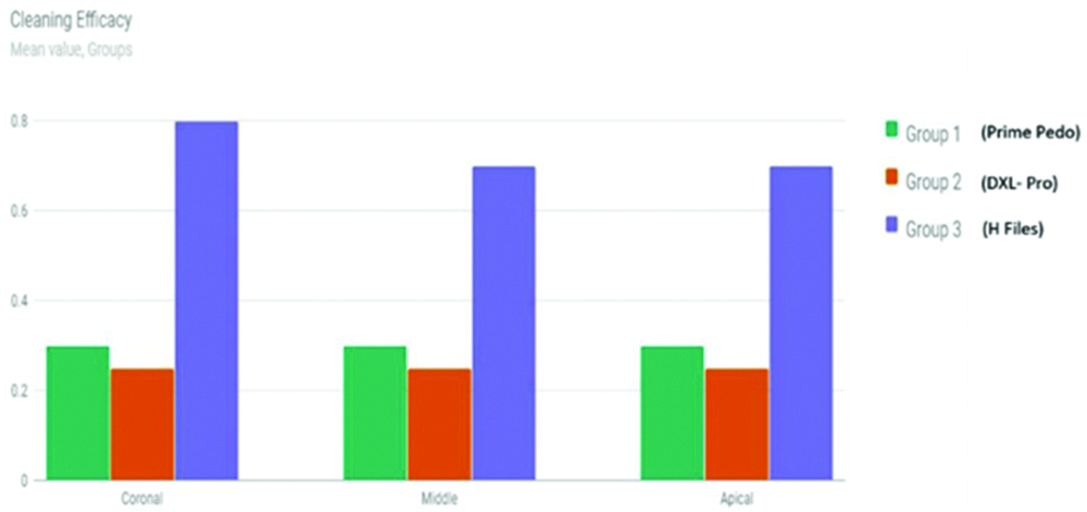
Cleaning efficacy at coronal, middle and apical third of root canal.
| Groups | | Coronal | Middle | Apical | f | p-value |
|---|
| Group 1 (Prime Pedo) | Mean | 0.3 | 0.3 | 0.3 | 0 | 1 |
| Standard deviation | 0.47 | 0.47 | 0.47 | |
| Group 2 (DXL-Pro) | Mean | 0.25 | 0.25 | 0.25 | 0.006 | 0.994 |
| Standard deviation | 0.444 | 0.444 | 0.444 | |
| Group 3 (H Files) | Mean | 0.8 | 0.7 | 0.7 | 0.077 | 0.926 |
| Standard deviation | 0.768 | 0.801 | 0.801 | |
In intergroup comparison, a statistically significant difference in the cleaning efficacy of Group I (Prime PedoTM) (p<0.025) and Group 2 (DXL-ProTM) rotary files (p<0.012) as compared to Group III (H files) at the coronal third of root canal was found. At the apical third of the root canal there was a statistically significant difference in cleaning efficacy of Group I (Prime PedoTM) and Group II (DXL-ProTM) as compared to Group III (H files). However, at middle third, no difference in cleaning efficacy was found between two paediatric rotary files (Prime PedoTM, DXL-ProTM) and H files [Table/Fig-4].
Intergroup comparison results.
| Cleaning efficacy | | Mean difference | Standard deviation | p-value |
|---|
| Coronal | Group 1 (Prime Pedo) vs. Group 3 (H files) | -0.5 | 0.183 | 0.024* |
| Group 2 (DXL-Pro) vs. Group 3 (H files) | -0.55 | 0.183 | 0.012* |
| Group 1 (Prime Pedo) vs. Group 2 (DXL-Pro) | -0.05 | 0.183 | 1 |
| Middle | Group 1 (Prime Pedo) vs. Group 3 (H files) | -0.4 | 0.188 | 0.113 |
| Group 2 (DXL-Pro) vs. Group 3 (H files) | -0.45 | 0.188 | 0.6 |
| Group 1 (Prime Pedo) vs. Group 2 (DXL-Pro) | 0.05 | 0.188 | 1 |
| Apical | Group 1 (Prime Pedo) vs. Group 3 (H files) | -0.5 | 0.202 | 0.048* |
| Group 2 (DXL-Pro) vs. Group 3 (H files) | -0.55 | 0.202 | 0.025* |
| Group 1 (Prime Pedo) vs. Group 2 (DXL-Pro) | -0.05 | 0.202 | 1 |
Discussion
Chemo-mechanical preparation is an important part of pulpectomy procedure in primary teeth. It includes mechanical cleansing with instruments and chemical cleansing with irrigant. Cleaning efficacy is dependent on many factors like type of instrument, method of instrumentation and irrigating solution used [3]. Various studies have compared the cleaning efficacy of rotary and hand files in primary molars. There are various techniques to determine the cleaning efficacy of file systems such as canal staining and clearing technique, cone beam computed tomography, peripheral quantitative computed tomography, spiral computed tomography, and plain and contrast medium-enhanced digital radiography [12]. Canal staining and clearing method is non-destructive and allows for three-dimensional visualisation of root canal system [13]. Hence, this study was conducted to evaluate and compare cleaning efficacy of paediatric rotary files (Prime PedoTM, DXL-ProTM) and H files in root canals of primary molars using clearing technique.
The root canals were stained with India ink and cleared in methyl salicylate before instrumentation to evaluate the cleaning efficacy of the rotary files and H files. After instrumentation, ink remaining in coronal, middle and apical third of root canal was scored. H files displayed the highest mean scores of remainder ink in each third of root canal. Therefore, H files had a significantly lower cleaning efficacy, as compared to two paediatric rotary files (Prime PedoTM, DXL-ProTM). Conventional H files are made of stainless steel. Stainless Steel files are rigid and because of this the H files do not bend and follow the natural anatomy of root canals. This leads to inadequate instrumentation in significant area of root canals [14].
Prime PedoTM files used have a triangular cross-section, are heat treated and have controlled memory. Heat treated files are less prone to deformation and follow the original anatomy of the root canals. DXL-ProTM files have a convex triangular cross section, guiding non-cutting tip and controlled memory. The orifice enlarging file has a length of 16 mm. In this study, the better cleaning efficacy of rotary files can be attributed to the triangular cross-section and positive rake angle of Prime PedoTM and DXL-ProTM rotary files. This triangular cross-section also reduces the contact areas between the file and the dentin and reduces the stresses on the files [15]. Prime PedoTM and DXL-ProTM rotary files possess controlled memory which allows these files to be centred and follow original canal anatomy in primary molars. They have higher flexibility and potential fatigue resistance. Increased fatigue resistance might reduce fracture of rotary files in curved root canals of primary molars [11].
Among the two rotary files used in this study, Group II (DXL-ProTM) had a better cleaning efficacy as compared to Group I (Prime PedoTM), however the difference was not statistically significant.
Results of this study are similar to Pinheiro et al. in 2012 who concluded that rotary files had a better cleaning efficacy as compared to hand files [16]. Katge F et al., concluded that rotary and reciprocating systems used had a significantly better cleaning efficacy as compared to conventional H files [9]. Musale PK and Mujawar SA, evaluated cleaning capacity and instrumentation time of manual, hybrid and rotary instrumentation techniques in primary molars. They concluded that manual instrumentation with K files resulted in lowest cleaning efficacy as compared to rotary and hybrid technique [17].
The results of this study are contradictory to Silva LA et al., Bahrololoomi Z et al., and Moghaddam KN et al., who found no significant difference in cleaning efficacy between manual and other rotary systems [18-20]. Azar MR and Mokhtara M compared two files and K files for assessing their cleaning efficacy. The author concluded that there was no significant difference in cleaning efficacy of rotary and K files at coronal, middle and apical third of root canal [8]. This difference may be accredited to the degree of root canal curvature, number of files, instrumentation techniques, irrigation protocols and methods employed for cleaning evaluation. However, all the above authors have used rotary files used in permanent teeth for primary molars.
Jeevanandan G and Govindaraju L, conducted a randomised clinical trial which concluded that Kedo-S files resulted in better obturation quality as compared to H files [10]. As this was an invivo study only quality of obturation and success of pulpectomy was assessed. The cleaning efficacy of paediatric rotary file was not evaluated.
Rotary files used in this study provide advantage of controlled memory, super elasticity, greater taper along with shorter length which facilitates effective cleaning of the primary root canals and ease of operation.
Limitation
The study had certain limitations. Both maxillary and mandibular molars were included in the study. The root canal curvature of these molars varies, so this could be a confounding factor. Standardisation of the type of teeth should have been done. Sample size could have been larger.
Conclusion
Based on the results of present study, it was concluded that two paediatric rotary files (Prime PedoTM, DXL-ProTM) provide significantly better cleaning efficacy at the coronal and apical third of the root canals in primary molars. However, at the middle third all the three file systems had similar cleaning efficacy. Between two paediatric rotary files, DXL-ProTM showed better cleaning efficacy as compared to Prime PedoTM. However, the difference was not statistically significant. Paediatric rotary files used in this study provide superior cleaning efficacy and facilitate conical preparation of the canals which results in better quality of obturation. Clinical Significance: the increased taper, controlled memory and shorter length of the paediatric rotary files used in this study provide the clinician ease of operation as compared to conventional H files. Paediatric rotary files can thus be used in routine endodontic practice in primary teeth for faster and better instrumentation.
[1]. Pinkham JR, Casamassimo PS, Paediatric dentistryInfancy through adolescence 2005 PhiladelphiaWB Saunders Co.:390 [Google Scholar]
[2]. Fuks AB, Papagiannoulis L, Pulpotomy in primary teeth: review of the literature according to standardized criteriaEur Arch Paediatr Dent 2006 7:64-71.10.1007/BF03320817 [Google Scholar] [CrossRef]
[3]. Kennedy DB, Kennedy DB, Paediatric operative dentistry 1979 Wright:45 [Google Scholar]
[4]. Barr ES, Kleier DJ, Barr NV, Use of nickel-titanium rotary files for root canal preparation in primary teethPediatr Dent 2000 22:77-78. [Google Scholar]
[5]. Siqueira JF Jr, Araújo MC, Garcia PF, Fraga RC, Dantas CJ, Histological evaluation of the effectiveness of five instrumentation techniques for cleaning the apical third of root canalsJ Endod 1997 23:499-502.10.1016/S0099-2399(97)80309-3 [Google Scholar] [CrossRef]
[6]. Kuo C, Wang Y, Chang H, Application of Ni-Ti rotary files for pulpectomy in primary molarsJ Dent Sci 2006 1:10-15. [Google Scholar]
[7]. Crespo S, Cortes O, Garcia C, Perez L, Comparison between rotary and manual instrumentation in primary teethJ Clin Pediatr Dent 2008 32(4):295-98.10.17796/jcpd.32.4.l57l36355u60657618767460 [Google Scholar] [CrossRef] [PubMed]
[8]. Azar MR, Mokhtare M, Rotary Mtwo system versus manual K-file instruments: Efficacy in preparing primary and permanent molar root canalsIndian J Dent Res 2011 22(2):36310.4103/0970-9290.8428321891918 [Google Scholar] [CrossRef] [PubMed]
[9]. Katge F, Patil D, Poojari M, Pimpale J, Shitoot A, Rusawat B, Comparison of instrumentation time and cleaning efficacy of manual instrumentation, rotary systems and reciprocating systems in primary teeth: an in vitro studyJ Indian Soc Pedod Prev Dent 2014 32(4):311-16.10.4103/0970-4388.14095725231039 [Google Scholar] [CrossRef] [PubMed]
[10]. Jeevanandan G, Govindaraju L, Clinical comparison of Kedo-S paediatric rotary files vs. manual instrumentation for root canal preparation in primary molars: a double blinded randomised clinical trialEur Arch Paediatr Dent 2018 19(4):273-78.10.1007/s40368-018-0356-630003514 [Google Scholar] [CrossRef] [PubMed]
[11]. de Arruda Santos L, de Azevedo Bahia MG, de Las Casas EB, Buono VT, Comparison of the mechanical behavior between controlled memory and super elastic nickel-titanium files via finite element analysisJ Endod 2013 39(11):1444-47.10.1016/j.joen.2013.07.03024139271 [Google Scholar] [CrossRef] [PubMed]
[12]. Neelakantan P, Subbarao C, Subbarao CV, Comparative evaluation of modified canal staining and clearing technique, cone-beam computed tomography, peripheral quantitative computed tomography, spiral computed tomography, and plain and contrast medium-enhanced digital radiography in studying root canal morphologyJ Endod 2010 36(9):1547-51.10.1016/j.joen.2010.05.00820728725 [Google Scholar] [CrossRef] [PubMed]
[13]. Robertson D, Leeb IJ, Mckee M, Brewer E, A clearing technique for the study of root canal systemsJ Endod 1980 6(1):421-24.10.1016/S0099-2399(80)80218-4 [Google Scholar] [CrossRef]
[14]. Alodeh MHA, Doller R, Dummer PMH, Shaping of simulated root canals in resin blocks using the step-back technique with K-files manipulated in a simple in/out motionInt Endod J 1989 22:107-17.10.1111/j.1365-2591.1989.tb00908.x2634618 [Google Scholar] [CrossRef] [PubMed]
[15]. Versluis A, Kim HC, Lee W, Kim BM, Lee CJ, Flexural stiffness and stresses in nickel-titanium rotary files for various pitch and cross-sectional geometriesJ Endod 2012 38(10):1399-403.10.1016/j.joen.2012.06.00822980187 [Google Scholar] [CrossRef] [PubMed]
[16]. Pinheiro SL, Araujo G, Bincelli I, Cunha R, Bueno C, Evaluation of cleaning capacity and instrumentation time of manual, hybrid and rotary instrumentation techniques in primary molarsInt J Endod 2012 45(4):379-85.10.1111/j.1365-2591.2011.01987.x22188162 [Google Scholar] [CrossRef] [PubMed]
[17]. Musale PK, Mujawar SA, Evaluation of the efficacy of rotary vs. hand files in root canal preparation of primary teeth in vitro using CBCTEu Arch of Paediatr Dent 2014 15(2):113-20.10.1007/s40368-013-0072-123893606 [Google Scholar] [CrossRef] [PubMed]
[18]. Silva LA, Leonardo MR, Nelson-Filho P, Tanomaru JM, Comparison of rotary and manual instrumentation techniques on cleaning capacity and instrumentation time in deciduous molarsJ Dent Child 2004 71:45-47. [Google Scholar]
[19]. Bahrololoomi Z, Tabrizizadeh M, Salmani L, In vitro comparison of instrumentation time and cleaning capacity between rotary and manual preparation techniques in primary anterior teethJ Dent Tehran Uni of Med Sci 2007 4:59-62. [Google Scholar]
[20]. Moghaddam KN, Mehran M, Zadeh HF, Root canal cleaning efficacy of rotary and hand files instrumentation in primary molarsIran Endod J 2009 4:53-57. [Google Scholar]