Six Mesh Hernia Repairs in a Single Patient Done Simultaneously- A Case Report
JS Rajkumar1, Aanchal Kothari2, JR Anirudh3, Akbar Syed4, Aluru Jaykrishna Reddy5
1 Chief Surgeon, Department of General Surgery, Lifeline Institute of Minimal Access and Surgery, Chennai, Tamil Nadu, India.
2 Surgical Registrar, Department of General Surgery, Lifeline Institute of Minimal Access and Surgery, Chennai, Tamil Nadu, India.
3 Surgeon, Department of General Surgery, Lifeline Institute of Minimal Access and Surgery, Chennai, Tamil Nadu, India.
4 Surgeon, Department of General Surgery, Lifeline Institute of Minimal Access and Surgery, Chennai, Tamil Nadu, India.
5 Surgeon, Department of General Surgery, Lifeline Institute of Minimal Access and Surgery, Chennai, Tamil Nadu, India.
NAME, ADDRESS, E-MAIL ID OF THE CORRESPONDING AUTHOR: Aanchal Kothari, No. 47/3, New Avadi Road, Kilpauk, Chennai-600010, Tamil Nadu, India.
E-mail: docaanchal92@icloud.com
Bilateral hernias being done simultaneously is quite a common procedure. Sometimes, a para umbilical hernia is fixed at the same time as well. However, it is rare to fix more than three hernias of the abdominal wall simultaneously. This case report presents a 73-year-old gentleman who underwent mesh hernioplasty of the hiatus, an epigastric hernia, a paraumbilical hernia, a Spigelian hernia, and bilateral inguinal hernia repair as well, all done in a single session. On follow-up at one year, he was asymptomatic, with no complications attributable to the multiple hernia meshplasty. This case report is being published to reiterate that multiple abdominal wall deficiencies can be corrected in a single sitting, which is advantageous in terms of cost and the need for multiple admissions, especially for patients from remote areas.
Laparoscopy, Meshplasty, Multiple hernia
Case Report
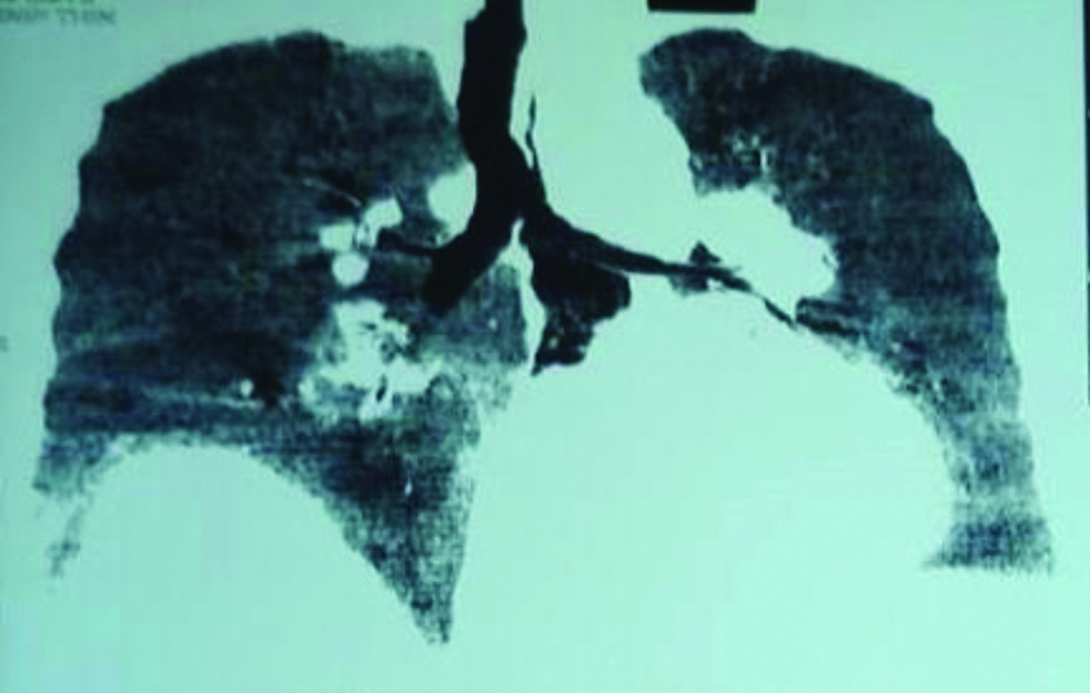
A 73-year-old gentleman presented with a history of recurrent attacks of breathlessness (postprandially), regurgitation of liquid and solid contents, heartburn, epigastric distress and chest discomfort, postprandially. There was also a complaint of two unsightly bulges in the upper abdomen. He gave history of bilateral inguinal hernia procedures, done sequentially, 10 years and 5 years prior to the presentation. He underwent a thorough investigation for his chest discomfort. Echocardiography and Electrocardiogram (ECG) were normal. Plain skiagram of the chest showed a large retro-cardiac soft tissue outline, suggestive of a large hiatus hernia. A Contrast enhanced computed tomography (CECT) of the chest and abdomen revealed a giant hiatus hernia, with a huge intrathoracic stomach [Table/Fig-1]. The abdominal CT also confirmed a large epigastric hernia, a small paraumbilical hernia, a moderate sized left Spigelian hernia, and a bilateral recurrent inguinal hernia, with a grand total of six parietal hernias. He underwent a detailed pre-operative work-up, and the anaesthesiologist, cardiologist, and the general physician opined that he was fit to undergo surgery for all six hernias under a general anaesthesia. However, it was clearly explained to the patient and attenders, that the primary procedure was the hiatal hernia repair. Only if he was absolutely stable, would rest of the procedures be performed.
Accordingly, he underwent laparoscopy with a five port approach. The omental adhesions to the epigastric port were first taken down with the sub-xiphoid port as optics and with the left midclavicular and the left anterior axillary ports used to perform the adhesiolysis. Then, through the epigastric defect, the 10 mm port was inserted to provide optics for the hiatus hernia defect repair. Through the conventional five ports of the upper GI surgeries, the hiatus was approached. The proximal half of the stomach was found to be completely intrathoracic, with a 6×7 cm defect in the diaphragm [Table/Fig-2]. The adhesions and the hernial sac were dissected off the crura of the diaphragm, and the lower oesophagus and the stomach where progressively brought down into the abdominal cavity. The large defect in the diaphragm was closed by approximation of the crura with 2-0 polypropylene sutures, followed by a Bard Crurasoft Mesh that completely overlapped and covered the crural repair with a “V”. In view of the severe reflux and the extensive diaphragmatic dissection, a Nissen’s fundoplication was also done. Using same ports, but looking towards the foot end of the patient, the Spigelian area was tackled [Table/Fig-3]. The omental contents were reduced, and the peritoneum was incised, and a flap raised. A 15×10 cm polypropylene mesh was put in to cover the Spigelian defect. With appropriate three ports, the bilateral inguinal defects were dissected out and a bilateral Transabdominal preperitoneal repair was performed [Table/Fig-4]. Finally, the epigastric [Table/Fig-5] and the para umbilical hernia [Table/Fig-6] were both tackled by simple Intraperitoneal on lay mesh (IPOM) technique, with tacker fixation after three midline transfascial sutures for each. Thus, the six abdominal wall hernias were all subjected to meshplasty in a single session.


Bilateral inguinal hernia.


The reduction of the stomach into the peritoneal cavity, crural approximation, mesh reinforcement of the diaphragm, and the fundoplication, were the cornerstone of his surgery. As he was very stable after this part of the procedure, with the anaesthetist’s consent, we proceeded with Spigelian and bilateral recurrent inguinal hernia repairs. In this particular patient, as the anaesthetic time was being extended, it was decided to supplement the transfascial fixation with tack fixations all around. Overall, two IPOM composite meshes were deployed, and one composite Crurisoft diaphragmatic Mesh was used for the hiatus hernia. The other three (the Spigelian and the two inguinal hernia defects) were repaired using simple polypropylene meshes and fixed using polypropylene sutures. Despite this, the cost of the three composite meshes, plus 2 sets of tacks, made the entire procedure an extremely expensive one. However, the patient had all his abdominal wall defects bridged at the same time, and retrospectively it has proved to be a good choice.
The total operating time was 320 minutes, and the patient was stable throughout the procedure. In view of his age, overnight ventilation was chosen, and the patient was extubated next morning. He had an asymptomatic post-operative period, and was discharged on the fourth post-operative day. At follow-up, on 3rd and 10th month post-operatively, he was completely asymptomatic, and has had no adverse events in relation to the mesh in sessions.
Discussion
Multiple abdominal wall surgeries usually indicate a primary collagen deficiency. Inadequate cross linking of collagen, with a large amount of type 3 collagen instead of type 1 collagen, is said to be the underlying connective-tissue problem in the formation of hernias [1].
Reports of multiple hernia repairs in a single patient are very rare [2]. Matsevych OY et al., published a case of six hernias: right sided direct and indirect hernia, prevascular (velpeau) hernias bilaterally and bilateral femoral hernias. The defects were all bridged over by a mesh that was placed in the extraperitoneal plane [3]. Yokoyama T et al., reported a case of seven hernia repair in a single patient that included: a left indirect inguinal, right direct inguinal, bilateral femoral, bilateral obturator and right spigelian hernia, for which a bilateral inguinal mesh repair was done, covering inguinal, femoral, obturator defects and a spigelian mesh repair was done as well [4]. Goldberger HA et al., also reported a case of multiple hernias (an incarcerated umbilical hernia with epigastric, ventral, spigelian and mesocolic hernias), rectified by connecting all the defects, to make it a single large defect, and then followed by a mesh hernioplasty [5]. Rashid F et al., published a case report of consecutive multiple hernia (right spigelian, epigastric and Congenital Diaphragmatic Hernia (CDH), aortic Stenosis and cognitive impairment in a patient inherited with Williams syndrome. Collagen deficiency is suspected in such patients. This patient underwent a laparoscopic hernia mesh repair of CDH [6]. Kim HA et al., reported a case of impacted obturator and inguinal hernia in a single patient, that was treated with a laparotomy resection and anastamosis of the perforated terminal ileum followed by a single prosthetic mesh repair of both inguinal and obturator hernia together [7].
Both tacker and suture mesh fixation were used here. Both methods of mesh fixation are no different in their outcomes. The only benefit while using tackers is a short operative period and less post-operative pain [8].
Conclusion
This case report is being published for its rarity, and to reiterate the fact that, with safe anaesthesia, up to six abdominal wall hernias can be repaired at the same time.
[1]. Friedman DW, Boyd CD, Norton P, Greco RS, Boyarsky AH, Mackenzie JW, Increases in type III collagen gene expression and protein synthesis in patients with inguinal herniasAnnals of Surgery 1993 218(6):75410.1097/00000658-199312000-000097710461 [Google Scholar] [CrossRef] [PubMed]
[2]. Yang X, Jiang L, Li Y, Liu J, Fan JK, Laparoscopic repair of multiple incisional hernias in a single midline incision by double composite meshJournal of Visualized Surgery 2018 4:58Published online 2018 Mar 2310.21037/jovs.2018.01.0110.21037/jovs.2018.01.0129682468 [Google Scholar] [CrossRef] [CrossRef] [PubMed]
[3]. Matsevych OY, Koto MZ, Becker JH, Multiple concurrent bilateral groin hernias in a single patient; A case report and a review of uncommon groin hernias: A possible source of persistent pain after successful repairInternational Journal of Surgery Case Reports 2016 29:204-07.10.1016/j.ijscr.2016.11.01927871011 [Google Scholar] [CrossRef] [PubMed]
[4]. Yokoyama T, Kobayashi A, Shimizu A, Motoyama H, Miyagawa S, Laparoscopic repair for a previously unreported form of ventral hernia on the right iliac fossa in an elderly emaciated womanHernia 2015 19(5):841-43.10.1007/s10029-013-1180-x24218077 [Google Scholar] [CrossRef] [PubMed]
[5]. Goldberger HA, Panebianco RR, Multiple hernia: Case of incarcerated umbilical hernia associated with spigelian, epigastric, ventral, and mesocolic herniasThe American Journal of Surgery 1938 42(2):423-28.10.1016/S0002-9610(38)91212-7 [Google Scholar] [CrossRef]
[6]. Rashid F, Chaparala R, Ahmed J, Iftikhar SY, Atypical right diaphragmatic hernia (hernia of Morgagni), spigelian hernia and epigastric hernia in a patient with Williams syndrome: A case reportJournal of Medical Case Reports 2009 3(1):710.1186/1752-1947-3-719128471 [Google Scholar] [CrossRef] [PubMed]
[7]. Kim HA, Lee RA, Kim KH, Obturator hernia which was combined with inguinal hernia and hiatal herniaJournal of the Korean Surgical Society 2005 68(2):168-71. [Google Scholar]
[8]. Sajid MS, Parampalli U, McFall MR, A meta-analysis comparing tacker mesh fixation with suture mesh fixation in laparoscopic incisional and ventral hernia repairHernia 2013 17(2):159-66.10.1007/s10029-012-1017-z23138861 [Google Scholar] [CrossRef] [PubMed]