Extra Uterine Leiomyoma with “Amianthoid-like” Fibers
Santosh Govind Rathod1, Bharat R Sonwane2, Shubhada N Pore3, Rajan S Bindu4
1 Senior Resident, Department of Pathology, Government Medical College and Cancer Hospital, Aurangabad, Maharastra, India.
2 Associate Professor, Department of Pathology, Government Medical College and Cancer Hospital, Aurangabad, Maharastra, India.
3 Assistant Professor, Department of Pathology, Government Medical College and Cancer Hospital, Aurangabad, Maharastra, India.
4 Professor, Department of Pathology, Government Medical College and Cancer Hospital, Aurangabad, Maharastra, India.
NAME, ADDRESS, E-MAIL ID OF THE CORRESPONDING AUTHOR: Dr. Santosh Govind Rathod, Senior Resident, Department of Pathology, Government Medical College, Aurangabad, Maharastra, India.
E-mail: drsgrathod2007@gmail.com
Uterine leiomyoma is a benign smooth muscle neoplasm, typically arising in uterus and deep soft tissue. In the present case, tumour size of 7×4×3 cm identified on subserosal aspect of uterus. Grossly, tumour was well circumscribed, whitish nodule and on cut surface whitish whirls were seen. Microscopic examination showed a cellular spindle cell population with numerous eosinophilic amianthoid-like fibers. Immunohistochemically, the tumour showed positivity for H-caldesmon, desmin, alpha smooth-muscle actin, and negativity for CD10. The finding of amianthoid-like fibers in leiomyoma is very rare. Amianthoid fibers are hyalinized collagen mats. It extends the histomorphological spectrum of leiomyomas. Amianthoid fibers are very characteristics of “intra nodal palisaded myofibroblastoma”. Presence of “Amianthoid-like fibers” creates histomorphologically similar picture between leiomyoma and “intra nodal palisaded myofibroblastoma”. Pathologist should be aware of such variant of leiomyoma that contains “Amianthoid-like fibers” to avoid confusion with other tumours.
Actin desmin, H-caldesmon, Palisaded myofibroblastoma
Case Report
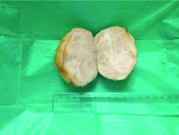
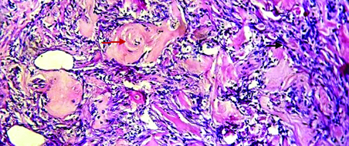
A 60-year-old menopause woman came with chief complaints of pain in abdomen, and uterine bleeding since two months. There was no significant family history. Medical history of hypertension was there. She was on regular antihypertensive drug beta-blocker atenolol 12.5 mg daily for three years. Patient underwent CECT of abdomen and pelvis. Heterogeneously enhancing lesion in pelvic region, measuring 70×40 mm in size noted. Patient underwent open abdominal surgery for removal of mass. Surgery was uneventful without postoperative complication. There was no recurrence in one-year follow-up of patient. One of the endometrial stromal tumour was considered as the top differential diagnosis. Grossly, tumour of size 7×4×3 cm noted, tumour was round and well-circumscribed, whitish whirls were seen on cut section [Table/Fig-1]. Microscopically, tumour showed interlacing fascicles of bland looking spindle shaped cell, eosinophilic cytoplasm, “cigar-shaped” nuclei and presence of numerous round to stellate-shaped, eosinophilic, acellular collagenous mats resembling “amianthoid-like fibers” [Table/Fig-2]. Immunohistochemical panel showed positivity of tumour cells for H-caldesmon, desmin and actin [Table/Fig-3a,b and c] and negative staining for CD10. Daignosis of leiomyoma with amianthoid like fiber was made. The blocks from case and control were cut and mounted on poly-lysin coated glass slides. Endogenous peroxidase activity was blocked by 0.3% hydrogen peroxide in methanol for 20 minutes. Heat induced antigen retrieval done by using buffer at pH 6. The antibodies confirmed the smooth muscle nature of tumour and diffusely expressed around amianthoid like fiber.
Tumour was round and well-circumscribed, whitish whirls seen on cut section.
H&E stain (20X) shows spindle shaped cell, eosinophilic cytoplasm, “cigar-shaped” nuclei (black arrow) and presence of numerous round to stellate-shaped, eosinophilic, acellular collagenous mats resembling “amianthoid-like fibers” (red arrow).
Diffuse H-caldesmon positivity around amianthoid like fiber, confirming smooth muscle nature of tumour (x10).
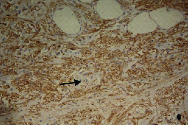
Diffuse desmin positivity around amianthoid like fiber, confirming smooth muscle nature of tumour (x10).
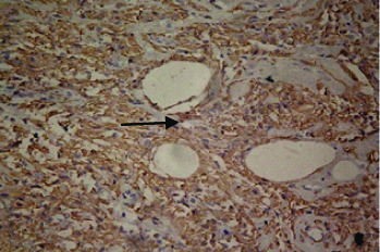
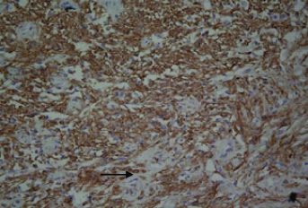
Diffuse actin positivity around amianthoid like fiber, confirming smooth muscle nature of tumour (x10).
Discussion
The present case was clearly of leiomyoma. Uterine leiomyoma are benign smooth muscle neoplasm of uterus and on rare occasion of deep soft tissue. Grossly, uterine leiomyoma appears as well circumscribed whitish nodule; on cut section, whitish whirls are seen. Histologically composed of interlacing fascicles of bland looking spindle shaped cell, abundant eosinophilic cytoplasm and “cigar-shaped” nuclei. In present case tumour showed presence of numerous round to stellate-shaped, deeply eosinophilic, acellular thick mats of collagen. These collagen mats have resemblance to “Amianthoid-like fibers” [1].
Amianthoid fibers are seen in variety of neoplastic and non-neoplastic condition [1]. They are characteristically seen in “Intranodal palisaded myofibroblastoma” [2-4]. They are also encountered in meningiomas [5], and benign soft tissue tumour [6]. Four cases of uterine leiomyoma with amianthoid like fibers are reported in the literature [7-9]. The first case by Bagwan IN et al., revealed fascicles of smooth muscle fibers set in myxoid stroma and at places amianthoid like fibers [7]. In case report by Zamecnick M et al., showed typical features of leiomyoma with amianthoid like fibers [8]. In case report by Longo F et al., showed features of atypical leiomyoma with amianthoid like fibers [9]. The present case, showed similar histological features as previously reported four cases.
The thickness of normal collagen fibrils ranges from 35 nm to 120 nm [1,7]. The thickness of gaint collagen (>200 nm) that constitutes amianthoid fibrils [1,7]. The thickness of amianthoid like fibers ranges between 120 nm to 200 nm. Hence, they are called as amianthoid like fibers [1,7]. Pathogenesis of amianthoid –like fiber in leiomyoma is unclear. Some author think that they are degenerative process of pre-existing collagen fibers [2,7]. However, according to some authors amianthoid like fibers are result of an active process of collagen secretion and deposition [6-10].
The finding of amianthoid like fibers in leiomyoma is very rare. Morphological and Immunohistochemically features were suggestive of leiomyoma. Diffuse expression of desmin, Alpha smooth muscle actin and H-caldesmon in tumour confirmed smooth muscle nature. At the same time tumour was negative for CD 10 marker, excluding the diagnosis of endometrial stromal tumour.
We are reporting a case of extra uterine leiomyoma with amianthoid like fibers. Our case report helps to widen the morphological spectrum of leiomyoma. Presence of amianthoid like fibers in leiomyoma is very rare, and creates close morphological similarity to that of “intra nodal palisaded myofibroblastoma” [3,4]. Intra nodal palisaded myofibroblastoma is a benign mesenchymal tumour which arises from smooth muscle and myofibroblasts. It is characterised by the proliferation of haemosiderin-laden histiocytes, spindle cells, and amianthoid like fibers in lymph node [3,4]. If lesion had been in inguinal region, it would have been diagnosed as “intra nodal palisaded myofibroblastoma”. It poses a great diagnostic challenge to the pathologist and they should be aware of such variant of leiomyoma.
Conclusion
Uterine leiomyoma may present with deeply eosinophilic thick collagen band, called as amianthoid-like fibers inside a spindle cell proliferation. Creating, a morphological resemblance to that of palisaded myofibroblastoma. Pathologist should aware of such variant of leiomyoma to avoid confusion with other tumours.
[1]. Eyden B, Tzaphlidou M, Structural variation of collagen in normal and pathological tissue: Role of electron microscopyMicron 2001 32:287-300.10.1016/S0968-4328(00)00045-7 [Google Scholar] [CrossRef]
[2]. Suster S, Rosai J, Intranodal hemorrhagic spindle-cell tumour with “amianthoid” fibers. Report of six cases of a distinctive mesenchymal neoplasm of the inguinal region that simulates Kaposi’s sarcomaAmerican Journal of Surgical Pathololgy 1989 13:347-57.10.1097/00000478-198905000-00002
[Google Scholar] [CrossRef]
[3]. Sonwane BR, Zadke PM, Swami SY, Palisaded myofibroblastoma of lymph nodeIndian J Pathol Microbiol 2008 51:413-14.10.4103/0377-4929.4254618723976 [Google Scholar] [CrossRef] [PubMed]
[4]. Nguyen T, Eltorky MA, Intranodal palisaded myofibroblastomaArch Pathol Lab Med 2007 131:306-10. [Google Scholar]
[5]. Chuaqui R, Gonzalez S, Torrealba G, Meningothelial meningiomas with “amianthoid” fibers: Case report with ultra structural studyPathology Research and Practice 1992 188:890-93.10.1016/S0344-0338(11)80249-9 [Google Scholar] [CrossRef]
[6]. Connly CE, Crystalline collagen production by an in usual benign soft tissue tumour (“amiantoma”)Histopathology 1981 5:11-20.10.1111/j.1365-2559.1981.tb01762.x7216173 [Google Scholar] [CrossRef] [PubMed]
[7]. Bagwan IN, Moss J, Fisher C, El-Bahrawy M, Amianthoid-like fibres inleiomyomaHistopathology 2008 53:606-09.10.1111/j.1365-2559.2008.03123.x18783466 [Google Scholar] [CrossRef] [PubMed]
[8]. Zamecnick M, Kascak P, Uterine leiomyoma with amianthoid–like fibersCesk Patol 2011 47:125-27. [Google Scholar]
[9]. Longo F, Musumeci G, Parenti R, Vecchio G, Magro G, Atypical cell leiomyoma of uterus with amianthoid-like fibers: A case reportOA Case Reports 2013 2(14):137 [Google Scholar]
[10]. Harkin JC, Webb SV, Soft-tissue tumour with abnormal amianthoid like collagen fibersArch Pathol Lab Med 1990 114:1281-82. [Google Scholar]