White Spot Lesions (WSLs) are presented as an enameled area with an opaque aspect [1]. The early diagnosis of these lesions, when cavitation has not occurred yet, it allows conservative interventions [1-3]. The use of a resin infiltrant has been cited as a promising treatment, in which a low viscosity resin applied to WSLs allows an efficient infiltration within the demineralised enamel [4,5]. The resin infiltrant promotes an obstructive effect, hindering the progression of lesions by preventing the diffusion pathways for cariogenic acids [4-6].
Initially, the infiltration concept was developed primarily for initial proximal caries [4,5]. However, some authors have transferred this concept to buccal surfaces, since the demineralised surface typically presents with a white appearance, which might be masked with a resin infiltrant [7,8]. It is composed of a hydrophobic resin with a refractive index close to sound enamel, favouring the aesthetics of areas affected by lesions [7,8]. These characteristics make the use of resin infiltrant a conservative treatment since there is no mechanical preparation of the enamel structure, such as when using the microabrasion technique [3].
The oral environment constantly suffers the actions caused by different challenges which may interfere with the integrity of the tooth structure or restorative materials. Toothbrushing leads to wear when the enamel is weakened, e.g., when affected by WSLs [9]. Another situation that occurs in the oral cavity is related to pH alterations, responsible for the demineralisation-remineralisation process [10]. Furthermore, restorative materials and tooth structure undergo degradation over time and artificial ageing has the potential of degrading organic matrix [11,12].
The literature is scarce on studies regarding colour changes and surface gloss of unpolished infiltrated WSLs submitted to different challenges.
Therefore, the present study aimed to evaluate the colour change and surface gloss of white spot lesions after application of unpolished resin infiltrant submitted to different challenges.
The null hypothesis of the current study was that the different challenges do not influence the colour changes and surface gloss of unpolished infiltrated WSLs.
Materials and Methods
Sample Preparation
The present in-vitro study was conducted from June 2014 to September 2014. This study was related to the Araçatuba School of Dentistry, São Paulo State University (UNESP), Araçatuba, SP, Brazil. Sound permanent bovine incisors obtained from steers aged 24 to 30-month-old were collected and stored in 0.1% thymol solution, after obtaining approval of the local Institutional Review Board (protocol #2014-00897). Specimens without caries, cracks or any other defect were selected and then cleaned using slurry pumice and brush. The sample size was calculated considering a maximum acceptable error of 5%, power of 80%, considering a minimum detectable difference in means and expected standard deviation of residuals, which resulted in at least six samples for each group, using Sigma Plot (version 2.0) software. Fifty bovine enamel/dentin were used in this in vitro study and divided into five groups (n=10): WSL (W-control), infiltrated WSL (IW), infiltrated WSL submitted to tooth brushing (IWb), infiltrated WSL submitted to pH cycling (IWc), and infiltrated WSL submitted to artificial ageing (IWa). Next, discs containing enamel and dentin were cut using a cylindrical diamond tip with a 5.7 mm diameter (DinserFerramentasDiamantadas, São Paulo, SP, Brazil) under constant irrigation.
The dentin surfaces were flattened and polished using 320-, 400-, 600-, 800-, and 1200-grit sandpaper (Buehler, LakeBluff, IL, USA) with a polishing machine (Politriz Aropol E, Arotec Indústria e Comércio Ltda., Cotia, São Paulo, SP, Brazil) under water cooling at low speed (200 rpm). Final polishing was performed using felt disks and 1 μm diamond paste (Extec Corp, Enfield, CT, USA).
Initial Colour Measurements
Colour analysis was performed using a reflection spectrophotometer (UV Visible, Model UV-2450, Shimadzu, Kyoto, Japan), following the Commission International de L’Eclairage L*a*b* system (CIELAB) [13]. A black silicone mould was used to allow standard positioning of the specimen during the readings. Three measurements were obtained for each specimen, and the average was calculated. The difference in initial and final values of L* (∆L), a* (∆a), and b* (∆b) were determined and the overall change in colour.
(∆E) was calculated using the following formula [13,14]:
∆E=[(∆L)2+(∆a)2+(∆b)2]1/2Samples were dried with absorbent paper, leaving the surface hydrated without water excess in all colour and gloss analyses.
Initial Surface Gloss
The surface gloss analysis was performed using a gloss meter (In Curve, Rho point Instrumentation, East Sussex, UK), assessing a 2 mm × 2 mm square area under 60 degrees of incidence. Measurements were expressed in surface Gloss Unit (GU). An opaque surface was assigned 0 Gloss Units (GU) and a gloss surface with 1567 refractive index had 100 GU [15]. Three measurements were made by rotating the specimen 90° around its centre. The average of three measurements was determined. The [Table/Fig-1] shows the experimental design.
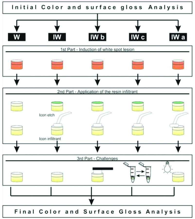
Induction of White Spot Lesion in Bovine Enamel
Next, two layers of acid-resistant nail varnish (Risqué, Barueri, São Paulo, SP, Brazil) were applied to leave only the enamel surface to be induced for development of an artificial WSLs. Then, specimens were placed individually in demineralising solution (1.3 mM/lCa (NO3)2×4H2O and 0.78 mM/l NaH2PO4H2O in 0.05 M/l acetate buffer, 0.03 μgF/mL, pH 5.0, 32 mL/block) for 24 hours at 37°C [16]. Subsequently, the specimens were removed from the solution and thoroughly washed with deionised water for three minutes.
Application of the Resin Infiltrant
The resin infiltrant (Icon, DMG, Hamburg Germany) was applied on the specimens of IW, IWb, IWc, and IWa groups and light cured at 1100 mW/cm2 using an LED device (Kavo, Poly Wireless, Joinville, SC, Brazil), following the manufacturer’s instructions [Table/Fig-2]. All specimens were not submitted to polishing. All the specimens were stored for seven days at 37°C in 100% relative humidity.
Composition and application steps of resin infiltrant according to the manufacturer’s instructions.
| Composition | Application steps | Batch no. |
|---|
| Icon-etch: (HCl15%) pyrogenic silicic acid, surface-active substances, | Apply Icon-Etch. Let it set for 2 minutes Rinse off with water for 30 seconds. Air dry | 711766/ |
| Icon-dry: 99% ethanol | Apply Icon-Dry. Let it set for 30 seconds. Air dry | 708255/ |
| Icon-infiltrant: methacrylate-based resin matrix, initiators, and additives | Apply Icon-Infiltrant. Let it set for 3 minutes. Light-cure for 40 seconds.Apply Icon-Infiltrant. Let it set for 1 minute. Light-cure for 40 second | 702131 |
Toothbrushing
Ten specimens of the IWb group were submitted to toothbrushing (10,000 cycles) on the enamel surface using a mechanical brushing machine (Elquip Máquina de Escovação, São Carlos, SP, Brazil), with a slurry of dentifrice and water (1:3 w/w, Colgate Total 12, Colgate-Palmolive, São Paulo, SP, Brazil, 1450 ppm as NaF) [16].
pH-Cycling Model
Ten specimens from the IWc group were individually submitted to a pH cycling model at 37°C for seven days [12]. The specimes were immersed in 35.5 mL of desmineralizing solution for six hours, alternated with immersion in 17.75 mL of demineralising solution for 18 hours for five days. Next, the specimens were kept for two more days in remineralising solution to complete seven days of the pH cycling [16].
Accelerated Artificial Ageing
Ageing of the IWa group was performed in a UV-accelerated ageing chamber (EQUV, Equilam, Diadema, SP, Brazil), ten specimens were subjected to a total of 252 hours of ageing and 168 hours of UVB irradiation with a 313 nm emission peak with condensation of distilled water saturated with oxygen promoting 100% humidity [12,13].
Final Evaluation of the Colour and Surface Gloss
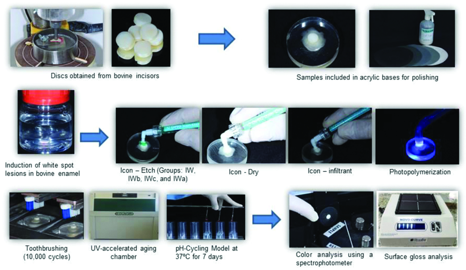
The samples were submitted to the final colour and surface gloss analyses in the same manner as previously described. All the experiment was performed in the local laboratory [Table/Fig-3].
Sequence of in vitro experiments.
Statistical Analysis
Statistical analyses were performed with Sigma Plot 12.0 software. Data were submitted to ANOVA and Tukey’s test for multiple comparisons (p<0.05). Normal distribution of data for the final surface gloss and delta a were not confirmed with the Shapiro-Wilk test; therefore, Kruskal-Wallis and Dunn’s tests were applied (p<0.05).
Results
The [Table/Fig-4] provides values of the deltas regarding colour analysis. When analysing the ∆L values; the lWb group showed a significant difference from the W, IW and IWc groups; however, its results were statistically similar to the IWa group (p>0.05). It was noted that IWa group suffer high alterations for ∆a and ∆b. When considering the ∆E all groups were similar (p>0.05).
Means and standard deviations of colour analysis.
| Groups | ∆L | ∆a | ∆b | ∆E |
|---|
| W | 8.47±8.86A | 0.22±0.43AB | -2.66±1.60C | 10.81±6.24A |
| IW | 4.32±8.33AB | -0.28±0.88BC | 0.56±1.86BC | 7.95±4.94A |
| IWb | -11.40±5.79C | 0.99 ±0.69AB | 0.41±1.73BC | 11.08±3.99A |
| IWc | 1.75±9.69AB | 2.43±2.17 A | 2.56±1.52AB | 8.69±2.64A |
| IWa | -2.56±4.54BC | 2.37±1.05 C | 9.32±4.04A | 11.18±3.05A |
Different letters within columns indicate statistical differences among groups (p< 0.05)*
∆E, ∆L and ∆b-ANOVA and Tukey’s test; ∆a-Dunn’s test
The [Table/Fig-5] demonstrates that the W group showed a statistically significant difference with the IW, IWb and IWc groups forthe final gloss (p<0.05). When considering the difference between the final and initial surface gloss values, there was a statistical difference for the W group from all other groups (p<0.05). Statistical differences were also observed for all groups in which the resin infiltrant was applied when initial and final values were compared (p<0.05).
Means and standard deviations of surface gloss.
| Groups | Initial gloss | Final gloss | Gloss difference |
|---|
| W | 84.62±11.91 Aa | 78.68±14.21 Aa | -5.94±17.19 A |
| IW | 84.30±12.72 Aa | 14.33±7.48 Bb | -69.97±18.38 B |
| IWb | 86.10±14.08 Aa | 10.49±7.42 Bb | -75.61±17.36 B |
| IWc | 80.96±15.28 Aa | 7.10±3.43 Bb | -73.86±15.68 B |
| IWa | 91.62±10.50 Aa | 20.36±6.26 ABb | -71.26±11.16 B |
Capital letters indicate differences within lines and lowercase letters indicate differences among columns (p<0.05)
Surface Gloss-Kruskal Wallis Test
Discussion
Colour and surface gloss analyses are used to evaluate the performance of resinous materials in aesthetic regions [14-17]. The decrease of surface gloss and colour changes may indicate alterations in material properties due to the oral environment conditions, interfering in the longevity of the resin restorations [18,19]. It is worth noting that the protocol used to create the artificial WSL in this study may produce a lesion with depth of approximately 120 μm [20,21]. Moreover, lesions were etched with hydrochloric acid as recommended for natural lesions, which results in erosion of 30-40 μm of the enamel [22], leaving sufficient lesion depth to be infiltrated.
Challenges proposed in the present study resulted in some significant changes in both colours and surface gloss. When analysing the colour, the ∆L results revealed that the IWa and IWb groups showed a greater loss of luminosity. Artificial ageing may promote the oxidation of single and double carbon bonds [23]. Such changes in the resin infiltrant matrix may be responsible for the loss of luminosity. The abrasive cycles used in this study are equivalent to one year of toothbrushing [24]. The resin infiltrant is composed of Triethylene Glycol Dimethacrylate (TEGDMA), which present low mechanical resistance [25]; therefore, the process of abrasion may result in a rougher material surface with significant loss of luminosity, since resin infiltrant also showed increased wear after 20,000 cycles of brushing [24].
It can be highlighted that artificial ageing resulted in high ∆a and ∆b values. Artificial ageing simulates the long-term effects of the environmental conditions [11,12,23]. In this context, the resin matrix of the infiltrant with TEGDMA molecules [4], which have a hydrophilic characteristic, are susceptible to absorption of water contacting the tooth surface [14].
Based on the present findings, all ∆E were superior than 3.3, which is higher than the acceptable threshold in dentistry [24]. Infiltrated enamel specimens had an overall colour change higher than the clinically acceptable delta after immersion in solutions [14]. The resin infiltrant was also affected by erosive and abrasive challenges, probably due to its composition. In this context, the structural configuration of the TEGDMA molecule seems to be susceptible to degradation, because of its hydrophilicity and the absence of strong secondary intermolecular interactions, such as hydrogen bonding [25-27]. Another factor to be considered is that the resin infiltrant has no filler in its matrix [17,28], which seem to contribute to its weakness, especially against mechanical or physical challenges [7,29].
However, a highly evident reversal of staining effects caused by artificial ageing was found using polishing paste with a prophy cup [7]. Repolishing the infiltrated lesions may minimise the staining effect [15]. It was also found that polishing infiltrated lesions may remove the oxygen inhibition layer and reduce the surface porosity, stabilising the masking effect [30].
Regarding surface gloss values, all the groups that received the resin infiltrant presented differences when initial and final surface gloss values were compared. One of the purposes, of the use of resin infiltrant is to mask WSLs, as this material has a light refractive index (1.52) that is close to enamel (1.62 to 1.65) [4]. However, this study observed that all groups in which the infiltrant was applied had a significant reduction in gloss. It is supposed that water can be present, especially in the interior of the WSL, thus representing an incomplete infiltration of the material in these regions [24], causing an alteration in gloss since water has a light refractive index of 1.33. Another factor that may be related to changes in surface gloss may be due to the simple difference in the values of light refractive indices of the resin infiltrant and enamel.
Differences in gloss of the W group when compared to infiltrate and submitted to challenges groups was expected. An enamel surface gloss is maintained when submitted toothbrushing, while the gloss surface of composites tends to further decrease under the same challenge over time; the presence of abrasives may have contributed to loss of surface gloss when resin infiltrant was used [15,18]. The pH cycling promotes a change in the light refraction of the substrate or the material indexes, since the demineralisation process leads to the formation of porosities on the sample surface, thereby modifying the transmission of light [30]. Artificial ageing may also degrade the monomer present in the resin infiltrant, altering the surface gloss.
Limitation
As a limitation of the present study, it was not possible to use a control side in the same specimen surface as a control, as other studies have done [16,27]. This is not possible with the spectrophotometer and gloss meter devices used in this study. Additionally, the surface gloss results might help to understand the behavior of this material under the tested protocols; however, it might not be extrapolated to clinical conditions in which a layer of saliva covering teeth might hinder the perception of surface gloss reduction by observers.
Conclusion
Unpolished resin infiltrant reduce the surface gloss of enamel independently if it is challenged. The differences in the mean colour and gloss changes were similar for all challenges.
Different letters within columns indicate statistical differences among groups (p< 0.05)*
∆E, ∆L and ∆b-ANOVA and Tukey’s test; ∆a-Dunn’s test
Capital letters indicate differences within lines and lowercase letters indicate differences among columns (p<0.05)
Surface Gloss-Kruskal Wallis Test