Introduction
Odontomas are one of the most common odontogenic tumours and are classified as benign odontogenic tumours. According to radiographic, microscopic and clinical features there are two main types of odontomas: compound and complex.
Aim
To analyse the prevalence of benign odontogenic tumours compatible with odontomas in digital panoramic radiographs in a Brazilian population.
Materials and Methods
This retrospective study was conducted from January 2015 to December 2015 and analysed 2,167 digital panoramic radiographs from the database of a Specialised Dental Radiological Clinic in Belo Horizonte, Brazil.
Results
Seven images presented lesions compatible with odontomas, resulting in a prevalence rate of 0.32%. Three images (42.9%) were compatible with compound odontomas and 4 (57.1%) with complex odontomas. Regarding gender, the majority of odontomas were found in female patients (71.4%). The age of the patients diagnosed with odontomas ranged from 7 to 37 years and the main clinical manifestation observed was their association with non-erupted teeth. Regarding the location of the lesions, compound odontomas were mainly observed in the incisor region (66.6%), whereas complex odontomas were found in the posterior region of the molars (50.0%).
Conclusion
Data presented in this study reinforce the importance of panoramic digital radiographic images during dental practice for incidental diagnoses of odontogenic tumours, such as odontomas, since these lesions are in most cases, asymptomatic.
Introduction
According to the latest World Health Organisation (WHO) 2017 classification of odontogenic tumours and cysts, odontomas are classified as benign odontogenic tumours [1]. These types of tumours have a mixed (Epithelial-Mesenchymal) origin and are formed by odontogenic soft and hard tissues [2]. Odontomas are considered to be developmental anomalies, which show slow and limited growth. The aetiology of odontomas is unknown, although different factors, such as trauma, infections, or genetic mutations, have been identified as probable aetiological factors [3,4].
Odontomas are the most common odontogenic tumours, accounting for approximately 75.9% of these lesions [5]. Authors have reported, however, that the prevalence of odontomas varies worldwide: ranging from 6.7% (China) to 67% (United States) to 50.4% (Brazil) [6].
According to radiographic, microscopic and clinical characteristics there are two main types of odontomas: complex odontomas and compound odontomas. In complex odontomas, the dental tissues are well-formed but appear in a disordered arrangement. In the case of compound odontomas the dental tissues are normal but their size and conformation are altered, giving rise to multiple and small denticles [7].
In general, compound odontomas are more frequent than complex odontomas [4,8,9], most of which are diagnosed in the second decade of life [4]. As regards localisation, complex odontomas are mostly located in the mandible, premolar and molar regions while compound odontomas are more frequently found in the maxilla, more especifically in the incisor and canine regions [8-10].
In most cases, odontomas are asymptomatic and are often identified in routine imaging tests [4,10] or when investigating other events, such as a delay in the exfoliation of deciduous teeth, an ectopic positioning of permanent teeth or a possible association with included or retained teeth [9]. The main clinical manifestation reported in the literature is the retention of permanent teeth [8-10].
Radiographically, compound odontomas appear as multiple radiopaque structures similar to small teeth, also called denticles surrounded by a radiolucent halo [8]. In complex odontomas, the radiopaque element appears as irregular and disordered masses with no resemblance to dental structures. Based on radiographic findings, differential diagnoses made as ameloblastic fibroma, ameloblastic fibro-odontoma, and odontoameloblastoma [11].
According to Hidalgo-Sánchez O et al., odontomas can be manifestations of hereditary anomalies and diseases, such as basal cell nevus, Gardner and Hermann syndromes, as well as hereditary adenomatosis and Tangier diseases [4].
The recommended treatment for odontomas is their surgical removal through the principles of tooth extraction. This surgical procedure separates the bone from the fibrous connective tissue capsule. These odontomas are usually easily removed. Momesso GA et al., recently described a novel approach removing a compound odontoma using the Piezosurgery technique [12].
Panoramic radiography is a primary examination that is widely used in the clinical diagnosis of various maxillofacial diseases [13]. Although the use of computed tomography has shown better results in relation to the clarity and accuracy of the changes, the high cost of the technique and the low availability in different regions of the country make it difficult to use. Considering that panoramic radiography is able to assist in the diagnostic conclusion of lesions located in the buccomaxillofacial complex, making it possible to visualise bone changes in the early stages [14], this technique has been used more frequently in dental practices.
Therefore, this study sought to analyse the prevalence of mixed odontogenic tumours through digital panoramic radiography obtained from patients who received dental care at a Specialised Dental Radiology Clinic in Belo Horizonte, Brazil, during the year of 2015.
Materials and Methods
Sampling and Data Collection
A retrospective imaging study of 2,167 digital panoramic radiographic images of patients were obtained from a Specialised Dental Radiology Clinic in Belo Horizonte, Brazil, from January to December 2015. The samples were selected regardless of ethnicity, and all personal information of these patients was kept confidential during the study. This study’s inclusion criteria were: images with gender identification (female or male), patients’ age and acceptable diagnostic quality standards.
Analysis of Samples and Diagnostic Criteria
Panoramic radiographic images were taken directly from the clinic’s database and evaluated. All images were analysed by the same observer in order to avoid interpretation bias. The diagnosis of compound odontomas was performed by observing radiopaque structures compatible with small teeth (denticles), while complex odontomas were identified by observing an irregular radiopaque mass. Both types of lesions were surrounded by a radiolucent halo.
Statistical Analysis
The collected data were arranged in tables to obtain the prevalence rate of mixed odontogenic tumour compatible with odontomas regarding gender, tumour type, and location. The prevalence rate was obtained by the ratio between the number of identified lesions and the total number of analysed samples; the results were expressed in terms of a percentage.
Results
A total number of 2,167 panoramic radiographs were analysed. The [Table/Fig-1] shows the number and gender predilection of lesions compatible with odontomas. It was found a greater prevalence for the female gender. The age of patients diagnosed with odontomas ranged from 7 to 37 years. According to the information obtained during data collection, female patients with these lesions were 7, 11, 20, 35, and 37 years of age, whereas male patients with these lesions were 16 and 22 years of age.
Prevalence of odontomas in panoramic radiographs from a Specialised Dental Radiology Clinic in Belo Horizonte, Brazil.
| Gender | Number | Odontomas | Prevalence (%) |
|---|
| Female | 1197 (55.2%) | 5 (71.4%) | 0.42 |
| Male | 970 (44.8%) | 2 (28.6%) | 0.21 |
| Total | 2167 | 7 | 0.32 |
The distribution of odontomas was presented considering the type of lesion, gender and location. Of the seven radiographs that presented lesions corresponding to odontomas, three images (42.9%) were compatible with compound odontomas and 4 (57.1%) with complex odontomas. Regarding compound odontomas, 66.6% of the lesions (2/3) were identified in female patients and 33.3% (1/3) in male patients. After analysing the images compatible with complex odontomas, 75.0% appeared in female patients (3/4), while 25.0% appeared in male patients (1/4) [Table/Fig-2].
Distribution of odontomas according to type, gender, and location.
| Location | Compound | Complex |
|---|
| Female | Male | Total (%) | Female | Male | Total (%) |
|---|
| Incisor | 1 | 1 | 2 (66.6) | 1 | 0 | 1 (25.0) |
| Premolar | 0 | 0 | 0 | 1 | 0 | 1 (25.0) |
| Molar | 1 | 0 | 1 (33.3) | 1 | 1 | 2 (50.0) |
| Total (%) | 2 | 1 | 3 (42.9) | 3 | 1 | 4 (57.1) |
Compound Odontomas were tabled according to the location they were observed [Table/Fig-2], 66.6% of which (2/3) were observed in the lower and upper incisor regions [Table/Fig-3,4] and 33.3% in the maxillary molar region (1/3) [Table/Fig-5].
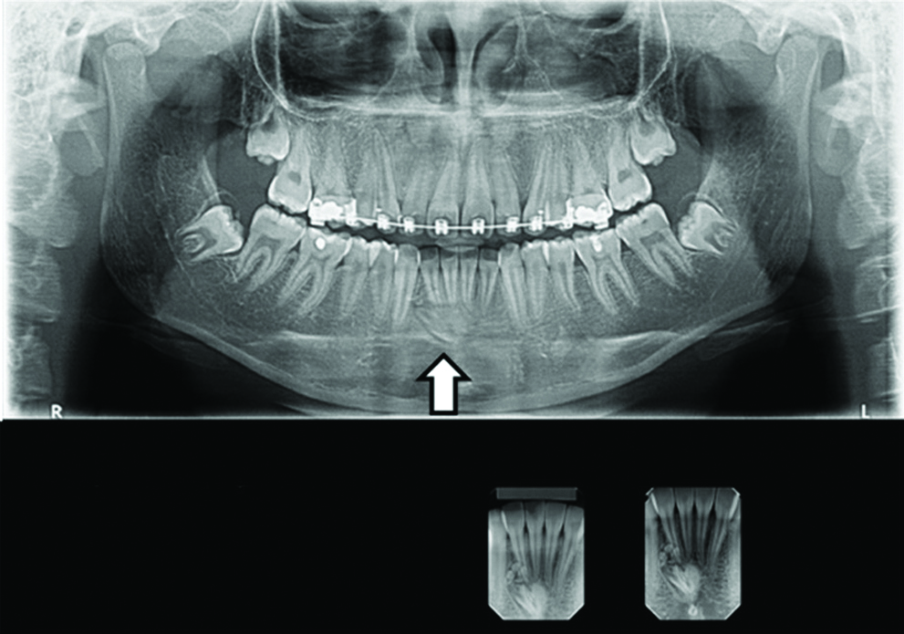
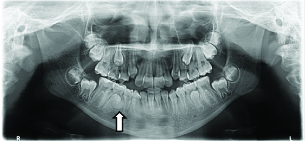
Panoramic and periapical radiograph suggesting a compound odontoma on the right side of the mandible, below the incisors (arrow).
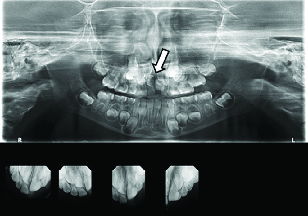
Panoramic and periapical radiograph suggesting of a compound odontoma on the right side of the maxilla, associated with the non-erupted central incisor (arrow).
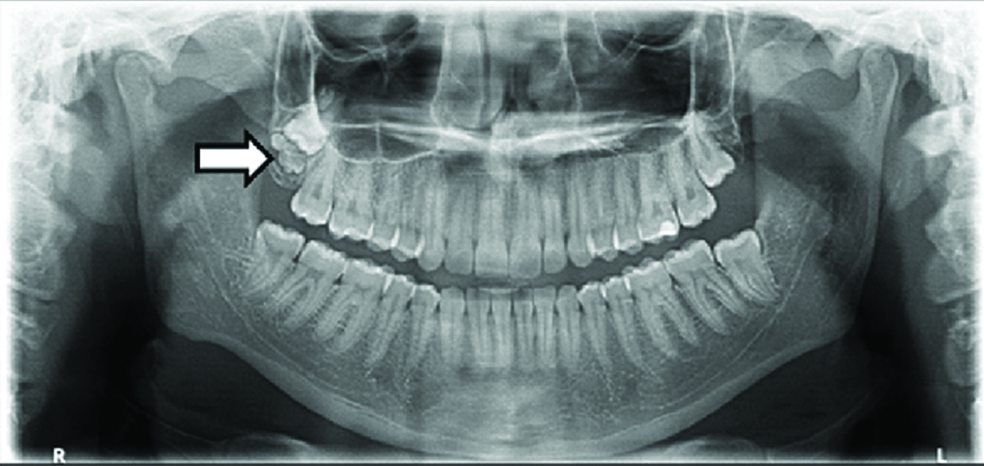
Panoramic radiograph suggesting of a compound odontoma appearing on the right side of the maxilla, associated with the non-erupted third molar tooth (arrow).
Complex odontomas were observed in the incisor region of the maxilla [Table/Fig-6], in the premolar region of the mandible [Table/Fig-7], and in the lower and upper third molars [Table/Fig-8,9]. However, the posterior region of the molars was the area most affected by complex odontomas (50.0%, 2/4) [Table/Fig-2].
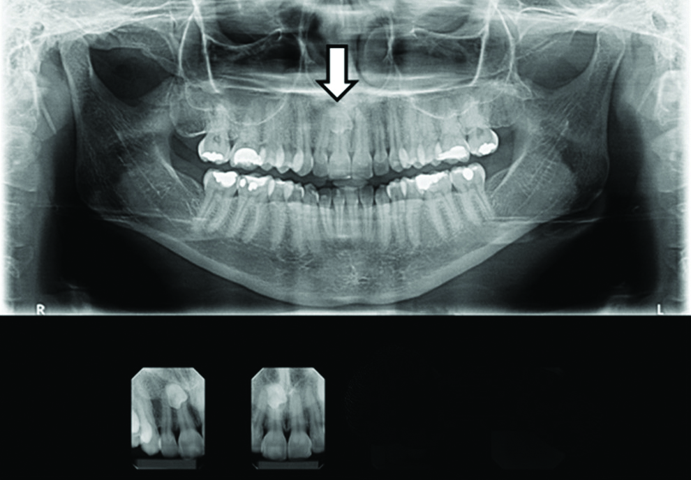
Panoramic and periapical radiograph suggesting a complex odontoma on the right side of the maxilla, near the apex of the incisor (arrow).
Panoramic and periapical radiograph suggesting a complex odontoma on the right side of the mandible, associated with the second non-erupted premolar (arrow).
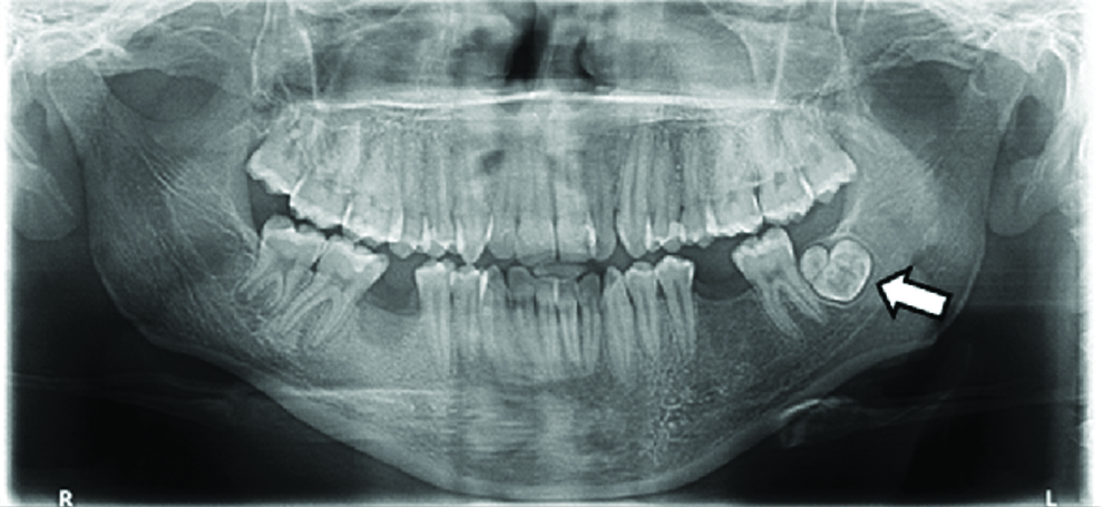
Panoramic radiograph of a 22-year-old male patient suggesting a complex odontoma on the left side of the mandible (molar region) (arrow).
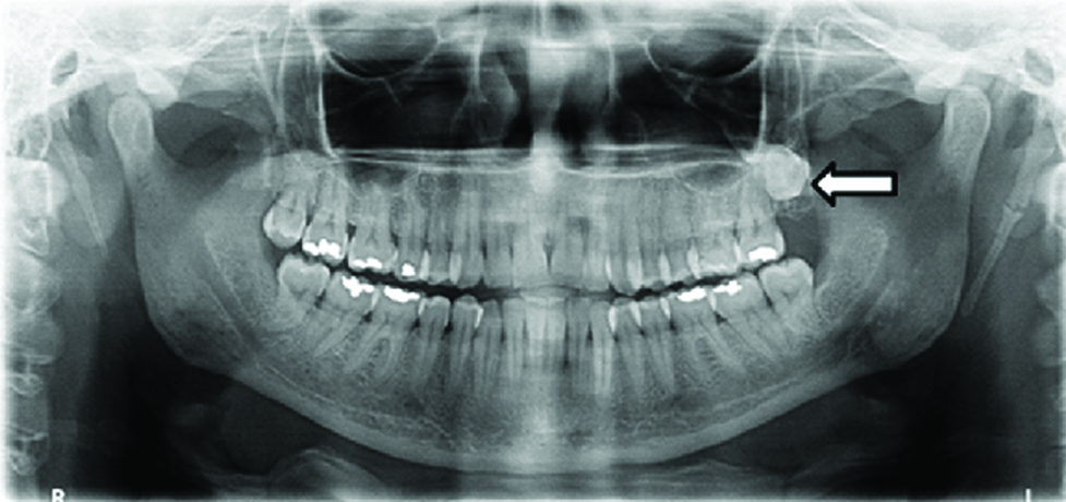
Panoramic radiograph suggesting a complex odontoma on the left side of the maxilla (molar region) (arrow).
Discussion
According to the 2017 WHO classification of odontogenic tumours and cysts, odontomas are classified as benign odontogenic tumours [1]. Odontomas correspond to a large percentage of all types of odontogenic tumours. Most studies identify these lesions as one of the most prevalent among all types of odontogenic tumours [15-17]. In a study conducted by da Silva LP et al., who analysed 289 odontogenic tumours, found that 11.4% of the tumours were diagnosed as odontomas [18]. In other similar cases, authors found a higher prevalence, as shown in [Table/Fig-10] [19-23].
Retrospective studies of odontogenic tumours.
| Literature review | N | Total | % | Male | Female | Maxilla | Mandible |
|---|
| Osterne RL et al., [22] | 36 | 185 | 19,46 | 12 | 24 | 20 | 14 |
| Servato JP et al., [19] | 178 | 431 | 41,3 | 96 | 82 | 105 | 53 |
| Da Costa DO et al., [23] | 37 | 201 | 18,4 | 26 | 11 | 19 | 15 |
| Martins-Filho PR et al., [21] | 22 | 66 | 33,3 | 10 | 12 | - | - |
| da Silva LP et al., [18] | 33 | 289 | 11,4 | - | - | 22 | 11 |
| Medeiros WKD et al., [20] | 89 | 247 | 36,0 | 40 | 49 | - | - |
| Total | 395 | 1419 | 26,64 | - | - | - | - |
Abbreviations: not specified=NS; Number of odontoma cases from the total number of analysed odontogenic tumours=N
Although odontomas are clinically characterised as asymptomatic, the pressure exerted by them can generate pain, devitalization, and dental resorption [24]. These lesions may also cause interference in tooth eruption, delay, or blockage of ectopic eruption movements, displacement and malformation of neighbouring teeth and diastema [25]. Therefore, the diagnosis of these odontogenic tumours, normally diagnosed in routine radiographs, is important to prevent or minimise the effects and enable the correct treatment of these lesions [26].
After analysing the obtained images, a prevalence of 0.32% of lesions compatible with odontomas was found. These results corroborate with findings reported by other authors, who observed a prevalence of 0.38% of odontomas after the analysis of 44,458 exams obtained over 30 years in Sri Lanka [15]. However, another study found a slightly lower prevalence of odontomas (0.27%) when evaluating the presence of odontogenic tumours in 33,354 radiographs collected between 1985 and 2006 in China [25].
In the present study, considering the type of odontoma, complex lesions presented a higher prevalence rate when compared to compound odontomas (57.1% and 42.9%, respectively), which is in agreement with other studies [9,25]. Data from the literature are controversial on this topic, since some studies have demonstrated the higher prevalence of compound odontomas [4,9,10].
According to the literature, the prevalence of odontomas increases with advancing age and is mainly found after the second decade of life [4,8]. These data corroborate with the findings observed in the present study, in which only one patient diagnosed with odontomas was in the first decade of life. In a study conducted by Soluk Tekkesin M et al., after analysing 160 odontomas, researchers obtained the mean age of the tumour of 27.9 years, observing a higher prevalence in patients between 10 and 19 years of age [27].
Regarding gender, the prevalence of odontomas was higher in female patients (71.4%). Although some studies did not identify a difference in relation to gender [4,10], a higher prevalence among women was also described in some previous studies [12,25]. In the present study, however, the prevalence of both odontogenic tumours was higher in female patients (66.6% and 75.0%, respectively).
Compound odontomas were most frequently identified in the incisor region, while complex lesions occurred mainly in the posterior region of the molars, corroborating with the higher prevalence described in the literature [4,8,10]. Compound odontomas are usually associated with non-erupted teeth, presenting a predilection for the anterior region, whereas in complex odontomas, odontogenic tissues are arranged in a disordered manner, constituting primary or immature dentin as the predominant component and predilection for the posterior region [4,9,10]. The results found in the present study confirm those from the literature, in which the images compatible with compound odontomas were associated with non-erupted teeth and in complex lesions, the presence of an irregular radiopaque mass was observed in the molar region.
From the radiographic characteristics presented by the odontogenic lesions it was possible to identify seven images compatible with odontomas, representing a prevalence of 0.32% of the analysed sample. Of these lesions, three presented characteristics that were compatible with compound odontomas and four with complex lesions. In both tumours, female patients presented the highest prevalence rate.
Limitation
Considering that the radiographic examination is a two-dimensional view of three-dimensional structures, this may well represent a limitation of this study, since the gold standard exam for the diagnoses of pathologies and anomalies is the Cone Beam Computed Tomography (CBCT), the availability of which is limited to metropolitan areas.
Conclusion
The data presented in this paper reinforce the theory that the use of panoramic digital radiographic images during dental practice aids in the diagnosis of odontomas. This study also showed that complex odontomas tend to be slightly more frequent than compound odontomas. The main goal of this study was to make epidemiological research of odontomas. It would be interesting in a further survey the complementation involving all odontogenic tumours in a similar population.
Abbreviations: not specified=NS; Number of odontoma cases from the total number of analysed odontogenic tumours=N
[1]. Wright JM, Vered M, Update from the 4th Edition of the World Health Organization Classification of Head and Neck Tumours: Odontogenic and Maxillofacial Bone TumorsHead Neck Pathol 2017 11(1):68-77.10.1007/s12105-017-0794-128247226 [Google Scholar] [CrossRef] [PubMed]
[2]. Soluk Tekkesin M, Wright JM, The World Health Organization Classification of Odontogenic Lesions: A Summary of the Changes of the 2017 (4th) EditionTurk Patoloji Derg 2018 34(1) [Google Scholar]
[3]. Patil S, Ramesh DNSV, Kalla AR, Complex odontoma: report of two unusual casesBraz J Oral Sci (online) 2012 11(4):509-12. [Google Scholar]
[4]. Hidalgo-Sánchez O, Leco-Berrocal MI, Martínez-González JM, Metaanalysis of the epidemiology and clinical manifestations of odontomasMed Oral Patol Oral Cir Bucal 2008 13(11):E730-34. [Google Scholar]
[5]. Buchner A, Merrell PW, Carpenter WM, Relative frequency of central odontogenic tumors: A study of 1,088 cases from Northern California and comparison to studies from other parts of the worldJ Oral Maxillofac Surg 2006 64(9):1343-52.10.1016/j.joms.2006.05.01916916667 [Google Scholar] [CrossRef] [PubMed]
[6]. Fregnani ER, Fillipi RZ, Oliveira CR, Vargas PA, Almeida OP, Odontomas and ameloblastomas: variable prevalences around the world?Oral Oncol 2002 38(8):807-08.10.1016/S1368-8375(02)00050-7 [Google Scholar] [CrossRef]
[7]. Kramer IRH, Pindborg JJ, Shear M, Histological typing of odontogenic tumours 1992 2th ednBerlinSpringer-Verlag Berlin Heidelberg10.1007/978-3-662-02858-2 [Google Scholar] [CrossRef]
[8]. MacDonald-Jankowski DS, Odontomas in a Chinese populationDentomaxillofac Radiol 1996 25(4):186-92.10.1259/dmfr.25.4.90842719084271 [Google Scholar] [CrossRef] [PubMed]
[9]. Bereket C, Çakır-Özkan N, Şener İ, Bulut E, Tek M, Complex and compound odontomas: Analysis of 69 cases and a rare case of erupted compound odontomaNiger J Clin Pract 2015 18(6):726-30.10.4103/1119-3077.15420926289508 [Google Scholar] [CrossRef] [PubMed]
[10]. An SY, An CH, Choi KS, Odontoma: a retrospective study of 73 casesImaging Sci Dent 2012 42(2):77-81.10.5624/isd.2012.42.2.7722783475 [Google Scholar] [CrossRef] [PubMed]
[11]. White SC, Weissman DD, Relative discernment of lesions by intraoral and panoramic radiographyJ Am Dent Assoc 1977 95(6):1117-21.10.14219/jada.archive.1977.0200271674 [Google Scholar] [CrossRef] [PubMed]
[12]. Momesso GA, de Sousa CA, de Lima VN, Bonardi JP, Coléte JZ, Ponzoni D, Removal of compound odontoma with piezosurgery techniqueJ Craniofac Surg 2017 28(2):584-85.10.1097/SCS.000000000000310328278140 [Google Scholar] [CrossRef] [PubMed]
[13]. Choi JW, Assessment of panoramic radiography as a national oral examination tool: review of the literatureImaging Sci Dent 2011 41(1):01-06.10.5624/isd.2011.41.1.121977466 [Google Scholar] [CrossRef] [PubMed]
[14]. Bondemark L, Jeppsson M, Lindh-Ingildsen L, Rangne K, Incidental findings of pathology and abnormality in pretreatment orthodontic panoramic radiographsAngle Orthod 2006 76(1):98-102. [Google Scholar]
[15]. Siriwardena BS, Tennakoon TM, Tilakaratne WM, Relative frequency of odontogenic tumors in Sri Lanka: Analysis of 1677 casesPathol Res Pract 2012 208(4):225-30.10.1016/j.prp.2012.02.00822439972 [Google Scholar] [CrossRef] [PubMed]
[16]. Bekiroglu N, Mete S, Ozbay G, Yalcinkaya S, Kargul B, Evaluation of panoramic radiographs taken from 1,056 Turkish childrenNiger J Clin Pract 2015 18(1):8-12. [Google Scholar]
[17]. AlSheddi MA, AlSenani MA, AlDosari AW, Odontogenic tumors: analysis of 188 cases from Saudi ArabiaAnn Saudi Med 2015 35(2):146-50.10.5144/0256-4947.2015.14626336021 [Google Scholar] [CrossRef] [PubMed]
[18]. da Silva LP, Serpa MS, Tenório JR, do Nascimento GJF, de Souza-Andrade ES, Veras-Sobral AP, Retrospective study of 289 odontogenic tumors in a Brazilian populationMed Oral Patol Oral Cir Bucal 2016 21(3):e271-75.10.4317/medoral.2102926827068 [Google Scholar] [CrossRef] [PubMed]
[19]. Servato JP, de Souza PE, Horta MC, Ribeiro DC, de Aguiar MC, de Faria PR, Odontogenic tumours in children and adolescents: a collaborative study of 431 casesInt J Oral Maxillofac Surg 2012 41(6):768-73.10.1016/j.ijom.2012.02.02122446071 [Google Scholar] [CrossRef] [PubMed]
[20]. Medeiros WKD, da Silva LP, Santos PPA, Pinto LP, de Souza LB, Clinicopathological analysis of odontogenic tumors over 22 years period: Experience of a single center in northeastern BrazilMed Oral Patol Oral Cir Bucal 2018 23(6):e664-71. [Google Scholar]
[21]. Martins-Filho PR, de Santana Santos T, Piva MR, da Silva HF, da Silva LC, Mascarenhas-Oliveira AC, A multicenter retrospective cohort study on pediatric oral lesionsJ Dent Child (Chic) 2015 82(2):84-90. [Google Scholar]
[22]. Osterne RL, Brito RG, Alves AP, Cavalcante RB, Sousa FB, Odontogenic tumors: a 5-year retrospective study in a Brazilian population and analysis of 3406 cases reported in the literatureOral Surg Oral Med Oral Pathol Oral Radiol Endod 2011 111(4):474-81.10.1016/j.tripleo.2010.10.01821239191 [Google Scholar] [CrossRef] [PubMed]
[23]. da-Costa DO, Maurício AS, de-Faria PA, da-Silva LE, Mosqueda-Taylor A, Lourenço SD, Odontogenic tumors: a retrospective study of four Brazilian diagnostic pathology centersMed Oral Patol Oral Cir Bucal 2012 17(3):e389-94.10.4317/medoral.1763022143740 [Google Scholar] [CrossRef] [PubMed]
[24]. Batra P, Duggal R, Kharbanda OP, Parkash H, Orthodontic treatment of impacted anterior teeth due to odontomas: a report of two casesJ Clin Pediatr Dent 2004 28(4):289-94.10.17796/jcpd.28.4.f2114718u87u471215366614 [Google Scholar] [CrossRef] [PubMed]
[25]. Luo HY, Li TJ, Odontogenic tumors: a study of 1309 cases in a Chinese populationOral Oncol 2009 45(8):706-11.10.1016/j.oraloncology.2008.11.00119147397 [Google Scholar] [CrossRef] [PubMed]
[26]. Santos LA, Lopes LJ, Roque-Torres GD, Oliveira VF, Freitas DQ, Complex odontoma: A case report with micro-computed tomography findingsCase Rep Dent 2016 2016:358475110.1155/2016/358475127293913 [Google Scholar] [CrossRef] [PubMed]
[27]. Soluk Tekkesin M, Pehlivan S, Olgac V, Aksakallı N, Alatli C, Clinical and histopathological investigation of odontomas: review of the literature and presentation of 160 casesJ Oral Maxillofac Surg 2012 70(6):1358-61.10.1016/j.joms.2011.05.02421840103 [Google Scholar] [CrossRef] [PubMed]