Chronic osteomyelitis of the long bones in children is one of the oldest diseases known. For orthopaedic surgeons in a developing country, it still remains a major challenge to treat such patients. The utmost priority remains to urgently diagnose and treat acute osteomyelitis as it can reduce the burden of this condition. We present a case report of an 11-year-old child with chronic osteomyelitis of the fibula and its subsequent management. The child hails from a low socio-economic background with limited access to healthcare facilities. Chronic osteomyelitis in children affects various long bones but involvement of the fibula has rarely been reported in literature.
Debridement, Developing countries, Sequestrectomy, Sinus tract
Case Report
An 11-year-old male child from a village in Indo-Bangladesh border, son of a manual labourer and holding a BPL (Below Poverty Line) card, presented to the orthopaedic out-patient department with a discharging sinus along with an exposed piece of bone over the lower lateral aspect of his right leg since seven months. The patient was apparently well prior to that, when he first developed an itching and burning sensation at the same site of his right leg followed by an insidious onset of swelling around that region with redness and high grade fever. He was first treated by a traditional healer who applied some herbal preparation over the swelling, following which he developed a discharging sinus leading to an exposed bone with a non-healing wound. He was subsequently taken to a general practitioner, who prescribed some medications as well as daily dressing of the wound. With the condition of the wound worsening and the child’s overall health deteriorating, he was eventually referred to our centre for further management.
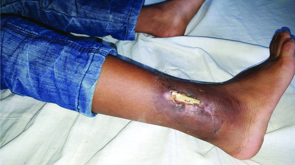
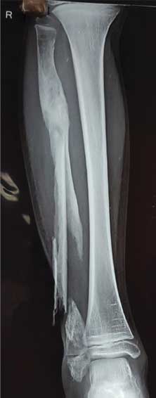
The child was of small built and appeared malnourished. At the time of presentation, his condition was stable. While questioning his parents regarding his medical history, it was found out that he had never completed his vaccination schedule. On examination of the affected limb, the fibula was exposed, straddling on a contaminated wound with irregular borders and an unhealthy bed of granulation tissue. Tenderness was present on deep palpation along the lower third of fibula associated with purulent discharge [Table/Fig-1]. Movements at the knee joint remained unaffected while ankle motion was restricted terminally. A radiograph of the involved extremity was obtained which depicted a segmental pathological fracture of the fibula. The presence of a large sequestrum was evident with features of thickening and sclerosis of the remaining fibular shaft and a pathological fracture distally [Table/Fig-2]. Routine blood investigations were sent which showed as: Hb: 9.2 gm%, TLC: 13800/cumm, ESR: 55 mm after one hour and the C-Reactive Protein was 10 mg/L.
Clinical photo of the leg showing the exposed fibula.
X-ray of the leg showing large sequestrum with features of thickening and sclerosis of the remaining fibular shaft and a pathological fracture distally.
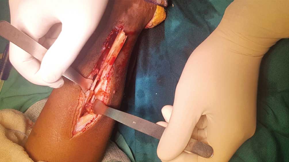
A diagnosis of chronic osteomyelitis of the right fibula was confirmed after clinical and radiological correlation. The differential diagnosis of Ewing’s Sarcoma and osteosarcoma was made. After obtaining consent from the child’s parents, he was taken for surgery. The fibula was exposed through a lateral approach and the freelying dead piece of bone (sequestrum) was excised followed by debridement and excision of the sinus tract [Table/Fig-3]. Debrided tissue along with a portion of the bony sequestrum was sent for histopathological examination. Purulent material from the sinus tract was sent for culture and sensitivity. The wound was thoroughly washed with copious amounts of normal saline and was closed over a negative suction drain. This drain was removed only after 48 hours. The culture and sensitivity report revealed growth of the organism Staphylococcus aureus with sensitivity to the following antibiotics- Erythromycin, Gentamycin, Cefixime and Clindamycin. Postoperatively, dressing of the wound was done on the 2nd day, 7th day and at final removal of sutures on the 14th day. He received intravenous antibiotic Gentamycin for two weeks and after discharge was kept on oral antibiotic (Cefixime+Clavulanic acid) for four weeks. Histopathological examination of the sequestrum corresponded with features of chronic osteomyelitis.
Intra-operative image showing the fibula with the sequestrum through the lateral approach.
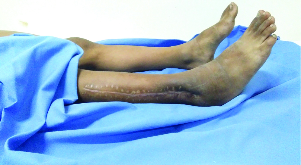
The child was followed up at regular intervals for a period of more than two years. He showed excellent recovery with overall healing of the wound with normalisation of his blood parameters. He was symptom free and was able to perform all his daily activities [Table/Fig-4].
Follow-up clinical image after two years.
Discussion
Chronic osteomyelitis is defined as long-standing infection of the bone characterised by growth of microorganisms, presence of a dead bone with surrounding unhealthy granulation tissue, inflammation and discharging sinus [1]. Chronic osteomyelitis continues to remain a major challenge in developing countries. Chronic osteomyelitis involving the extremities remains a major cause of morbidity and disability in children in India. The prolonged treatment affects the ambulatory status, functional mobility, and independence [2].
Chronic osteomyelitis may result from inadequately treated acute haematogenous osteomyelitis or more commonly from a contiguous source of infection. The contiguous spread might trail treatment of open fracture, internal fixation of fractures, or prosthetic replacements [3].
With the miscellaneous problems, it is necessary that a multidisciplinary effort is needed for successful treatment. The team should consist of orthopaedic surgeons, microbiologists, nutritionists, and a psychologist if needed [4].
The objective of management in chronic osteomyelitis is whole abolition of contamination though conserving the soft-tissue envelope, healing of bone segment, and preservation of limb length and function.
The treatment plan should be based on individual’s requirement after assessing the extent of disease, microbiological study and the patient’s physiological status.
Chronic haematogenous pyogenic osteomyelitis in children, though rarely seen in the western countries, remains a major cause of morbidity and disability in children living in developing countries. Chronic osteomyelitis and low socio-economic status are closely related. Poor dental and skin hygiene, host deficiencies and inadequate healthcare provision are the major factors that predispose children to the chronic osteomyelitis [5].
The diagnosis of chronic pyogenic osteomyelitis is in most cases straight forward as the history and the radiological picture is usually diagnostic. However, in some patients, different conditions may present with similar clinical features. Therefore, while evaluating suspected osteomyelitis, other differential diagnosis should also be considered like primary tumours (Ewing’s sarcoma, Fibrous dysplasia, Osteosarcoma) as well as non-pyogenic osteomyelitis (fungal, tubercular) [6]. Scintigraphy is the most sensitive and specific investigation for the diagnosis of acute osteomyelitis and the evaluation of activity of chronic osteomyelitis. Computed tomography can provide confirmation of the presence of sequestra [7]. However, in our patient clinical findings of exposed bone, discharging sinus, radiological presence of sequestrum, blood parameters suggesting infection, were all in favour of the diagnosis of chronic osteomyelitis.
In adults, Staphylococcus aureus is the most common organism isolated [8]. In infants, the most frequent pathogens are Staphylococcus aureus, Streptococcus agalactiae, and Escherichia coli. Staphylococcus aureus, Streptococcus pyogenes, and Haemophilus influenzae are most commonly isolated organisms in children more than one year of age [9]. After age 4, the incidence of H. influenzae as a cause of osteomyelitis has dramatically decreased with the emergence of H. influenzae vaccine [10,11].
The treatment strategy as in all cases of extremity chronic osteomyelitis remains unchanged. Treatment of extremity generally includes radical sequestrectomy, management of dead space, soft tissue reconstruction and the restoration of bone stability. The curative approach to chronic osteomyelitis has the following goals-to eliminate infection and achieve excellent functional outcome [12].
Studies on adult patients have shown that the most frequent type of extremity was traumatic osteomyelitis following an open injury due to a road traffic accident. Chronic osteomyelitis was more common in adult males and most commonly the lower limbs, with tibia, femur, calcaneus, and toes being the usual sites in order of involvement [13]. In our case, the cause of chronic osteomyelitis in this child was due to neglected treatment of an acute osteomyelitis which was of haematogenous origin. Chronic osteomyelitis involving only the fibula, is rare with only a few cases reported in literature [14-16].
A sequestered fibula may be removed without hesitation provided the ipsilateral tibia remains uninvolved and continues to support the weight of the body. Dissection of the fibula through the plane lying in between the peroneal muscle anteriorly and the soleus posteriorly with careful attention to the surrounding neurovascular structures would facilitate the removal of the infected fibula.
A fibular sequestrectomy alone was performed on our patient and because of an intact lateral malleolus; there was no requirement for any bony reconstruction procedure. Various studies emphasise that only distal resection of fibula warranted reconstruction since the lateral malleolus is a part of ankle stability [17,18]. At two years of follow-up, our patient was bearing full weight, having full range of motion at the adjacent joint and had no residual deformity of the operated limb.
Conclusion
Chronic haematogenous osteomyelitis affects various long bones of the body. However, our case report highlights the rarity of fibular involvement in chronic osteomyelitis. A high index of suspicion of this uncommon location should be borne in mind while treating such patients. Chronic osteomyelitis continues to affect many children in developing countries especially those from the low socio-economic status and who do not have access to modern healthcare facilities. There is a great need to educate the healthcare personnel and the population, to enable early detection of acute osteomyelitis and the need for prompt treatment. Early and appropriate treatment of acute osteomyelitis at the outset is the most effective means in reducing the burden of chronic osteomyelitis as the resultant chronicity poses an enormous challenge to the treating surgeon.
[1]. Mouzopoulos G, Kanakaris NK, Kontakis G, Obakponovwe O, Townsend R, Giannoudis PV, Management of bone infections in adults: The surgeon’s and microbiologist’s perspectivesInjury 2011 42(5):S18-S23.10.1016/S0020-1383(11)70128-0 [Google Scholar] [CrossRef]
[2]. Masini BD, Waterman SM, Wenke JC, Owens BD, Hsu JR, Ficke JR, Resource utilization and disability outcome assessment of combat casualties from Operation Iraqi Freedom and Operation Enduring FreedomJ Orthop Trauma 2009 23:261-66.10.1097/BOT.0b013e31819dfa0419318869 [Google Scholar] [CrossRef] [PubMed]
[3]. Calhoun JH, Manring MM, Shirtliff M, Osteomyelitis of the long bonesSeminPlast Surg 2009 23(2):59-72.10.1055/s-0029-121415820567728 [Google Scholar] [CrossRef] [PubMed]
[4]. Pande K, Optimal management of chronic osteomyelitis: Current perspectivesOrthopaedic Research and Reviews 2015 (7):71-81.10.2147/ORR.S50753 [Google Scholar] [CrossRef]
[5]. Jones HW, Beckles VLL, Akinola B, Stevenson AJ, Harrison WJ, Chronic haematogenous osteomyelitis in childrenJ Bone Joint Surg Br 2011 93-B:1005-10.10.1302/0301-620X.93B8.2595121768620 [Google Scholar] [CrossRef] [PubMed]
[6]. Arkader A, Myung KS, Stanley P, Mascarenhas L, Ewing sarcoma of the tibia mimicking fibrous dysplasiaJ PediatrOrthop B 2013 22:222-27.10.1097/BPB.0b013e32834dfe4d22094991 [Google Scholar] [CrossRef] [PubMed]
[7]. Tumeh SS, Tohmeh AG, Nuclear medicine techniques in septic arthritis and osteomyelitisRheum Dis Clin North Am 1991 17:559-83. [Google Scholar]
[8]. Lew DP, Waldvogel FA, OsteomyelitisN Engl J Med 1997 336:999-1007.10.1056/NEJM1997040333614069077380 [Google Scholar] [CrossRef] [PubMed]
[9]. Song KM, Sloboda JF, Acute hematogenous osteomyelitis in childrenJ Am Acad Orthop Surg 2001 9:166-75.10.5435/00124635-200105000-00003 [Google Scholar] [CrossRef]
[10]. De Jonghe M, Glaesener G, Type B Haemophilus influenzae infections. Experience at the Paediatric Hospital of LuxembourgBull Soc Sci Med Grand Duche Luxemb 1995 132:17-20. [Google Scholar]
[11]. Blyth MJ, Kincaid R, Craigen MA, Bennet GC, The changing epidemiology of acute and subacute haematogenous osteomyelitis in childrenJ Bone Joint Surg Br 2001 83:99-102.10.1302/0301-620X.83B1.083009911245548 [Google Scholar] [CrossRef] [PubMed]
[12]. Walter G, Kemmerer M, Kappler C, Hoffaman R, Treatment algorithms for chronic osteomyelitisDtsch Arztebl Int 2012 109(14):257-64.10.3238/arztebl.2012.025722536302 [Google Scholar] [CrossRef] [PubMed]
[13]. Jiang N, Ma Y, Jiang Y, Zhao X, Xie G, Hu Y, Clinical characteristics and treatment of extremity chronic osteomyelitis in southern ChinaMedicine 2015 94(42):1-7.10.1097/MD.000000000000187426496345 [Google Scholar] [CrossRef] [PubMed]
[14]. Khurana A, Chhawra S, Gupta R, Kumar S, Osteomyelitis of fibula: rare case with various differential diagnosisJ Orthop Case Rep 2017 7(3):59-62. [Google Scholar]
[15]. Singh J, Kalia A, Virk J, Isloated chronic osteomyelitis of fibula in a child: A case report and review of literatureInt J Orthop Sci 2017 3(4):923-26.10.22271/ortho.2017.v3.i4m.126 [Google Scholar] [CrossRef]
[16]. Singh G, Roy AV, Isolated osteomyelitis of fibula following contact with Urticadioica (BichuGhaas) in a childSch J Med Case Rep 2014 2(4):297-98. [Google Scholar]
[17]. Capanna R, Van Horn JR, Biagini R, Ruggieri P, Bettelli G, Campanacci M, Reconstruction after resection of the distal fibula for bone tumorActa Orthop Scand 1986 57:290-94.10.3109/174536786089943943538754 [Google Scholar] [CrossRef] [PubMed]
[18]. Yadav SS, Ankle stability after resection of the distal third of the fibula for giant cell lesions: report of two casesClin Orthop 1981 155:105-07.10.1097/00003086-198103000-00019 [Google Scholar] [CrossRef]