Multicystic Left Kidney and Contralateral Pelvic Kidney with Ectopic Ureter and Renal Failure in a Young Male: A Rare Association
Pritesh Jain1, Debansu Sarkar2, Dilip Kumar Pal3
1 Senior Resident, Department of Urology, Institute of Postgraduate Medical Education and Research, Kolkata, West Bengal, India.
2 Associate Professor, Department of Urology, Institute of Postgraduate Medical Education and Research, Kolkata, West Bengal, India.
3 Professor, Department of Urology, Institute of Postgraduate Medical Education and Research, Kolkata, West Bengal, India.
NAME, ADDRESS, E-MAIL ID OF THE CORRESPONDING AUTHOR: Dr. Dilip Kumar Pal, 244, AJC Bose Road, Kolkata-700020, West Bengal, India.
E-mail: urologyipgmer@gmail.com
Multicystic Dysplastic Kidney (MCDK) is a relatively common cystic disease of kidney which may be associated with various urogenital abnormalities. Here, authors encountered a case of 16-year-old male who presented with left side abdominal pain and lump. On evaluation, he was found to be growth-retarded with chronic renal failure. Imaging studies revealed left MCDK with contralateral pelvic kidney with ectopic ureter opening in prostatic urethra. This patient also had calculus in right ectopic ureter near its opening in prostatic urethra. The calculus was surgically removed; however, he required dialysis as there was no improvement in renal function. This conglomeration of anomalies is extremely rare and a dilemma exists not only in diagnosis but also in therapeutic decision making. Every case of MCDK or ectopic ureter should be further investigated to detect associated abnormalities, for timely management.
Multicystic dysplastic kidney, Ureteral diseases, Urinary calculi
Case Report
A 16-year-old male presented with complaints of gradually increasing pain and swelling in left upper quadrant of abdomen, since 24 months. Pain was chronic, dull aching and non-radiating with no specific aggravating or relieving factors. He also had complaints of decrease in urine output, anorexia and nausea from last few months. For the pain, he was taking medications without prescription. Gradually, he developed abdominal lump for which he visited a local practitioner who referred him to higher centre after initial evaluation by blood test and ultrasound study. He denied any history of hypertension, diabetes, jaundice, fever, haematuria or voiding symptoms.
General examination revealed a malnourished growth-retarded male (Height-141 cm, weight- 32 kg, BMI-16.1) with pallor. Blood pressure was 156/94 mmHg. A palpable lump of approximately 10 cm×12 cm size, non-tender, soft to firm in consistency with lobulated margins was present in left upper quadrant of abdomen. External genitalia and digital rectal examination were unremarkable.
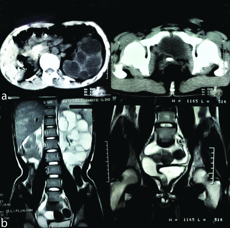
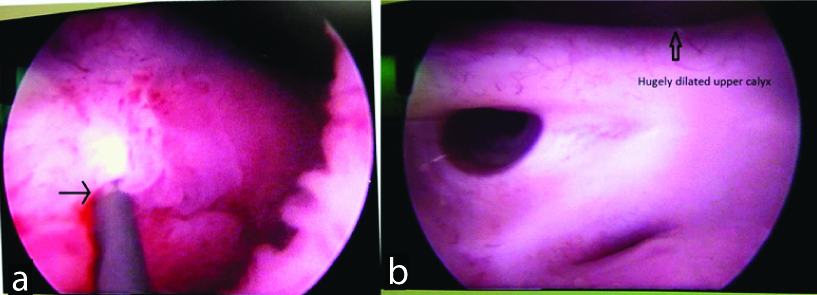
His haemoglobin was 7.2 g/dL with normal differential leukocytes counts. Renal function tests were grossly abnormal with serum urea level of 164 mg/dL, creatinine 9.2 mg/dL, serum Na+ 128 meq/dL and serum K+ 5.4 meq/dL. Urine analysis revealed Grade-3 proteinuria with plenty of pus cells however urine culture was sterile. Ultrasound of Kidney Ureter Bladder (KUB) showed possibility of left side unilateral fused kidney with multiple non-communicating cysts filled with echogenic floating material along with insertion of grossly dilated right ureter in posterior urethra with calculus in terminal part of ectopic ureter. Non-Contrast Computed Tomography (NCCT) scan revealed enlarged left kidney with multiple non-communicating cysts in non-dilated renal pelvis. Right kidney could not be localised in right renal fossa however a dilated cystic structure was found in right side of pelvis with possible insertion near bladder neck with a calculus near its terminal part [Table/Fig-1a]. To further delineate anatomy, Magnetic Resonance Imaging (MRI) was done which revealed enlarged left kidney with multiple non-communicating cysts. Right kidney was found in right side of pelvis near bladder with gross hydronephrosis and thinned out parenchyma. Dilated ureter of right kidney was found inserting in posterior urethra [Table/Fig-1b]. Cystourethroscopy revealed right ureter opening in prostatic urethra, with normal bladder with normally situated left ureteric opening [Table/Fig-2a]. Right ureter could be negotiated without difficulty even with 17 FrCystoscope which showed multiple dilated calyces with stone in one of the calyx [Table/Fig-2b].
a) NCCT KUB images (axial view) depicting left kidney with multiple non-communicating cysts with non-dilated renal pelvis and right ureter opening distal to bladder neck with calculus in terminal part (arrow); b) MR Urogram images (coronal view) depicting left multicystic dysplastic kidney and right hydronephrotic pelvic kidney with ectopic ureter opening in prostatic urethra (arrow).

a) Endoscopic view of urethra showing guide wire in ectopic right ureter opening at prostatic urethra (arrow); b) Cystoscopic view of right kidney showing multiple calyces (Upper calyx was hugely dilated with a calculus in it).

The stone was fragmented with pneumatic lithoclast. A double J stent was placed in left kidney however there was no significant improvement in function or anatomy at six weeks follow-up. Patient could not be weaned off dialysis so he was enlisted for renal transplant.
Discussion
MCDK is among common cystic kidney diseases with an incidence of 1 per 1000 to 4000 newborns [1]. Patients with MCDK frequently manifest contralateral urinary abnormalities like Ureteropelvic Junction Obstruction (UPJO) and Vesico-Ureteral Reflux (VUR) [2]. This disease is a form of renal dysplasia characterized by the presence of multiple, non-communicating cysts separated by dysplastic parenchyma and the absence of a normal pelvicalyceal system. Male-to-female ratio of 1.48:1 has been described in literature [3]. Left kidney is involved in 55% of cases, and right kidney in 45% whereas bilateral MCDK occurs in about 20% of prenatally diagnosed cases. Embryological basis of this anomaly is thought to be due to abnormal induction of the metanephric mesenchyme by the ureteral bud [4]. Timing of injury to ureteric bud and its effect on ureteric branching decides final structure of the dysplastic kidney [4,5]. Most cases of unilateral MCDK undergo spontaneous involution or it may persist without any change and rarely may increase in size with time [6].
Unilateral MCDK is usually asymptomatic and can remain undetected into adulthood. It may be discovered during an investigation for urinary tract infection, voiding dysfunction, hypertension or when diagnostic imaging studies are performed to investigate a non-urinary problem [7]. Clinically, it may present as an abdominal mass which is mobile, non-tender, ballotable, irregular in shape and might trans-illuminate [6,7]. In the present case, initial symptoms of patient were pain and abdominal lump for which he consulted to physician. Associated anomaly of contralateral kidney is common and seen in up to 39% of patients [2]. Common malformations of opposite kidney are UPJO, VUR, renal agenesis, dysplasia, hypoplasia, ectopic kidney etc., [2,8].
Imaging modalities used in evaluation of such cases include USG to characterise anatomy of bilateral renal system, voiding cystourethrogram to detect VUR. In cases of confusion between hydronephrotic kidney and MCDK, a dimercaptosuccinic acid renogram is useful and demonstrates absence or very poor function in later. Direct cyst puncture with installation of contrast material is also helpful in differentiating UPJO from MCDK [9]. Pelvic ectopia with ectopic ureter in a male is itself a very rare anomaly and its association with MCDK as presented in the present case is extremely rare. MR urogram is very useful in suspected cases to better delineate anatomy and identifying course of ureter [10]. In the present case it was MRI which helped us to identify pelvic position of opposite kidney and associated ectopic ureter opening.
MCDK requires regular follow-up in asymptomatic patients. Nephrectomy is required in symptomatic patients or when associated hypertension is present. Pelvic ectopic kidney usually are asymptomatic and doesn’t require intervention however when hydronephrosis due to associated ectopic ureter is present ureteroneocystostomy may be considered to prevent deterioration of function [10,11]. Li J et al., studied cases of single system ectopic ureter and associated anomalies, they found that congenital dysplasia although rare may be seen in such cases [12]. This case with extremely rare conglomeration of anomalies had not been described previously in literatures as far as authors’ knowledge.
Conclusion
Pelvic kidney associated with ectopic ureter in a male is a rare anomaly and rarer is its association with cystic kidney. This case highlights an extremely rare association of bilateral renal anomalies and diagnostic and therapeutic challenges in management of such cases.
[1]. Kalyoussef E, Hwang J, Prasad V, Barone J, Segmental multicystic dysplastic kidney in childrenUrology 2006 68(5):121110.1016/j.urology.2006.06.02417095057 [Google Scholar] [CrossRef] [PubMed]
[2]. Atiyeh B, Husmann D, Baum M, Contralateral renal abnormalities in multicystic-dysplastic kidney diseaseJ Pediatr 1992 121:65-67.10.1016/S0022-3476(05)82543-0 [Google Scholar] [CrossRef]
[3]. Robson WL, Leung AK, Thomason MA, Multicystic dysplasia of the kidneyClin Pediatr (Phila) 1995 34(1):32-40.10.1177/0009922895034001067720326 [Google Scholar] [CrossRef] [PubMed]
[4]. Mackie GG, Stephens FD, Duplex kidneys: a correlation of renal dysplasia with position of the ureteral orificeJ Urol 1975 114(2):274-80.10.1016/S0022-5347(17)67007-1 [Google Scholar] [CrossRef]
[5]. Pope JC 4th, Brock JW 3rd, Adams MC, Stephens FD, Ichikawa I, How they begin and how they end: classic new theories for the development and deterioration of congenital anomalies of the kidney and urinary tract, CAKUTJ Am Soc Nephrol 1999 61:2018-28. [Google Scholar]
[6]. Onal B, Kogan BA, Natural history of patients with multicystic dysplastic kidney-what follow-up is needed? J Urol 2006 176:1607-11.10.1016/j.juro.2006.06.03516952700 [Google Scholar] [CrossRef] [PubMed]
[7]. Al-Khaldi N, Watson AR, Zuccollo J, Twinning P, Rose DH, Outcome of antenatally detected cystic dysplastic kidney diseaseArch Dis Child 1994 70:520-22.10.1136/adc.70.6.5208048824 [Google Scholar] [CrossRef] [PubMed]
[8]. Mathiot A, Liard A, Eurin D, Dacher JN, [Prenatally detected multicystic renal dysplasia and associated anomalies of the genito-urinary tract]J Radiol 2002 83:731-35. [Google Scholar]
[9]. Whittam BM, Calaway A, Szymanski KM, Carroll AE, Misseri R, Kaefer M, Ultrasound diagnosis of multicystic dysplastic kidney: is a confirmatory nuclear medicine scan necessary?J Pediatr Urol 2014 10(6):1059-62.10.1016/j.jpurol.2014.03.01124909606 [Google Scholar] [CrossRef] [PubMed]
[10]. Peters CA, Mendelsohn C, Ectopic Ureter, Ureterocele, and Ureteral Anomalies. Wein AJ, Kavoussi LR, Partin AW, Peters CA, editorsCampbell-Walsh Urology 2016 PhiladelphiaElsevier Saunders:3087-96. [Google Scholar]
[11]. Prieto J, Ziada A, Baker L, Snodgrass W, Ureteroureterostomy via inguinal incision for ectopic ureters and ureteroceles without ipsilateral lower pole refluxJ Urol 2009 181(4):1844-48.10.1016/j.juro.2008.12.00419233406 [Google Scholar] [CrossRef] [PubMed]
[12]. Li J, Tingze HU, Minghe W, Xuewu J, Shaoji C, Lugang H, Single ureteral ectopia with congenital renal dysplasiaJ Urol 2003 170:558-59.10.1097/01.ju.0000076000.82388.8012853829 [Google Scholar] [CrossRef] [PubMed]