The treatment of congenital agenesis of mandibular incisors is complex, and the absence of these teeth can cause functional, aesthetic, and psychological problems. This paper reports the treatment of an Angle Class I malocclusion associated with agenesis of both mandibular central incisors. Among various treatment alternatives, the chosen therapeutic approach was to consolidate space between the first and second premolars to make room for further implant and prosthesis placement. This orthodontic-prosthetic approach provided balanced occlusion, dental aesthetic, periodontal health, improved self-esteem, and a satisfactory smile to the patient. The treatment outcomes have remained stable during follow-up of 3 years and 1 month.
Case Report
A nine-years and one-month-old male patient came for orthodontic consultation referred by his pediatric dentist because of the absence of both permanent mandibular central incisors [Table/Fig-1a-c]. The general medical and dental history was unremarkable with no pathologic findings. The extraoral examination showed a well-balanced and symmetric face. On intraoral examination he presented with Angle Class I malocclusion, mixed dentition, 3.0 mm of overjet, deep overbite (70%), and mandibular midline diastema of 8.0 mm between the lateral incisors [Table/Fig-1d-g]. A panoramic radiograph showed agenesis of both mandibular central incisors [Table/Fig-1h].
Phase I. 9.1years-old. a) Lateral view; b) Frontal view; c) Frontal view with anterior teeth; d) Intraoral right view; e) Intraoral frontal view; f) Intraoral left view; g) occlusal view showing absence of both incisors; h) Panoramic radiograph.

The orthodontic treatment was planned and the informed consent was obtained from the patient’s parents after explaining the treatment plan and before starting the dental procedures.
During Phase I, diastema between both mandibular lateral incisors was reduced with standard edgewise 0.022”×0.028” brackets (Morelli, Sorocaba, Brazil) bonded on both lateral incisors. A 0.016”×0.022” stainless steel rectangular wire (Morelli, Sorocaba, Brazil) was used for applying a reciprocal force generated by an elastic chain (Morelli, Sorocaba, Brazil), to maintain alveolar bone height and width and to improve the patient’s self-esteem [Table/Fig-2a-f]. The patient returned for Phase II orthodontic treatment at 12 years and 10 months of age with a deep overbite and a generalised diastema in the mandibular arch [Table/Fig-3a-h].
Phase I. Biomechanics for reduction the mandibular midline diastema. a,d) Initial diastema closing stage; b,e) Reduction of the diastema; c,f) Final stage with the diastema closed.

Phase II, 12.10 years-old. a) Lateral view; b) Frontal view c) Frontal view with anterior teeth; c) Intraoral right view; e) Intraoral frontal; f) Intraoral left view; g) Mandibular teeth with uniform diastema and absence of both central incisors; h) Panoramic radiograph showing uniform diastema.

In Phase II, the treatment objectives were to achieve optimal alignment and levelling of teeth, maintain a balanced soft-tissue profile, and establish a proper occlusal relationship with normal overjet and overbite. The lingual cusp of the first lower premolar was slightly reshaped and canine height was reduced with a tapered shaped diamond bur (Komet, USA). The diastemas in the mandibular arch were closed by moving teeth forward, and consolidating space around the premolars, lateral incisors, and other teeth to make room between the premolars, for further dental implant and prosthesis placement.
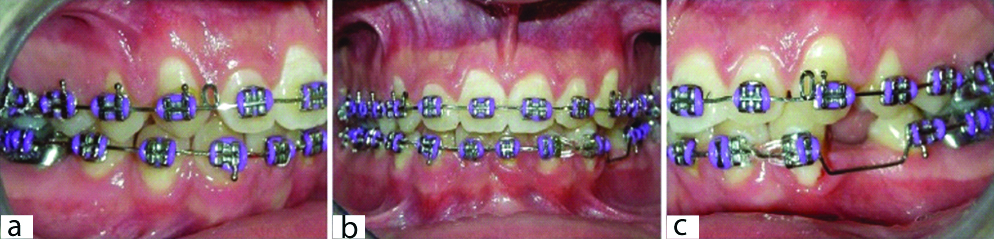
During Phase II, a standard fixed edgewise orthodontic appliance with 0.022”×0.028” slots was used. Alignment and levelling were performed using NiTi, SS arches, and 0.019”×0.025” SS arches with open coil springs between the mandibular left first and second premolar to open space for implant-prosthesis rehabilitation [Table/Fig-4a-c].
Progress of phase II treatment. Intraoral view of fixed appliance and the biomechanics applied into the phase II.
In Phase I, the mandibular lateral incisors was moved closer, despite the diastema, which remained stable until the complete permanent dentition eruption. During Phase II, a good aesthetic outcome was obtained by aligning and levelling the anterior teeth by ensuring good intercuspation of posterior teeth with overjet of 3.0 mm and an overbite of 50% [Table/Fig-5a-h]. After 40 months of effective active treatment, the appliance was debonded and a mandibular canine-to-canine and between both left premolars [Table/Fig-5f,g] retainer was bonded to stabilise mandibular incisor alignment (0.032-in stainless steel) and retain the space for implant placement (0.019 ×0.025-in stainless steel rectangular wire). A maxillary wraparound with 0.036-in stainless steel round wire retainer was used to maintain the alignment and levelling while waiting for implant-prosthesis placement [Table/Fig-5h]. Panoramic radiograph showed aspects of normality of the dental roots and no pathological findings as well as enough space for implant placement [Table/Fig-5i].
Post-treatment, 18.8 years-old. a) Lateral view; b) Frontal view; c) Frontal view with anterior teeth; d) Intraoral right view; e) Intraoral frontal view; f) Intraoral left view; g) Mandibular teeth with space between both left premolars; h) Wraparound type retainer; i) Panoramic radiograph.

The patient was satisfied with his facial aesthetic and harmonious occlusion with long-term stability has been observed during follow-up of 3 years and 1 month [Table/Fig-6a-g]. Space was rehabilitated with an implant as a “third” premolar inserted at 20 years 1 month of age [Table/Fig-6g,h]. The patient was referred to an oral surgeon for third molar extractions, but the patient refused to get the extractions done [Table/Fig-6h]. The superimposition of cephalometric tracings from Phase I to post-treatment reveals proportional downward and forward growth of the face [Table/Fig-7], (black, blue and red lines). No such growth was observed when post-treatment [Table/Fig-7], (red line) and follow-up [Table/Fig-7], (green line) cephalometric tracings were superimposed.
21.9 years-old. Follow-up. a) Lateral view; b) Frontal view; c) Frontal view with anterior teeth; d) Intraoral right view; e) Intraoral frontal; f) Intraoral left view; g) Mandible with prosthesis as a third premolar; h) Panoramic radiograph with implant-prosthesis.

Superimposition. Phase I, black lines (9,1-years), Phase II, blue (12,10-years), Posttreatment, red (18,8-years), Follow-up (21,9 years-old).

Discussion
Restorative and orthodontic procedures are the treatment strategies for missing mandibular incisors to improve aesthetic and function [1]. This paper illustrates an orthodontic-implant-prosthesis approach of a patient who presented with agenesis of both mandibular central incisors.
The loss or absence of permanent teeth can cause functional, aesthetic and psychological problems. Most orthodontists have treated at least one patient with a mandibular incisor that was either missing or severely damaged by injury or disease. In these situations, removing it remains the best choice for the patient [2].
One of the most important factors for the successful treatment of patients with agenesis is multidisciplinary intervention that involves close work by a committed team (including a general dental practitioner, paediatric dentist, orthodontist, prosthodontist and implantodontist). In this context, each professional contributes expertise to achieve optimal results for the patient [3], like in the present clinical case.
The diastema between both mandibular lateral incisors in Phase I was reduced to maintain alveolar bone height and width and to improve the patient’s self-esteem. A standard edgewise 0.022”×0.028” brackets were bonded on both lateral incisors and applied a reciprocal force generated by an elastic chain.
According to Zhang R et al., there are three common orthodontic treatment modalities for a patient with agenesis of mandibular incisors [4]. One approach involves creating space for a fixed prosthesis or implant rehabilitation. Another choice is removal of the maxillary incisors or premolars to reduce the patient’s severe overjet by retracting the anterior teeth. The third option is to close the spaces by moving the mandibular canines and the posterior teeth forward, which differs from our alternative treatment of opening space between the mandibular left first and second bicuspid; which has not been previously reported in the literature.
The application of different orthodontic treatment alternatives to preserve growth and good development of dentition of paediatric patient can produce satisfactory results without compromising any additional future treatment [5]. In this case, the patient’s Class I dental relationship and pleasant profile allowed for treatment without extraction on the maxillary arch. The patient’s parents accepted the orthodontic treatment plan, which offered excellent results.
The preservation of space and maintenance of alveolar bone height for prosthetic replacement in a more posterior space away from the esthetic area (incisors area), particularly for young patients, is an important point to be considered in the treatment plan [6]. Given this consideration, a fixed space retainer was bonded to the buccal surfaces of both premolars until growth ceased. Despite the disadvantage of waiting until adulthood to place the dental implant and prosthesis, the patient and his parents accepted the treatment plan. Recently, the semi-permanent replacement of missing anterior incisors using a mini-implant-retained prosthesis has been described [7].
In cases involving the absence of mandibular incisors, if the widths of the maxillary anterior teeth cannot be sufficiently reduced, an overjet will remain; this issue can be ameliorated by establishing contact between the lower incisor edges and the lingual surfaces of the maxillary incisors [1]. In this case, the open coil springs applied between the premolars helped increase angulation of the mandibular incisors and reduce the overjet. According to Barros SE et al., in the absence of discrepancies related to tooth size, a missing mandibular incisor negatively affects immediate anterior guidance; therefore, the clinician must perform dental compensation to achieve satisfactorily functional occlusion for the patient [8].
Careful treatment planning is important in cases involving a missing mandibular incisor because of both immediate and long-term adverse implications must be addressed. In case of missing two mandibular incisors, therapeutic approach raises certain concerns for the clinician, including treatment complexity, the risk of diastema recurrence, and the difficulty of achieving high quality aesthetic results because of the risk of tooth movement across the midline. In this case, the orthodontic treatment time was extended because the first panoramic radiograph indicated a longer time for root movement than for crown movement. Starting the treatment during early mixed dentition also delays the overall treatment time because certain stages could be started only after the eruption of permanent teeth.
The patient in this case report expressed satisfaction with the final aesthetic outcomes, which was achieved by a careful and detailed treatment plan and an orthodontic-implant-prosthesis approach.
Conclusion
The orthodontic treatment of a child patient who presented with agenesis of both permanent mandibular central incisors was performed by opening spaces between the left premolars. Good results were achieved using an orthodontic-prosthetic approach that provided well balanced occlusion, dental aesthetic and periodontal health, as well as improved the patient’s self-esteem and satisfaction with his smile.
[1]. Zarow M, D’Arcangelo C, D’Amario M, Marzo G, Conservative approach for the management of congenital bilateral agenesis of permanent mandibular incisors: case report and literature reviewEur J Paediatr Dent 2015 16:154-58. [Google Scholar]
[2]. Kokich VG, Shapiro PA, Lower incisor extraction in orthodontic treatment. Four clinical reportsAngle Orthod 1984 54:139-53. [Google Scholar]
[3]. Nunn JH, Carter NE, Gillgrass TJ, Hobson RS, Jepson NJ, Meechan JG, The interdisciplinary management of hypodontia: background and role of paediatric dentistryBr Dent J 2003 194:245-51.10.1038/sj.bdj.480992512658298 [Google Scholar] [CrossRef] [PubMed]
[4]. Zhang R, Bai Y, Li S, Use of Forsus fatigue-resistant device in a patient with Class I malocclusion and mandibular incisor agenesisAm J Orthod Dentofacial Orthop 2014 145:817-27.10.1016/j.ajodo.2013.08.02124880853 [Google Scholar] [CrossRef] [PubMed]
[5]. Nagaveni NB, Umashankara KV, Congenital bilateral agenesis of permanent mandibular incisors: case reports and literature reviewArchives of Orofacial Sciences 2009 4:41-46. [Google Scholar]
[6]. Zachrisson BU, Rosa M, Toreskog S, Congenitally missing maxillary lateral incisors: canine substitutionPoint. Am J Orthod Dentofacial Orthop 2011 139:434:436:438passim10.1016/j.ajodo.2011.02.00321457853 [Google Scholar] [CrossRef] [PubMed]
[7]. Ciarlantini R, Melsen B, Semipermanent replacement of missing maxillary lateral incisors by mini-implant retained pontics: A follow-up studyAm J Orthod Dentofacial Orthop 2017 151:989-94.10.1016/j.ajodo.2016.12.01228457277 [Google Scholar] [CrossRef] [PubMed]
[8]. Barros SE, Janson G, Torres FC, de Freitas MR, de Almeida RR, Class I malocclusion treatment: influence of a missing mandibular incisor on anterior guidanceAm J Orthod Dentofacial Orthop 2010 138:109-17.10.1016/j.ajodo.2008.02.03020620841 [Google Scholar] [CrossRef] [PubMed]