An Innovative Technique to Evaluate Quantitative Pupillary Light Reflex by Dynamic Pupillometry using Infrared Videography
AV Siva Kumar1, R Padmavathi2, KN Maruthy3, B Sowjanya4, K Mahesh Kumar5
1 Ph.D Scholar, Department of Physiology, Sri Ramachandra Medical College and Research Institute, Sri Ramachandra Institute of Higher Education, Chennai, Tamil Nadu, India.
2 Professor and Head, Department of Physiology, Sri Ramachandra Medical College and Research Institute, Sri Ramachandra Institute of Higher Education, Chennai, Tamil Nadu, India.
3 Professor and Head, Department of Physiology, Narayana Medical College, Nellore, Andhra Pradesh, India.
4 Professor, Department of Biochemistry, Narayana Medical College, Nellore, Andhra Pradesh, India.
5 Assistant Professor, Department of Physiology, Sri Ramachandra Medical College and Research Institute, Sri Ramachandra Institute of Higher Education, Chennai, Tamil Nadu, India.
NAME, ADDRESS, E-MAIL ID OF THE CORRESPONDING AUTHOR: Dr. R Padmavathi, Professor and Head, Department of Physiology, Sri Ramachandra Medical College and Research Institute, Sri Ramachandra Institute of Higher Education (SRIHER), Chennai-600116, Tamil Nadu, India.
E-mail: rpadmavathi@sriramachandra.edu.in
Introduction
An innovative approach was designed to quantify the pupillary light reflex using infrared and white light illumination. This method is convenient to detect pupil edges, area and the diameter of pupil contour recorded in different ambient light conditions.
Aim
To establish newer methodology, to quantify pupillary light reflex using image analysis.
Materials and Methods
A standard web camera was modified as an Infrared camera to capture the real time pupil response to various intensities of illuminations. Pupillary response to a flash of light was video graphed and split into video frames. These images of the pupil thus obtained were subjected to an image analysing software. This methodology also provides a solution to exclude infra-red LED reflection within the pupil circle. The orientation of pupil within iris can be appreciated with this protocol.
Results
The steps were tailored to measure various parameters of pupillary light reflex like minimum, maximum and mean pupil diameters. It also facilitates to calculate constriction and dilation velocities. The capture, cleavage and offline analysis of these video frames were done using all open source softwares.
Conclusion
This simple, user-friendly, innovative technology can be used for quantifying the pupil response which can be used as an indicator for autonomic dysfunction.
Autonomic nervous system, Baseline pupil diameter, Image J, Light-emitting diode, Maximum constriction velocity
Introduction
The pupil is the central aperture of the iris that controls intensity of the light falling on the retina [1]. The pupil is controlled by sphincter pupillae and dilator pupillae [2]. The bright light and accommodation declines pupil size mediated by parasympathetic nerves [3]. The other stimulants like cognitive load and dark light cause dilation of pupil [4-6]. So the pupil size provides potential information in the diagnosis of the patient [7]. The measurement of the pupil’s reaction to light serves as a non-invasive tool in the field of neuroscience [8]. The initial pupillometers were time consuming, low precision due to the low frame rate [9].
But, recently the automated pupillometers were evolved with high precision, reliability. However, these pupillometers are very expensive. The accuracy of the pupillary measurements increased over the years due to availability of cameras with good resolution and frame rate. With the advancement of optics, capturing good quality images has become easy. These cameras can also be used to capture pupil. The present study was aimed establish novel methodology to quantify pupillary light reflex.
Materials and Methods
Instrument Design
The PC based infra-red pupillometer was developed from a web camera (Technotech ZB V90 WEBCAM-640×380). The camera was dissected to remove the infra-red filter to capture pupil in darkness under infrared illumination. The camera was fixed to a virtual reality box to create darkness. It is the easiest way to eliminate confounding factors related to illumination. Later the camera was surrounded with two 5 mm infra-red light emitting diodes 850 nm (5 milliwatt- for continuous illumination) to provide infra-red illumination in darkness for which pupil does not respond. A white LED (5 milli watt for 2000 milli second flash) was also fixed near the camera to produce a flash of white light stimulus. The camera was fixed at a spatial distance of seven centimetres from the anterior curvature of the eye which does not initiate accommodation reflex. The frame rate of the camera is 30 frames/sec with a resolution of 33.3 milli sec/frame. The system was connected to a microcontroller based electronic circuitry to control the intensity of Infra-red LED for continuous illumination and to provide two seconds white light flash. The electronic circuitry was powered through the USB port of the computer. As the system works with 5 volt DC and its electronic circuit does not come in contact with the body surface and it is very safe to use.
Methodology
The modified flexible virtual reality box fits for everyone and also creates darkness. The recordings cannot be influenced by illumination of the surroundings due to the closed box. The real-time pupil examination can be seen on a large computer screen which is not possible in portable pupillometers. The video recording was done using Debut software (version 5.09, NCH software). It is free software for non-commercial use and was used in the current study for recording of the real-time pupillometry. It is user-friendly and easy to navigate with a logical layout. This software can modify the quality and resolution of the recording video offline. However, it does not change the dimensions of pupil throughout video frames. These video recordings were split into jpeg images using video splitter software (Video to jpeg converter Ver.5.0.101.201). Further, these images were subjected to image analysis software (ImageJ ver.1.43u National Institute of Health, USA). Image J: It is a Java-based image processing software developed at the National Institutes of Health (NIH) and the Laboratory for Optical and Computational Instrumentation. Image J was developed with an open source layout that can be customise with recordable macro java plugins for image processing and analysis. Image J built-in development environment has made it a popular platform for processing of images. The video frames obtained from recording pupil response were analysed in a batch using Image J software by customising java based macro plugin [10].
Steps of Image Processing
Images can be analysed either individually or as a batch of images. These pictures for pupil can be measured either in pixels or in physical units with known distances as fixing the scale which provides the area and diameter of the pupil. The following steps can be followed in order to identify and measure pupil dimensions [Table/Fig-1].
Shows the steps of image processing for pupil area.
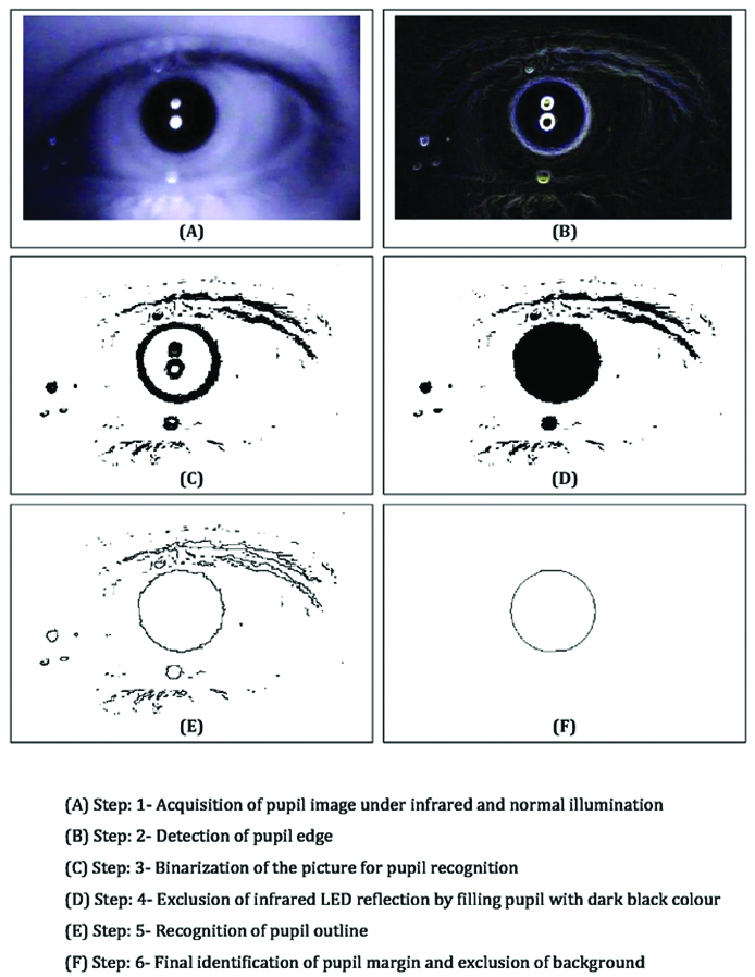
The video frame or image was selected for analysis which contains pupil, iris and its background. Automatic pupil size determination was not possible unless the threshold of the image was adjusted as per the requirement. Because it is the prerequisite for detection of the pupil edges even when the image is with low threshold and pupil’s contour is quite dark. However, usually, the pupil can be demarcated from iris due to image acquired under of infrared light illumination which increases the contrast of the picture background (Step-1).
The pupil margins can be detected using the command in the software “finding edges” which establishes the clear outline of the pupil. The perimeter of pupil identification is tedious when the subject’s eye is not positioned during acquirement process of pupillary light reflex. The simple edge detector was used to highlight sharp changes in the intensity of the active photograph or selected region (Step-2).
The LED reflection within the user interface can be eliminated by applying the portion of interest that binarizes the image. This is an essential step to evacuate the effect of different colours in the pupil. However, it does not interfere in the absolute pupil size (Step-3).
The binarized pupil circle can be filled with black colour in the selected region to avoid LED reflections which are also known as Purkinje phenomenon (Step-4).
The fine edges of the pupil can be detected by applying a command “find outlines” which gives definite pupil edges. However, still, the outline of the pupil was not smooth due to fluctuations of the threshold in pupil contour (Step-5).
The pupil outlines can be precisely analysed by eliminating the background noise, normalises the pupil shape and procures absolute pupil outline. This can be accessed by applying the command “Analyse particles with proper circularity (0-1) (Step-6).
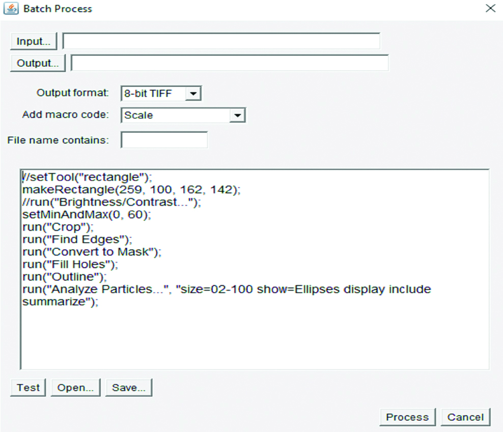
Automatic Batch Processing
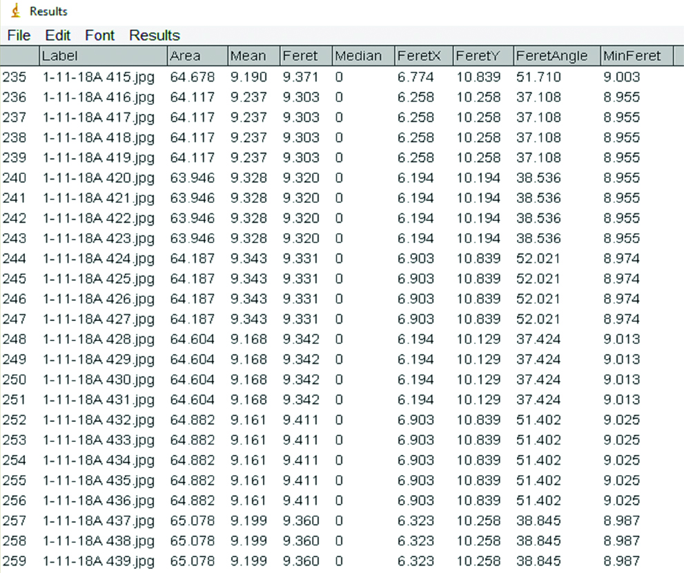
Image J software can also be exercised to measure a batch of arrayed pupil images using a macro plugin program as per the requirement. All the steps followed earlier were programmed as a javascript macro plugin to analyse the area and diameter of entire cluster of pupil pictures from pupillary light reflex [Table/Fig-2]. However, automatic processing is required to provide a predetermined area of an image of an entire video to eliminate the inter-subject variability of pupil dimensions. The results can be shown after in a separate window of the program either in the area or in diameter of the pupil [Table/Fig-3].
The macro plugin program for automatic batch analysis.
Result sheet of Automatic Image analysis with different parameters.
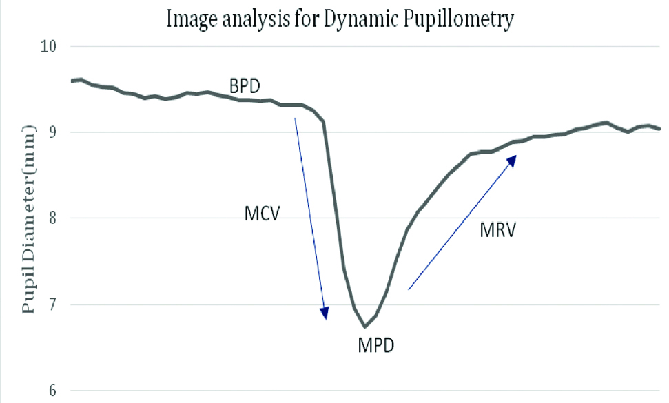
Once the video frames were analysed by the automatic batch processing through macro code plug-in the program, the results can be transferred to an excel sheet and graph can be plotted. These results can assist to identify the vital parameters like Minimum Pupil Diameter (MPD), Baseline Pupil Diameter (BPD). The time-based parameters like Maximum Constriction Velocity (MCV), Maximum Redilation Velocity (MRV) can be calculated based on the frame rate and change in diameter of each frame [Table/Fig-4].
Pattern of pupillary light reflex.
Discussion
The aim of the current study was to establish a simple technique to evaluate pupillary light reflex under infrared and white light illumination. Pupillary examination varies with the skill of the examiner and can result in differences in the management of the patient of pupils. Determination of pupil diameter is the vital diagnostic tool, especially in brain injuries. Litvan I et al., had used an objective scale that reported interrater variability of pupil measurement [11]. But this method is useful only in the presence of visible light and is inefficient to measure in low illumination. The usage of infrared light improves the quality of pupil acquisition in total darkness and does not initiate pupillary light reflex. Quantitative infrared based pupillometry can produce reliable, accurate pupil measurements which are higher than the information obtained through the clinical examination by the physician [12]. Du R et al., and Fountas KN et al., introduced the automated pupillometer (Neur Optics, Inc., Irvine, CA) which is a hand-held, portable device with liquid crystal display screen and has an option of video recording for pupil. The accuracy and precision of the device are much higher compared to the manual examination [13,14]. Johnson EL et al., also used a camera that automatically captures the pupil at 7 cm distance, with a temporal resolution of 120 Hz, to evaluate task-evoked pupil dilation among children who were subjected to short term memory tasks [15]. However, these automated pupillometers are highly expensive. Our study demonstrates to eliminate artefacts of image processing which was the major obstacle for quantitative pupil response through videography. Our study is similar to Lin X et al., study in which they developed a technique to analyse accurate elliptical contour of the pupil from the images taken by a special infrared camera. They also illustrated the protocol that deals with the Purkinje phenomenon [16]. However, they measured the pupil in pixels instead of physical units. Iacoviello D et al., also established image processing of pupil after video graphed using binarization of each image and analysed in Matlab toolbox [17]. However, he focused to obtain pupil noise using a neural network in the resting state of pupil recorded from participants. Leal G et al., had developed a pupillometer to detect the pupil variations in time and the output signal expresses features variation in time. They also converted the pupil responses into a frequency which is similar to frequency domain analysis of heart rate variability that describes autonomic balance. The authors of this study stated physical units are not required to evaluate ANS activity [18]. But most of the clinicians may not understand the technical aspects of the pupil response if it is provided in pixels or frequencies. The physical units like minimum and maximum pupil diameter and velocities of constriction and dilation provide the ideal and straight forward results which will be helpful for clinicians and researchers. The current study described the methodology of pupil identification for physical units using the easy and robust technique. This method is inexpensive, simple and also eliminates the examiner and ambience bias.
Limitation
The limitation of this method is that the data is processed offline due to which real time results are not possible. We are planning to make a second version of the same in which the results are displayed in real time and the stored data can be analysed offline in detail for more information.
Conclusion
This newly developed technique with a modified webcam and by using freely available software makes the dynamic pupillometry a very useful cost-effective tool wherein the pupil response can be quantified. Different static and dynamic parameters of the pupil are useful in various clinical departments like ophthalmology, neuropsychology and psychiatry, for evaluation of pupillary response. It also helps to assess the autonomic balance, optic neuritis, ocular hypertension and neuronal activity in head injuries. We believe that by making pupillometry more accessible to diagnose for the above clinical situations.
[1]. James B, The pupilsIn: Ophthalmology: Investigation and Examination Techniques 2007 :123-27.10.1016/B978-0-7506-7586-4.50015-52013695 [Google Scholar] [CrossRef] [PubMed]
[2]. Mathôt S, Pupillometry: Psychology, physiology, and functionJournal of Cognition 2018 1(1):1-23.10.5334/joc.18 [Google Scholar] [CrossRef]
[3]. Nowak W, Zarowska A, Szul-Pietrzak E, Misiuk-Hojło M, System and measurement method for binocular pupillometry to study pupil size variabilityBiomed Eng Online 2014 (1):13-69.10.1186/1475-925X-13-6924899167 [Google Scholar] [CrossRef] [PubMed]
[4]. Binda P, Pereverzeva M, Murray SO, Pupil size reflects the focus of feature-based attentionJ Neurophysiol 2014 112(12):3046-52.10.1152/jn.00502.201425231615 [Google Scholar] [CrossRef] [PubMed]
[5]. Binda P, Gamlin PD, Renewed attention on the pupil light reflexTrends Neurosci 2017 40(8):455-57.10.1016/j.tins.2017.06.00728693846 [Google Scholar] [CrossRef] [PubMed]
[6]. van der Wel P, van Steenbergen H, Pupil dilation as an index of effort in cognitive control tasks: A reviewPsychon Bull Rev 2018 25(6):2005-15.10.3758/s13423-018-1432-y29435963 [Google Scholar] [CrossRef] [PubMed]
[7]. Vinik AI, Maser RE, Mitchell BD, Freeman R, Diabetic autonomic neuropathyDiabetes Care 2003 26(5):1553-79.10.2337/diacare.26.5.155312716821 [Google Scholar] [CrossRef] [PubMed]
[8]. Hall C, Chilcott R, Eyeing up the future of the pupillary light reflex in neurodiagnosticsDiagnostics 2018 8(1):pii: E1910.3390/diagnostics801001929534018 [Google Scholar] [CrossRef] [PubMed]
[9]. Fazari GM, Testing the validity of pupillometer technology against traditional drug screening instrumentsFed Probation 2011 75(3):37-44. [Google Scholar]
[10]. Rasband W, Ferreira T, ImageJ user guideUser Guide Image J 2014 :138-39. [Google Scholar]
[11]. Litvan I, Saposnik G, Maurino J, Gonzalez L, Saizar R, Sica RE, Bartko JJ, Pupillary diameter assessment: need for a graded scaleNeurology 2000 54(2):530-31.10.1212/WNL.54.2.53010668738 [Google Scholar] [CrossRef] [PubMed]
[12]. Boev AN, Fountas KN, Karampelas I, Boev C, Machinis TG, Feltes C, Quantitative pupillometry: Normative data in healthy pediatric volunteersJ Neurosurg: Pediatrics 2005 103(6):496-500.10.3171/ped.2005.103.6.049616383247 [Google Scholar] [CrossRef] [PubMed]
[13]. Du R, Meeker M, Bacchetti P, Larson MD, Holland MC, Manley GT, Evaluation of the portable infrared pupillometerNeurosurgery 2005 57(1):198-202.10.1227/01.NEU.0000163425.79170.CB15987563 [Google Scholar] [CrossRef] [PubMed]
[14]. Fountas KN, Kapsalaki EZ, Machinis TG, Boev AN, Robinson JS, Troup EC, Clinical implications of quantitative infrared pupillometry in neurosurgical patientsNeurocritical Care 2006 5(1):55-60.10.1385/NCC:5:1:55 [Google Scholar] [CrossRef]
[15]. Johnson EL, Miller Singley AT, Peckham AD, Johnson SL, Bunge SA, Task-evoked pupillometry provides a window into the development of short-term memory capacityFrontiers in Psychology 2014 5:21810.3389/fpsyg.2014.0021824659980 [Google Scholar] [CrossRef] [PubMed]
[16]. Lin X, Craig J, Klette G, Klette R. Accurately Measuring the Size of the Pupil of the Eye. IVCNZ 2003, At Massey University, Volume: 221-26. https://www.researchgate.Net/publication/37986916_Accurately_Measuring_the_Size_of_the_Pupil_of_the_Eye [Google Scholar]
[17]. Iacoviello D, Analysis of pupil fluctuations after a light stimulus by image processing and neural networkComput Math with Appl 2007 53(8):1260-70.10.1016/j.camwa.2006.05.022 [Google Scholar] [CrossRef]
[18]. Leal G, Neves C, Vieira PM, Pupillometry: development of equipment for studies of autonomic nervous systemIn Doctoral Conference on Computing, Electrical and Industrial Systems 2012 Feb 27 Berlin, HeidelbergSpringer:553-562.10.1007/978-3-642-28255-3_61 [Google Scholar] [CrossRef]