Robot Assisted Nephron-sparing Surgery for Renal Leiomyoma
Vinit Kumar Singh1, Altaf Khan2, Muhammed AP Manzoor3, M Mujeeburahiman4
1 Postgraduate, Department of Urology, Yenepoya Medical College, Mangaluru, Karnataka, India.
2 Associate Professor, Department of Urology, Yenepoya Medical College, Mangaluru, Karnataka, India.
3 Research Associate, Yenepoya Research Centre, Yenepoya University, Mangaluru, Karnataka, India.
4 Professor, Department of Urology, Yenepoya Medical College, Mangaluru, Karnataka, India.
NAME, ADDRESS, E-MAIL ID OF THE CORRESPONDING AUTHOR: Dr. Altaf Khan, Associate Professor, Department of Urology, Yenepoya Medical College, Mangaluru-575018, Karnataka, India.
E-mail: draltafkhan@gmail.com; manzoorapkdy@gmail.com
Leiomyomas are benign mesenchymal tumour originating from muscle cell. Renal leiomyomas an unusual primary renal tumour of the kidneys originating from smooth muscle cell. Here, authors describe a rare case of renal leiomyoma in a 46-year-old female patient who presented with recurrent haematuria and flank pain. Authors report their first experience in robot-assisted nephron-sparing surgery for renal leiomyoma. Easier resection and suturing was a potential advantage of this approach. The patient improved after tumour removal and the prognosis is excellent without recurrence after six months postsurgically. Authors also discuss the summary of clinical features of renal leiomyomas.
Muscle cell, Primary renal tumour, Resection
Case Report
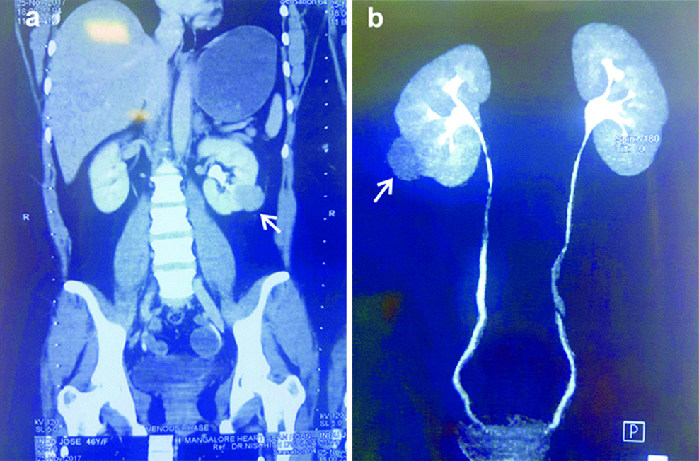
A 46-year-old woman presented to the Department of Urology with complaints of pain in left flank. The patient underwent a baseline assessment including a detailed medical history, physical examination, urinalysis, complete blood count, ultrasonography of abdomen and CT urogram. All routine blood investigation results were normal. Ultrasonography detected a mass in the left kidney 4 cm×3 cm occupying the posterolateral aspect of the lower pole of the left kidney. CT urogram demonstrated a more precise heterogeneously enhancing well circumscribed, predominantly exophytic mass lesion in posterolateral aspect of lower pole of the left kidney [Table/Fig-1a,b].
(a and b) Contrast-enhanced CT scan showing well-defined, exophytic masslesion in posterolateral aspect of lower pole of the left kidney (arrow).
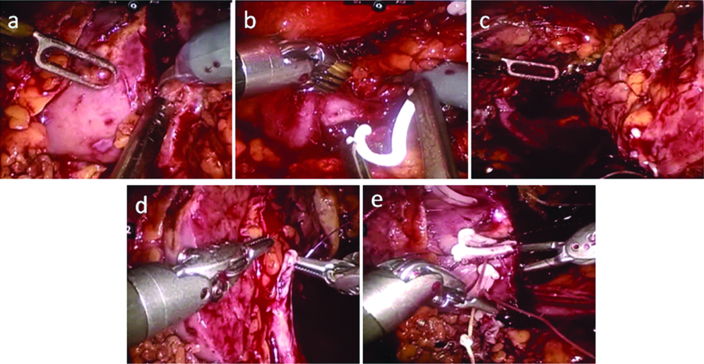
The patient underwent left nephron-sparing surgery with assistance of the da Vinci robotic system [Table/Fig-2a-e]. This robotic set enabled the authors to perform surgery through a few small incisions. The main features include the 3D high definition view of patient’s body. The total operation time was 90 minutes and the intraoperative blood loss was around 50 mL. The visual analogue score for pain was 2 (Visual analogue scale is a psychometric scale for response to severity of pain). Tramadol (50 mg) was required for one day. No intraoperative or postoperative complications were observed. Patient was discharged on third postoperative day.
Nephron-sparing surgery with assistance of the da Vinci robotic system: (a) Cutting the tumour with 5 mm of normal margin; (b) Clamping the segmental artery; (c) Tumour with normal parenchyma is seen on the right side; (d) Repair of the opened collecting system; (e) renorrhaphystitch.
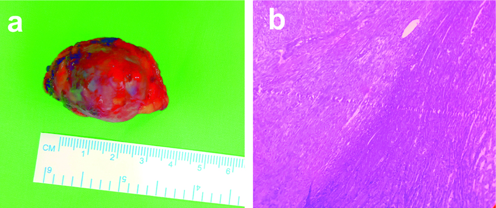
Gross examination of the specimen demonstrated a 4 cm×3 cm globular tissue mass [Table/Fig-3a]. Outer surface was capsulated, soft glistening with fibrofatty tissue attached to one area. Cut surfaces revealed a well-circumscribed pale white area measuring 2.7 cm×2.2 cm×1.5 cm, normal looking area measuring 0.5 cm seen. Microscopically, the tumour showed a circumscribed neoplasm composed of interlacing fascicles of fairly uniform spindle-shaped cell [Table/Fig-3b]. No mitotic figures were seen in the sections examined. Necrosis and haemorrhage were absent and many mast cells were seen. Morphological features and histological features were consistent with renal leiomyoma. The patient was followed-up for six months with abdominal CT which showed no signs of local recurrence and metastases.
(a) Renal leiomyoma gross appearance showing well circumscribed tumour; (b) Microscopic image reveals well circumscribed neoplasm composed of interlacing fascicles of fairly uniform spindle shaped cell.
Discussion
Here, authors report a case of renal leiomyoma in a female patient who underwent robot-assisted nephron-sparing surgery. Authors also discuss the summary of clinical and histological features of renal leiomyomas. Leiomyomas are benign mesenchymal tumour originating from muscle cell. Renal leiomyomas are one such example and are classified into capsular, subcapsular or subpelvic type, which is originating from smooth muscle cells in the renal capsule, tunica media of the renal cortical vasculature or muscularis of the renal pelvis respectively [1] Advances in technology and modern analytical tools such as ultrasonography and CT scan have allowed early recognition of renal tumours. In the present case, contrast-enhanced CT showed a heterogeneously enhancing well circumscribed, predominantly exophytic mass lesion in posterolateral aspect of lower pole of the left kidney. The patient underwent left robot assisted nephron-sparing surgery and tumour was completely excised and histopathological examination confirmed the diagnosis of renal leiomyoma. Macroscopically renal leiomyoma is a well-defined, solid encapsulated mass and elastic consistency [2].
Robotic assisted surgeries are having advantage due to its significant improvements in visibility and manipulation compared to conventional laparoscopic surgery [3]. In addition, robot assisted surgeries are safe, feasible and have less perioperative complications compared to laparoscopic and open surgeries [4]. Moreover, robotic assisted surgeries have minimum blood loss, conversion rates are lower and less hospital stay. The major limitations of laparoscopic surgery are restricted range of motion of the laparoscopic instruments, and poor ergonomic positioning of the surgeon [5] Radical nephrectomy and kidney-sparing surgery are recommended treatments for large and smaller leiomyoma respectively [6]. Nephron-sparing surgery has high success rate and less operative morbidity and mortality rate [7]. For small size tumours, nephron-sparing surgery can be performed using robot assisted approach. This has potential advantages such as easier excision, suturing and less warm ischaemia time. In addition, lesser blood loss and shorter hospital stay. The indicates robotic assisted nephron-sparing surgeries performed in India and abroad. Robotic assisted nephron-sparing surgeries for renal leiomyoma are very less from India [Table/Fig-4] [8-14].
Robotic assisted nephron sparing surgery performed in India and other countries [8-14].
| Case series/Reference | Country | Outcome |
|---|
| Robot-assisted partial nephrectomy in renal masses over 4 cm [8] | France | Median warm ischemia time: 23 min (10-59).Median operation TIME: 180 min (110-425)Estimated blood loss: were:100 cc (0-2500)Median hospital length of stay: 5 days |
| Robotic-assisted transperitoneal nephron-sparing surgery (n=37) [9] | Italy | Mean warm ischemia time: 21.5 minMean total procedure time: 149.2 min.Mean estimated blood loss: 187.1 mL.Mean hospital stay was 4.4 days. |
| Robotic nephron sparing surgery in cases of high renal nephrometry scores (n=18) [10] | India | Mean operative time: 73.61±52.66 minMean estimated blood loss: 363.89±296.45 mL.Mean hospital length of stay: 5.39±1.91 days |
| Robotic-assisted laparoscopic partial nephrectomy (n=13) [11] | USA | The mean operative time was 215 minutes (range 130 to 262), and the mean blood loss was 170 mL (range 50 to 300). The mean warm ischemia was 22 minutes (range 15 to 29),The length of hospital stay averaged 4.3 days (range 2 to 7). |
| Robot-assisted Partial Nephrectomy (n=100) [12] | United States | Median tumour size was 2.8 cm (range, 1.0-8). Mean warm ischemia time was 25.5 minutes (range, 0-53). |
| Robotic assisted Partial Nephrectomy (n=48) [13] | United States | The mean operative time was 152 minutes. The mean warm ischemia times were 14 min. The estimated blood loss was 122 mL |
| Robot-Assisted Partial Nephrectomy [14] | USA | Mean tumour size was 2.87 cm.Mean total operative time was 210 min.Mean ischemic time was 23.9 min.Mean estimated blood loss was 131.5 mL. |
Conclusion
Robot-assisted nephron-sparing surgery is a safe and potential approach for the management of small renal masses. However, care should be taken to rule out the associated recurrence and a long-term follow-up is recommended.
Declaration of patient consent: Written informed consent was obtained from the patient for publication of this case report and any accompanying images.
[1]. Larbcharoensub N, Limprasert V, Pangpunyakulchai D, Sanpaphant S, Wiratkapun C, Kijvikai K, Renal Leiomyoma: A Case Report and Review of the LiteratureUrol Case Rep 2017 13:3-5.10.1016/j.eucr.2017.03.01828417074 [Google Scholar] [CrossRef] [PubMed]
[2]. Mitra B, Debnath S, Pal M, Paul B, Saha TN, Maiti A, Leiomyoma of kidney: an Indian experience with literature reviewInt J Surg Case Rep 2012 3:569e7310.1016/j.ijscr.2012.07.01522940698 [Google Scholar] [CrossRef] [PubMed]
[3]. Köckerling F, Robotic vs. standard laparoscopic technique–what is betterFront Physiol 2014 1:1510.3389/fsurg.2014.0001525593939 [Google Scholar] [CrossRef] [PubMed]
[4]. Azhar RA, Gill IS, Aron M, Robotic nephron-sparing surgery for renal tumours: Current statusIJU 2014 30(3):27510.4103/0970-1591.13566725097313 [Google Scholar] [CrossRef] [PubMed]
[5]. Marano A, Choi YY, Hyung WJ, Kim YM, Kim J, Noh SH, Robotic versus laparoscopic versus open gastrectomy: a meta-analysisJ Gastric Cancer 2013 13:136-48.10.5230/jgc.2013.13.3.13624156033 [Google Scholar] [CrossRef] [PubMed]
[6]. Nakib G, Mahgoub N, Calcaterra V, Pelizzo G, Renal leiomyoma in pediatric age: a rare case report with review of the literatureJ PediatrSurg Case Rep 2017 27:43-46.10.1016/j.epsc.2017.09.012 [Google Scholar] [CrossRef]
[7]. Uzzo RG, Novick AC, Nephron sparing surgery for renal tumours: indications, techniques and outcomesJ Urol 2001 166(1):6-18.10.1016/S0022-5347(05)66066-1 [Google Scholar] [CrossRef]
[8]. Masson-Lecomte A, Yates DR, Bensalah K, Vaessen C, de la Taille A, Roumiguie M, Robot-assisted laparoscopic nephron sparing surgery for tumours over 4 cm: operative results and preliminary oncologic outcomes from a multicentre French studyEJSO 2013 39(7):799-803.10.1016/j.ejso.2013.03.00723566551 [Google Scholar] [CrossRef] [PubMed]
[9]. Ceccarelli G, Codacci-Pisanelli M, Patriti A, Ceribelli C, Biancafarina A, Casciola L, Robotic-assisted transperitoneal nephron-sparing surgery for small renal masses with associated surgical procedures: surgical technique and preliminary experienceUpdates Surg 2013 65(3):183-90.10.1007/s13304-013-0209-023619828 [Google Scholar] [CrossRef] [PubMed]
[10]. Bora GS, Mavuduru RS, Sharma AP, Devana SK, Kakkar N, Lal A, Initial experience of robotic nephron sparing surgery in cases of high renal nephrometry scoresIJU 2017 33(3):23010.4103/iju.IJU_331_1628717275 [Google Scholar] [CrossRef] [PubMed]
[11]. Gettman MT, Blute ML, Chow GK, Neururer R, Bartsch G, Peschel R, Robotic-assisted laparoscopic partial nephrectomy: technique and initial clinical experience with DaVinci robotic systemUrology 2004 64(5):914-18.10.1016/j.urology.2004.06.04915533477 [Google Scholar] [CrossRef] [PubMed]
[12]. Scoll BJ, Uzzo RG, Chen DY, Boorjian SA, Kutikov A, Manley BJ, Robot-assisted partial nephrectomy: a large single-institutional experienceUrology 2010 75(6):1328-34.10.1016/j.urology.2009.10.04020080290 [Google Scholar] [CrossRef] [PubMed]
[13]. Pierorazio PM, Patel HD, Feng T, Yohannan J, Hyams ES, Allaf ME, Robotic-assisted versus traditional laparoscopic partial nephrectomy: comparison of outcomes and evaluation of learning curveUrology 201 78(4):813-19.10.1016/j.urology.2011.04.06521802120 [Google Scholar] [CrossRef] [PubMed]
[14]. Benway BM, Bhayani SB, Rogers CG, Porter JR, Buffi NM, Figenshau RS, Robot-assisted partial nephrectomy: an international experienceEuropean urology 2010 57(5):815-20.10.1016/j.eururo.2010.01.01120116163 [Google Scholar] [CrossRef] [PubMed]