Definitive impressions play a vital role in the process of fabrication of prosthesis. Quality of the final prosthesis to a large extent depends on the accuracy of impression. Conventional Impression (CI) making with elastic impression materials is still the widely used technique for replicating the intraoral anatomy and to transfer this information to the dental laboratory for fabrication of indirect dental restorations. Demand of fixed prosthesis is increasing and manufacturing of Fixed Partial Denture (FPD) with intraoral Digital Impression (DI) techniques is now becoming an important part of Prosthodontics [1-3]. Digital impressions are receiving an ever-increasing popularity and acceptance from the clinicians when compared to conventional impressions. Digital impressions present with a benefit of three-dimensional pre-visualisation of the preparation, cost-effective and reduced working time [2]. Other advantages include elimination of tray selection procedure; minimising the risk of distortion and material consumption during impression making, pouring, disinfecting, and shipping to the dental laboratory; and enhanced patient comfort and acceptance [3,4]. These impressions can be stored electronically and communicated as digital information, and later on retrieved without distortion [3,5]. Digital impressions eliminate casts, wax-ups, investing, casting, and firing of ceramic restorations [1,2,6]. The major challenge encountered by prosthesis fabricated using digital impression technique is compromised marginal fit that can lead to plaque retention causing secondary caries, periodontal, and pulpal inflammation and washout of the luting agent resulting in loss of axial retention and rotation stability. As these possible consequences has to be considered, accuracy of digital impressions and dental restorations manufactured in a fully digitised work flow and CAD/CAM systems has to be evaluated in both in-vitro and in-vivo conditions [1,2,5]. The accuracy of DI and CI were compared based on either internal fit or marginal fit or both internal and marginal fit. Other technical measures to compare accuracy are superimposing virtual images of impressions and dies and these techniques are evaluated based on patient acceptances, operator preferences and time effectiveness [2,3,5].
The primary objective of this review was to generate a comprehensive over view on the comparative superiority of digital impression techniques based on accuracy, patient acceptance, operators’preference and time effectiveness. However, none of the studies in literature individually provided comprehensive overview on all these parameters.
Materials and Methods
This rapid review was performed in accordance with the PICO(S) approach (Patient or Population, Intervention, Control or Comparison, Outcome, and Study types). The Population, Intervention, Comparison, Outcome, Study framework was used to form the following search strategy. P=Edentulous and partially edentulous patients, preclinical models; I=Digital impression technique; C=Conventional impression technique; O=Accuracy, patient preference, operator preference and time effectiveness and S=Clinical and preclinical studies and flowchart was generated following PRISMA guidelines [7].
Information Sources and Search Strategy
An electronic search of articles published in English literature between 1980 and 2017 was undertaken on 12 January 2018. Data bases searched were PubMed, Medline and Cochrane via Ovid, along with additional hand searches. MeSH terms used were: Dental Impression Technique, accuracy, CAD CAM or Computer-Aided Design/computer-aided manufacturing, digital impressions, optical impression, CAD/CAM, intraoral scanner, impression scanner, Cost-Benefit Analysis and economic analysis, patient preference, operator preference [Table/Fig-1].
| 1 | Dental impression technique/or impression.mp. |
| 2 | Digital dentistry |
| 3 | CAD CAM.mp. or Computer-Aided Design/ |
| 4 | Digital impressions |
| 5 | CAD CAM |
| 6 | Intraoral scanner |
| 7 | Optical impression |
| 8 | Impression scanner |
| 9 | Cost-Benefit Analysis/or economic analysis. |
| 10 | Operator preference |
| 11 | Patient preference |
| 12 | 1 and 2 |
| 13 | 1 and 3 |
| 14 | 1 and 5 |
| 15 | 1 and 6 |
| 16 | 3 and 9 |
| 17 | 4 and 9 |
| 18 | 5 and 9 |
| 19 | 6 and 9 |
| 20 | 7 and 9 |
| 21 | 8 and 9 |
Selection of articles was done using Covidence software. In the first phase, two reviewers independently performed the titles and abstracts screening. A third independent reviewer resolved the conflicts present in first phase. Full text screening was performed by the same two reviewers independently by employing the selection criteria and another independent reviewer resolved the conflicts in full text screening. The reviewers collected the required information from the chosen articles. Following this, cross-checking procedure was performed by another independent reviewer to assure the completeness and precision of the collected information.
Study Eligibility Criteria
Inclusion criteria
Clinical, preclinical studies and randomised controlled trials
Studies which compared accuracy, patient preference and operator’s preference.
Articles published in English
Exclusion criteria
Articles published in languages other than English
Expert opinion and narrative reviews
Animal studies
Systematic reviews
Study Selection
Citations retrieved in the database searches were assessed in a two-stage review process. Both authors verified the eligibility of the potentially relevant articles and independently screened titles and abstracts to evaluate the articles for full-text reading. Any conflicts arising during the course of the procedure were resolved by a third reviewer.
Data Extraction
Data and information extracted from the included studies were: author, country, year of publication, funding for the study, study design, clinical or preclinical study, sample size, arch, jaw and type of prosthesis, brand of optical impression system and brand of conventional impression material, results, outcomes that is accuracy, patient preference, operator preference and time effectiveness and outcome measurement. Data collection was also done to assess the outcome. Outcome measurement included the mode of measurement and the outcome which comprised accuracy, patient outcome, operator outcome and cost.
Results
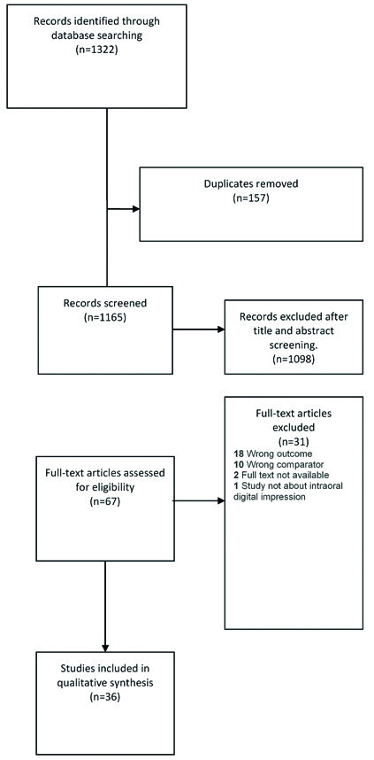
Initial electronic and manual searches yielded 1322 articles. After eliminating 157 duplicate references, 1165 studies were taken for title and abstract screening. After resolving the conflicts, 1098 articles were rejected. Remaining 67 articles were screened through full text among which 36 articles complied fully with inclusion criteria. A total of 31 articles were excluded during this stage. Reasons for exclusion were18 articles due to wrong outcome other than accuracy, patient preference, and operator preference, 10 because of wrong comparator where there was no comparison with conventional impression technique and two as full text was not available and one study was not about intraoral digital impression [Table/Fig-2].
Description of Included Studies
a) Accuracy
This systematic review included 36 articles. Among these 24 articles compared the accuracy of conventional and digital impressions. Three comparative studies were based on internal fit of restorations [8-10] and six studies compared accuracy based on marginal fit [11-16]. Six research studies assessed accuracy based on both marginal fit and internal fit [17-22]. Remaining nine studies compared the accuracy by precision of impressions and dies [3,5,23-29]. However, further assessment of accuracy readings revealed variation in accuracy measurement. Five of them compared accuracy using stereomicroscopy [8,11,12,15,16], four of them compared accuracy using replica method and stereomicroscopy [14,19,21,22], three studies used microscopic examination and computer software [9,17,20]. Dauti R et al., assessed marginal fit using optical microscope followed by scanning electron microscope [13]. Seelbach P et al., evaluated marginal fit and internal fit with 3D-coordinate measuring system using a traveling microscope with electronic data acquisition and digital micrometer heads [20]. Eleven studies compared accuracy based on superimposition of virtual images [3,5,10,18,23-29]. Among these twenty-four studies,16 of them reported that digital impressions are superior to conventional impressions. Both the techniques exhibited clinically acceptable level of accuracy [Table/Fig-3].
Summary of descriptive characteristics of articles with accuracy outcome.
| Study/Specimen ID | Parameter compared | Scan device and software | Accuracy measurement | Main Outcome |
|---|
| Lee SJ et al., [3] | Precision | i-Tero; Cadent iTero TM, | Superimposed with the STL data set and scaned impression data | Both showed acceptable values |
| Papaspyridakos P et al., [5] | Precision | TRIOS; 3 shape, | Superimposed with the STL data set and scaned impression data | Both showed acceptable values |
| Berrendero S et al., [8] | Internal fit | Ultrafast Optical Sectioning technology | Stereomicroscopeat magnification factor ×40, with a built-in charge-couple device camera and Image analysis software | DI better than CI |
| Cetik S et al., [9] | Internal fit | 3 shape trios | Microscopic examination and computer software | DI better than CI |
| Cho SH et al., [10] | Internal fit | Flex 3A; Otto Vision Technology | Superimposed with the STL data set and scaned impression data | Both showed acceptable values |
| Abdel-Azim T et al., [11] | Marginal fit | I trio | Stereomicroscope | DI better than CI |
| Abdel-Azim T et al., [12] | Marginal fit | Lava COS (3M ESPE), and iTero (Cadent) | Stereomicroscope | DI better than CI |
| Dauti R et al., [13] | Marginal fit | Lava cos | Optical microscope and a scanning electron microscope | Both showed acceptable values |
| Ashtiani RE et al., [14] | Marginal fit | Trios 3 IOS (3Shape), Ceramill map 400; Aman Gir back | Replica technique, stereomicroscopy | Both showed acceptable values |
| Pradı G et al., [15] | Marginal gap | Lava Chairside Oral Scanner, 3M ESPE | Stereomicroscopy | DI better than CI |
| Zarauz C et al., [16] | Marginal fit | TRIOS Pod system (3Shape, Copenhagen, Denmark | Stereomicroscope | DI better than CI |
| Almeida e Silva JS et al., [17] | Marginal fitInternal fit | Lava COS (3M ESPE) | Microscopic examination and computer software | DI better than CI |
| Malaguti G et al., [18] | Marginal gapInternal gap | Extra oral scanner-dental wing serie 7, intraoral scanner-MHT scanner 3d progress | Superimposed with the STL data set and scaned impression data | DI better than CI |
| Rödiger M et al., [19] | Marginal fitInternal fit | TRIOS system | Replica technique, camera integrated with light microscope | Both showed acceptable values |
| Seelbach P et al., [20] | Marginal fitInternal fit | Lava C.O.S., CEREC AC, and iTero | 3D-coordinate measuring system, with a traveling microscope with electronic data acquisition and also with digital micrometer heads | Both showed acceptable values |
| Su TS et al., [21] | Marginal fitInternal fit | Trios cart | Replica method, and steriomicroscopy | DI better than CI |
| Yun MJ et al., [22] | Marginal fitInternal fit | iTero | Replica method and measuring microscope | DI better than CI |
| Amin S et al., [23] | Precision | CEREC Omnicam, True Definition scanner 4.1., 3M ESPE | Superimposition | DI better than CI |
| Basaki K et al., [24] | Precision | 3 shapetrios | Superimposition | Both showed acceptable values |
| Ender A et al., [25] | Precision | CEREC Bluecam (CER; Sirona Dental Systems); CEREC Omnicam (OC; Sirona Dental Systems); Cadent iTero(ITE; Cadten Ltd); Lava COS (LAV; 3M ESPE); True Definition Scanner (T-Def; 3M ESPE); 3 Shape Trios (TRI; 3 Shape); and 3 Shape Trios Color (TRC; 3 Shape) | Superimposing using special diagnostic software | CI better than DI |
| Ender A et al., [26] | Precision | CEREC Bluecam (CER; Sirona Dental Systems); CEREC Omnicam (OC; Sirona Dental Systems) Cadent iTero (ITE; Cadten Ltd) Lava COS (LAV; 3M ESPE) | Superimposed with the STL data set and scaned impression data | DI better than CI |
| Ender A et al., [27] | Precision | True Definition Scanner (T-Def; 3 M ESPE); Lava COS Cadent iTero 3Shape Trios, 3 Shape Trios Color, CEREC Bluecam, and CEREC Omnicam (OC; Sirona Dental Systems). | Superimposed with the STL data set and scaned impression data | CI better than DI |
| Kamimura E et al., [28] | Precision | Lava COS, 3 M ESPE, Germany | Superimposed with the STL data set and scaned impression data | DI better than CI |
| Marghalani A et al., [29] | Precision | IOS (CEREC Omnicam; Dentsply Sirona), True Definition; 3 M ESPE | Superimposed with the STL data set and scaned impression data | DI better than CI |
b) Patient Preferences
Five articles compared digital and conventional impressions based on patient preference [30-34]. All of the massessed patient preference based on Visual Analogue Scale (VAS) and questionnaires. Among these five articles, four studies reported that digital impression was the preferred choice [30,31,33,34]. Benic GI et al., stated that both the impression techniques were equally acceptable [Table/Fig-4] [30].
Summary of descriptive characteristics of articles on the basis of Pateint’s preferences.
| Study/Specimen ID | Parameter compared | Scan device and software | Patient outcome measurement tool | Main outcome |
|---|
| Benic GI et al., [30] | Comfort | Lava (Lava COS; 3M ESPE), iTero (Align Technology Inc), and Cerec (CerecBluecam; Sirona Dental Systems GmbH) | visual analog scales (VAS | Both had similar results |
| Burhardt L et al., [31] | Gag reflex, queasiness, difficulty to breathe, discomfort, time perception, anxiety, experience of the powder used for digital impressions. | CEREC Omnicam, Lava C.O.S. | Perception Questionaire | DI preferred |
| Joda T et al., [32] | Patient’s subjective convenience level, anxiety, bad oral taste, nausea sensation, pain sensation during impression taking, patients’ satisfaction concerning convenience, speed | Trios 3 IOS (3 Shape) | VAS | DI preferred |
| Wismeije D et al., [33] | Preparation, Time involved Analogue, Taste, Bite registration, Impression tray/scan head, Gag reflex, Overall preference | Cadent Itero | Questionnaire | DI preferred |
| Yuzbasioglu E et al., [34] | Patient perception, treatment comfort, effectivness and clinical outcome | CEREC Omnicam, Sirona | Questionnaire | DI preferred |
c) Operator’s Preferences
Eight articles compared digital impression and conventional impression based on operator preference [30,32,35-40]. The variables used were time, operator preference and operator difficulty. Joda T et al., and Marti AM et al., compared conventional and digital impression based on time [37,40], Gjelvold B et al., assessed CI and DI based on time and operator difficulty [36]. Lee SJ et al., evaluated DI and CI based on operator preference and difficulty [38] and the remaining four articles compared all the three variables [30,32,35,39]. Seven articles reported that digital impressions were preferred by the operator [Table/Fig-5].
Summary of descriptive characteristics of articles based on operator’s preferences.
| Study/Specimen ID | Parameter compared | Scan device and software | Operator outcome measurement tool | Main outcome |
|---|
| Benic GI et al., [30] | Impression difficulty,TimeOperator comfort | Lava (Lava COS; 3M ESPE), iTero (Align Technology Inc), and Cerec (CerecBluecam; Sirona Dental Systems GmbH) | Impression difficutly-VAS.Time-From mixing to removal of impression from mouth,Operator comfort-VAS | CI preferred timeFor clinician perception of difficulty, the conventional impression and the digital impression with iTero revealed more favorable outcomes than the digital impression with Lava |
| Joda T et al., [32] | Time efficencyOperator difficultyOperator preference | Trios pod | VAS | DI preferred |
| Lee SJ et al., [35] | Time efficencyOperator difficultyOperator preference | I Tero cadent | VAS | DI preferred |
| Gjelvold B et al., [36] | DifficultyTime | Trios 3 IOS (3Shape) | VAS | DI preferred |
| Joda T et al., [37] | Time | Trios 3 IOS (3Shape) | VAS | DI preferred |
| Lee SJ et al., [38] | Difficulty levelOperator preference | I Tero cadent | VAS | DI preferred |
| Zitzmann NU et al., [39] | Level of difficultyEfficency of intraoral Scanning,Time | trios | VAS | DI preferred |
| Marti AM et al., [40] | Time | LAVA COS | VAS | Both CI and DI has similar results |
Discussion
The definitive impression plays a critical role in success and longevity of restorations. Various impression techniques have been followed to generate a definitive cast that ensures accurate clinical fit of prosthesis [3,11,12,17,23,24,32,40]. The present review critically evaluated the literature comparing the optical impression with conventional impression based on accuracy, patient preference and operator preference. The results show that the digital and conventional impressions vary in accuracy, patient preference and operator preference.
Accuracy
Accuracy of digital and conventional impressions can be measured based on precision of impressions [5,23-28,31,41,42] as well as precision of prosthesis which is fabricated from the impressions [8,33]. Accuracy can also be assessed by evaluating the die which has been made from the impression [9,10,30,32,40,43]. The precision of prosthesis can be measured by measuring the marginal fit [1-4,11,23,42], internal fit or both together. Various studies which compared the accuracy of digital and conventional impressions used stereomicroscopy, super imposition and replica technique for measurement [13,14,25-27,44,45].
The factors that have been documented to influence the marginal fit of a dental restoration are the preparation dimension, location of the finish line whether subgingival or supragingival, restorative material, fabrication method, impression material and technique. The marginal fit is the oretically represented by a gap-free transitionor a linear contact line between the restoration margin and the preparation [43]. Thus, digital impressions show superior results when compared with the conventional impressions.
According to literature, ideal marginal fit desirable for clinical success of full crowns has been widely discussed as 120 μm or less [46-52] whereas in CAD/CAM or copy-milling systems, the marginal opening has been reported to range between 60 μm and 300 μm [52-55]. Wider marginal gaps would provide a niche for oral pathogens and saliva, leading to complications like periodontal inflammations, secondary caries and cement dissolution which in turn reduce the lifespan of the restoration. The pressure generated during the cementation and the cement space factors that affect the fit of the prosthesis [18,28,29,38-40].
Among the 25 articles which compared digital impressions with conventional impressions 16 articles reported that digital impressions are superior to conventional impressions though all of them depicted the clinically satisfactory values for both. Conventional impressions reported slightly inferior values for internal fit; this could be due to the work flow of this technique. It requires the model production, making of restoration on it and then the actual processing. All these steps are eliminated in digital impression. As every step in the work flow contribute to error elimination of master model, coping fabrication reduced the errors. Conventional impressions are also associated with errors from contraction or expansion of impression and model materials. The less accurate values for marginal fit of digital impressions in comparison to internal fit mean values could be due to the variations in the methodologies and measurement techniques. Another reason could be due to the titanium powder accumulation at the finish line region since these areas bear more susceptibility for that.
Patient Outcomes
Evaluation of included studies which measured patient centered outcomes revealed that, patient preference is more for digital impression technique. Assessments were done based on VAS and customised questionnaires. Criteria for the assessment was patient comfort, gag reflex, queasiness, difficulty to breathe, discomfort, time perception, anxiety, taste irritation, experience of the powdering procedure used for digital impressions [16,34,35,37,44,46]. The VAS criteria addressed and measured patient outcome successfully, but there is lack of uniformity among studies. Validation of questionnaires was also not done.
Preference for digital impression is another indication that today’s patients have more concern on comfort. This is because the digital impressions are associated with reduced invasiveness [46]. Unacceptable conventional impressions require remaking of entire impression. However, with digital impression technique missing and unacceptable areas can be corrected by a segmental rescanning. This reduces working time and increases patient comfort.
Operator Outcomes
Among all the included articles, which reported on operator outcome preferred digital impression method [3,14,37,45]. The reasons may be the reduced procedure time, reduction in procedure steps and ease of use [16,39,40,41]. Operator centered outcome were measured for digital and conventional impressions by assessing working time, operator perception and procedure difficulty. Assessment was done using VAS and questionnaires reported that digital impressions require reduced time [3,37,40,45]. The work flow of digital impression technique took reduced time. Even though when a remaking was necessary, the time required for rescan of the digital impression was significantly less. Rescans were done mainly due to the difficulty in scanning the interproximal contact areas and in areas of reflection from light source.
Operator perception was measured on the level of difficulty in performing the procedure and was significantly lower for the digital impression technique. Manipulation and learning curve for the intra-oral scanner were less and they seem to be more user-friendly. Operators perceived that missing and unacceptable area can be corrected more easily with digital impressions while the conventional technique demanded remaking of entire impression [35].
Limitation
The results of the present study have to be interpreted with caution because of its limitations. The quality of the included studies varied greatly. Our electronic database search strategy identified 31 studies which were excluded after detailed review for various reasons. The most common reasons for exclusion were that the studies used wrong interventions other than digital impression technique. Because these studies did not contribute to the review, we may be missing important results. Due to heterogeneity of the included studies, meta-analysis could not be performed. Most of the studies had limited follow-up period and did not mention any specific outcome calibration criteria.
Conclusion
Multiple clinical and preclinical comparative studies had been reported on various aspects of DI and CI techniques. It is of utmost importance for the clinician to have a comprehensive overview on both the techniques to choose the best technique based on evidence. Compared to conventional impressions, digital impression possessed superior accuracy without any statistically significant difference. Patient and operator preference assessment favored digital impression technique with a higher level of acceptance and satisfaction.