Isoniazid Induced Toxic Epidermal Necrolysis in a HIV Positive Patient during Treatment for Extra Pulmonary Tuberculosis: A Case Report
Bhanukumar Muthaiah1, George Mathew Panachiyil2, Tirin Babu3, Allu Harshavardhini4
1 Professor, Department of General Medicine, JSS Medical College and Hospital, JSS Academy of Higher Education and Research, Mysuru, Karnataka, India.
2 Pharm D Intern, Department of Pharmacy Practice, JSS College of Pharmacy, JSS Academy of Higher Education and Research, Mysuru, Karnataka, India.
3 Pharm D Intern, Department of Pharmacy Practice, JSS College of Pharmacy, JSS Academy of Higher Education and Research, Mysuru, Karnataka, India.
4 Postgraduate, Department of General Medicine, JSS Medical College and Hospital, JSS Academy of Higher Education and Research, Mysuru, Karnataka, India.
NAME, ADDRESS, E-MAIL ID OF THE CORRESPONDING AUTHOR: Miss. Tirin Babu, Pharm D Intern, Department of Pharmacy Practice, JSS College of Pharmacy, JSS Academy of Higher Education and Research, Mysuru-570015, Karnataka, India.
E-mail: erumalat@yahoo.com
Toxic Epidermal Necrolysis (TEN) is characterised by extensive erythema, necrosis, and bullous detachment of the epidermis and mucous membranes. Drug induced TEN is commonly seen with sulphonamides, penicillins, other antibiotics, non-steroidal anti-inflammatory drugs, anticonvulsants etc., Human Immunodeficiency Virus (HIV) infected patients are more susceptible to these drug reactions than the general population.
Isoniazid (INH) remains one of the first line agent for Antitubercular Therapy (ATT) in HIV patients co-infected with tuberculosis. An alteration in the treatment of tuberculosis is recommended if the patient develops TEN on any first line antitubercular drugs. Here, we report a case of INH induced TEN in a HIV patient during treatment for extra pulmonary tuberculosis. She was non-compliant to the Antiretroviral Therapy (ART) drugs and was on ATT drugs (INH and ethambutol) for the past four months. She was admitted to the hospital with the complaints of extensive skin lesions of one-week duration which was diagnosed to be TEN clinically and also based on the biopsy report. The frequency of INH causing TEN was not defined by the clinical trial investigators, however this was suspected to be due to INH. The severe cutaneous adverse reaction subsided after the withdrawal of INH followed by the supportive therapy for skin lesions.
Acquired immune deficiency syndrome, Antitubercular treatment, Cutaneous reactions, Isonicotinylhydrazide
Case Report
A 60-year-old female, known case of HIV diagnosed eight months back, was admitted to the hospital with the complaints of skin lesions all over the body since one week. In addition to skin lesions, she had a history of fever and chills of one day.
The patient initially was on treatment in an out-patient clinic with two drug regimen of INH and ethambutol, and then was referred to the hospital for further management. The skin lesions started as painful erythematous macules, progressed into blisters followed by diffuse exfoliation of the skin involving bilateral lower extremities, back, abdomen, forearms, face and oral cavity within a few days before admission to the hospital [Table/Fig-1]. Her medication history revealed non-adherence to the ART from the past six months after the development of jaundice induced by Tenofovir, Lamivudine and Efavirenz (TLE) regimen. She was also on treatment for extra pulmonary tuberculosis with INH and ethambutol for the past four months and these drugs were dechallenged in the out-patient clinic in view of the skin lesions. There was no history of drug induced hypersensitivity reactions.
Erythematous maculopapular lesions, before treatment.
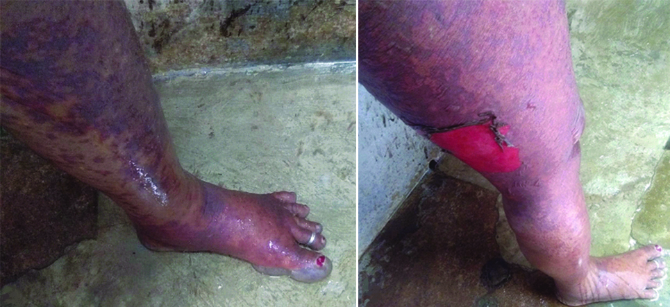
General examination revealed: temperature of 37°C, heart rate of 98 beats per minute, respiratory rate of 18 breaths per minute, blood pressure of 130/90 mmHg and SpO2 was 97% at room air. Systemic examination was normal. There were erythematous maculopapular lesions with bullous detachment of the skin involving about 90% of the body surface area including the back, buttocks, face, neck and limbs. Additionally, there was also involvement of oral and conjuctival mucosa. Nicholsky’s sign was positive over thigh skin.
Skin biopsy was taken from left thigh, which revealed necrotic epidermis overlying viable dermis. The superficial dermis showed dilated blood vessels and lymphocytic infiltrates, features consistent with TEN with regenerative epidermis. The CD4 count was 118 cells per cubic millimeter of blood. Routine blood investigations showed total leucocyte count of 2960 cells/cumm, platelet count of 0.266 lac/cumm and the peripheral blood smear impression was leukopenia with thrombocytopenia.
She had abnormal renal parameters which resolved to baseline after correction of hypovolaemia, because of fluid loss through extensive skin lesions. Radiograph of chest and electrocardiogram done were normal. She was managed conservatively with maintenance IV fluids, topical steroid (hydrocortisone cream 2.5%, two times a day), antibiotic (meropenem 1 gm IV, three times a day) along with appropriate wound care (framycetin 1% cream, two times a day). Fresh Frozen Plasma (FFP) and platelets were also transfused to the patient.
A final diagnosis of drug induced TEN was made based on history of ATT drugs exposure with a typical clinical manifestation of erythema, blistering and detachment of skin involving more than 30% of Body Surface Area (BSA) and mucosal involvement, supported by histological findings. In our case, INH was suspected to be the cause of TEN and the patient was restarted on ATT drugs with ethambutol and pyrazinamide without any recurrence of her cutaneous problems. The final diagnosis made was INH induced TEN in a HIV positive patient with extra pulmonary tuberculosis.
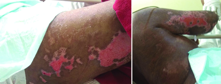
During the stay in the hospital, the woman improved steadily with above therapy [Table/Fig-2]. Skin lesions were resolved gradually and disappeared completely after four weeks.
Resolving lesions, during treatment.
Discussion
A number of factors favour INH as the probable cause of TEN: First, INH has more number of published studies of causing TEN in literature when compared to ethambutol [1-4]. Second, ethambutol is known to cause TEN, but in our case, patient was restarted ATT with ethambutol and pyrazinamide without any recurrence of her cutaneous problems. So, the TEN can be attributed to INH alone. INH induced TEN has been reported rarely in the literature [1,5-10]. Rechallenge was not attempted for INH because of fear of exacerbated skin reaction, as the reaction is potentially fatal.
Drugs are the most common causative agents for TEN. Other aetiological factors identified in TEN are infections, immunizations and immunosuppressive conditions etc., [5]. The following mechanisms make HIV infected patients more susceptible to TEN due to drugs. First, TEN is related to genetic differences in patient’s metabolite reactions to drugs. Second, multiple prior cutaneous drug reactions, glutathione deficiency and also increased number of abnormal polyclonally activates B cells in HIV patients. So, there is a need to rule out an underlying HIV infection in all patients having drug induced TEN [11].
The most effective management of INH induced TEN is the withdrawal of the culprit drug, monitoring of fluids and electrolytes balance, proper wound care, nutritional support and supportive systemic therapy [5]. In our case, patient developed TEN after four months of treatment with INH. Swami A et al., reported a case of INH induced TEN in a retro-positive patient in which patient started developing oral and mucosal ulcerations of TEN from 6th day of ATT and by day 12 of the ATT [5]. Another difference from our case is that it was not known that the patient was HIV positive at the time of hospital admission. To the best of authors knowledge this is the second reported case in India of TEN secondary to INH in a patient having HIV infection.
Assessment of ADR
The causality assessment was done using both World Health Organisation (WHO) causality assessment algorithm and Naranjo scale. Based on a temporal relationship and the reaction unlikely attributed to other concurrent diseases or medications, it was probable according to the WHO scale and same with Naranjo scale with a score of seven. The reaction was not predictable as frequency is not defined for the same. It is definitely preventable because there is a known treatment for the adverse drug reaction.
Conclusion
TEN, being the most severe cutaneous adverse drug reaction with high mortality risk, it is essential that the disease is properly managed. In our case, withdrawal of the culprit drug followed by the supportive therapy associated with the treatment of skin lesions were the most vital elements that led to healing and recovery of the patient from the fatal condition.
[1]. Patel T, Barvaliya M, Sharma D, Tripathi C, A systematic review of the drug-induced Stevens-Johnson syndrome and toxic epidermal necrolysis in Indian populationIndian Journal of Dermatology, Venereology, and Leprology 2013 79(3):38910.4103/0378-6323.11074923619444 [Google Scholar] [CrossRef] [PubMed]
[2]. Avakian R, Flowers F, Araujo O, Ramos-Caro F, Toxic epidermal necrolysis: A reviewJournal of the American Academy of Dermatology 1991 25(1):69-79.10.1016/0190-9622(91)70176-3 [Google Scholar] [CrossRef]
[3]. Chaudhary SC, Atam V, Gupta A, Arya R, Soni D, Ethambutol- induced toxic epidermal necrolysisThe Journal of the Association of Physicians of India 2011 59:391-92. [Google Scholar]
[4]. Criton S, Devi K, Sridevi P, Toxic epidermal necrolysis- A retrospective studyInternational Journal of Dermatology 1997 36(12):923-25.10.1046/j.1365-4362.1997.00100.x9466199 [Google Scholar] [CrossRef] [PubMed]
[5]. Swami A, Gupta B, Bhattacharjee P, Response to modified antitubercular drug regime and antiretroviral therapy in a case of HIV infection with disseminated tuberculosis with isoniazid induced toxic epidermal necrolysisCase Reports in Infectious Diseases 2012 2012:62670910.1155/2012/62670923259095 [Google Scholar] [CrossRef] [PubMed]
[6]. Heng M, Drug-induced toxic epidermal necrolysisBritish Journal of Dermatology 1985 113(5):597-600.10.1111/j.1365-2133.1985.tb02384.x4063192 [Google Scholar] [CrossRef] [PubMed]
[7]. Nanda A, Drug-induced toxic epidermal necrolysis in developing countriesArchives of Dermatology 1990 126(1):125-25.10.1001/archderm.1990.01670250131031 [Google Scholar] [CrossRef]
[8]. Lehloenya R, Dheda K, Cutaneous adverse drug reactions to anti-tuberculosis drugs: state of the art and into the futureExpert Review of Anti-infective Therapy 2012 10(4):475-86.10.1586/eri.12.1322512756 [Google Scholar] [CrossRef] [PubMed]
[9]. Yamashita H, Ueda Y, Takahashi Y, Mimori A, A Case of Stevens-Johnson Syndrome (SJS) Progressive Toxic Epidermal Necrolysis (TEN) onset during hyposensitization therapy for pulmonary tuberculosis complicated with dermatomyositisKansenshogaku Zasshi 2012 86(4):419-24.10.11150/kansenshogakuzasshi.86.41922991850 [Google Scholar] [CrossRef] [PubMed]
[10]. Scheid P, Kanny Ph, Trechot G, Rosner V, Menard O, Vignaud J, Anthoine D, Isoniazid-induced bullous skin reactionAllergy 1999 54(3):296-97.10.1034/j.1398-9995.1999.00049.x10321574 [Google Scholar] [CrossRef] [PubMed]
[11]. Dixit R, Chhabra N, Sharma S, George J, Sharma A, Toxic epidermal necrolysis caused by fluconazole in a patient with human immunodeficiency virus infectionJournal of Pharmacology and Pharmacotherapeutics 2012 3(3):27610.4103/0976-500X.9944523129968 [Google Scholar] [CrossRef] [PubMed]