The mesiodistal tooth sizes of maxillary and mandibular arches must have an ideal dimensional relationship to ensure proper interdigitation, overbite and overjet. Black was one of the first to investigate mesiodistal tooth dimensions [1]. Ballard M et al., studied discrepancies in mesiodistal tooth dimensions between right and left sides in models of 500 orthodontic patients. He concluded that discrepancy and asymmetry in mesiodistal tooth dimensions were present in 90% of the cases assessed [2].
An increase or decrease in the mesiodistal tooth dimensions may be a cause of malocclusion [3]. This discrepancy in size may exist within gender groups or Angle’s malocclusion groups. Thus, mesiodistal tooth dimensions of individual teeth may be associated with a specific malocclusion in one gender, more than the other. Identifying the exact location of this discrepancy may be beneficial in defining the treatment plan [4].
Malkoc S et al., studied dimensional variations of teeth among the different classes of malocclusion separately in males and females [4]. Males exhibited larger dimensions for maxillary canines, first premolars, second molars and mandibular canines in the Class II malocclusion group whereas females showed reduced dimensions for all maxillary teeth and mandibular central incisors, canines and first premolars in the Class III group. This difference in dimensional variation among males and females points to the existence of gender dimorphism of individual teeth. The authors have also stated that all mesiodistal widths were found to be statistically different according to gender dimorphism. However, the nature of dimorphism was not specified in their study.
There is a lack of knowledge about the interaction between gender, Angle’s classification and individual tooth size. Additional information is necessary to understand this relationship. This study intended to address this lacuna.
The aim of this study was to evaluate gender dimorphism in mesiodistal crown dimensions of individual teeth in Angle’s Class I, Class II division 1 and Class III malocclusion groups and compare the dimensions among the three Classes. It was also decided to compute Bolton’s ratios separately for males and females in the three classes studied.
Materials and Methods
This cross-sectional study was conducted on 308 study models obtained from departmental pretreatment orthodontic records at Government Dental College Kozhikode, Kerala, India, for a period of six months (from February-July 2017), based on the following inclusion criteria:
Good quality study models exhibiting a full complement of maxillary and mandibular permanent teeth excluding third molars of patients below 20 years.
Presence of Angle’s Class I molar relation on both sides with ANB angle of 0-4 were included in class I group; Angle’s Class II molar relation on both sides with an ANB angle greater than 4 degrees with proclination of upper anteriors and overjet >5 mm in the Class II group and Angle’s Class III molar relation on both sides with an ANB angle less than 0 degree in the Class III group [12].
Models of patients of Dravidian ethnicity.
Exclusion Criteria
Models exhibiting grossly carious, malformed or broken teeth, gross restorations, build-ups, crowns, onlays or class II restorations that could affect the mesiodistal tooth dimensions were excluded. Models with presence of severe occlusal or interproximal wear (as seen in older individuals) and severe crowding liable to interfere with computation of mesiodistal measurements were also not included.
Clearance was obtained from the institutional ethical committee and sample size was calculated using the formula 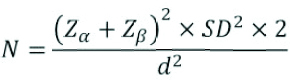 where Zα = 1.96 and Zβ = 0.84. SD was the standard deviation of the study group.
 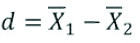 where
where  was the expected mean in Caucasians and
  was the expected mean in the study population. Accordingly the minimum sample size for the study was calculated to be 45 in each of the groups. The sample consisted of study models of 308 patients allocated to three malocclusion groups. This included 104 study models belonging to Class I; (53 males, 51 females), 104 models of Class II division 1 (54 males, 50 females), and 100 models with Class III malocclusion (50 males, 50 females).
Calibrations were made directly on unsoaped study models using a digital vernier caliper (Mitutoyo, Tokyo, Japan) with a 0.01 mm accuracy. Each tooth’s width was measured from the mesial to distal contact points at its greatest interproximal distance. The caliper beaks were placed at the buccal/facial surface and held perpendicular to the long axis of the tooth. The beaks were then closed until gentle contact was made with the contact points of the tooth. The measurements included the mesiodistal width of all 14 maxillary and mandibular teeth from the right second permanent molar to the left. The measurements were made as carefully as possible to avoid any damage to the casts. All the third molars were excluded. One investigator carried out all the measurements under natural light.
Each sample was measured twice by the operator and the average value was recorded. If there was a discrepancy >0.4 mm between the recordings, the measurements were repeated. To prevent any effects of fatigue, only 8-10 models were measured each day. All the measurements done from second molar to second molar were then noted on the data sheets.
In addition, the anterior and overall Bolton’s ratios were calculated for each Class of malocclusion in both males and females [13,14]. The standard Bolton’s ratios of 91.3±1.91% for overall ratio and 77.2±1.65% for anterior ratio were used as guidance values for comparison.
Statistical Analysis
The data was analysed using SPSS software. Mean and Standard Deviation (SD) for mesiodistal tooth dimensions and anterior and total Bolton’s ratios in all three groups for both sexes were calculated. Variation in tooth size between the two genders was analysed for the groups using Independent sample t-test. The Bolton’s ratios obtained were compared with the Bolton’s proposed norms for total and anterior Bolton ratios. ANOVA test was conducted to study variation in tooth size between Angle’s three groups of malocclusion. Post-Hoc Bonferroni test was used to compare mesiodistal tooth dimensions and Bolton’s ratios of the three malocclusion groups with each other for males and females.
One week later, 30 study models (15 males, 15 females) were randomly selected and re-examined by the same examiner. The intra-observer reliability evaluated using Intraclass Correlation Coefficient (ICC) ranged between 0.99 to 1.0 for different measures.
Results
Gender Dimorphism
[Table/Fig-1] shows mesiodistal dimensions of all individual teeth from central incisor to second molars for all Angle’s Classes of malocclusion in males and females respectively. In Angle’s Class I group, both upper and lower canines showed mesiodistal dimensions to be significantly larger in males than females (p<0.05, p<0.001). Maxillary central incisors (p<0.05) and mandibular first molars (p<0.05) were also larger in males. No significant variation was seen in the rest of the dentition. Angle’s Class II division 1 did not reveal gender dimorphism in any of the teeth, except for upper and lower canines (p<0.05, p<0.01) and upper second molars (p<0.05). In Angle’s Class III, statistically significant gender dimorphism was seen in almost all teeth except second premolars and upper central incisors with males exhibiting larger dimensions. The anterior Bolton’s ratio for Class III malocclusion group was also found to be significantly larger in males (p<0.001).
Comparison of mean values of mesiodistal dimensions of individual teeth, anterior and overall ratios between genders for Angle’s Class I, II and III malocclusion in males and females.
| Teeth | Gender | Angle’s Class I | Angle’s Class II | Angle’s Class III |
|---|
| Mean | SD | Sig. | Mean | SD | Sig. | Mean | SD | Sig. |
|---|
| Upper central incisor | Male | 9.20 | 0.38 | 0.043* | 8.86 | 0.46 | 0.378 | 8.52 | 0.37 | 0.748 |
| Female | 9.04 | 0.43 | 8.78 | 0.49 | 8.49 | 0.40 |
| Upper lateral incisor | Male | 7.55 | 0.47 | 0.715 | 7.14 | 0.57 | 0.974 | 7.37 | 0.49 | 0.000*** |
| Female | 7.51 | 0.48 | 7.14 | 0.46 | 7.01 | 0.46 |
| Upper Canine | Male | 8.23 | 0.44 | 0.038* | 8.09 | 0.40 | 0.016* | 8.08 | 0.47 | 0.000*** |
| Female | 8.05 | 0.42 | 7.89 | 0.43 | 7.65 | 0.40 |
| Upper First premolar | Male | 7.48 | 0.33 | 0.404 | 7.28 | 0.41 | 0.384 | 7.44 | 0.38 | 0.001*** |
| Female | 7.43 | 0.27 | 7.21 | 0.40 | 7.16 | 0.40 |
| Upper Second premolar | Male | 7.05 | 0.33 | 0.703 | 6.89 | 0.42 | 0.418 | 6.89 | 0.37 | 0.063 |
| Female | 7.07 | 0.34 | 6.82 | 0.37 | 6.74 | 0.42 |
| Upper First molar | Male | 10.46 | 0.55 | 0.433 | 10.26 | 0.47 | 0.493 | 10.26 | 0.49 | 0.010** |
| Female | 10.39 | 0.43 | 10.19 | 0.54 | 10.03 | 0.37 |
| Upper Second molar | Male | 9.75 | 0.50 | 0.431 | 9.73 | 0.42 | 0.044* | 9.64 | 0.49 | 0.025* |
| Female | 9.82 | 0.34 | 9.54 | 0.51 | 9.44 | 0.37 |
| Lower central incisor | Male | 5.69 | 0.31 | 0.575 | 5.44 | 0.36 | 0.173 | 5.59 | 0.37 | 0.024* |
| Female | 5.73 | 0.34 | 5.55 | 0.43 | 5.44 | 0.30 |
| Lower lateral incisor | Male | 6.32 | 0.37 | 0.397 | 6.05 | 0.40 | 0.778 | 6.26 | 0.44 | 0.000*** |
| Female | 6.26 | 0.35 | 6.02 | 0.41 | 5.87 | 0.35 |
| Lower canine | Male | 7.33 | 0.37 | 0.000*** | 7.12 | 0.44 | 0.004** | 7.23 | 0.37 | 0.000*** |
| Female | 7.05 | 0.33 | 6.88 | 0.39 | 6.71 | 0.34 |
| Lower first premolar | Male | 7.52 | 0.38 | 0.602 | 7.28 | 0.45 | 0.704 | 7.37 | 0.49 | 0.000*** |
| Female | 7.48 | 0.33 | 7.25 | 0.35 | 7.01 | 0.37 |
| Lower second premolar | Male | 7.49 | 0.40 | 0.492 | 7.21 | 0.39 | 0.756 | 7.30 | 0.41 | 0.097 |
| Female | 7.43 | 0.36 | 7.19 | 0.41 | 7.16 | 0.41 |
| Lower first molar | Male | 11.56 | 0.57 | 0.002** | 11.06 | 0.46 | 0.076 | 11.18 | 0.54 | 0.023* |
| Female | 11.21 | 0.51 | 10.89 | 0.47 | 10.94 | 0.51 |
| Lower second molar | Male | 10.34 | 0.50 | 0.869 | 9.78 | 0.50 | 0.597 | 10.29 | 0.52 | 0.000*** |
| Female | 10.35 | 0.37 | 9.72 | 0.50 | 9.93 | 0.34 |
| Anterior ratio | Male | 77.47 | 2.32 | 0.171 | 77.25 | 2.14 | 0.571 | 79.66 | 2.92 | 0.001*** |
| Female | 77.43 | 2.72 | 77.51 | 2.59 | 77.88 | 2.28 |
| Overall ratio | Male | 91.88 | 1.84 | 0.100 | 91.03 | 1.89 | 0.697 | 92.45 | 2.13 | 0.302 |
| Female | 91.27 | 1.88 | 91.18 | 2.17 | 92.01 | 2.11 |
p-value <0.05* <0.01** <0.001***
*Independent sample t-test
Variations among the Angle’s Malocclusion Groups
One-Way Analysis of Variance (ANOVA) was used to identify overall differences in mean values of mesiodistal dimensions of each tooth in Angle’s Class I, II and III malocclusion, the results of which are given in [Table/Fig-2]. When differences between groups were found to be significant, the Bonferroni test for multiple comparisons was applied [Table/Fig-3].
ANOVA test for males and females between Angle’s Class I, Class II division 1 and Class III malocclusion groups.
| Teeth | Male | Female |
|---|
| F | Sig. | F | Sig. |
|---|
| Upper central incisor | 35.526 | 0.000*** | 18.789 | 0.000*** |
| Upper lateral incisor | 8.219 | 0.000*** | 15.507 | 0.000*** |
| Upper Canine | 1.759 | 0.176 | 11.593 | 0.000*** |
| Upper First premolar | 3.994 | 0.020* | 7.889 | 0.001*** |
| Upper Second premolar | 2.874 | 0.059 | 10.087 | 0.000*** |
| Upper First molar | 2.882 | 0.059 | 8.045 | 0.000*** |
| Upper Second molar | 0.778 | 0.461 | 10.874 | 0.000*** |
| Lower central incisor | 7.203 | 0.001*** | 8.442 | 0.000*** |
| Lower lateral incisor | 6.519 | 0.002** | 13.302 | 0.000*** |
| Lower canine | 3.478 | 0.033* | 10.928 | 0.000*** |
| Lower first premolar | 4.275 | 0.016* | 9.134 | 0.000*** |
| Lower second premolar | 6.362 | 0.002** | 7.361 | 0.001*** |
| Lower first molar | 12.898 | 0.000*** | 6.177 | 0.003** |
| Lower second molar | 19.850 | 0.000*** | 30.633 | 0.000*** |
| Anterior ratio | 14.871 | 0.000*** | 0.445 | 0.642 |
| Overall ratio | 6.943 | 0.001*** | 2.426 | 0.092 |
p-value <0.05* < 0.01** <0.001***
*ANOVA
Post-Hoc Bonferroni analysis for multiple comparisons to assess mesiodistal tooth size between Angle’s Class I, Class II division 1, and class III malocclusion groups among males and females.
| Dependent Variable | (I) Molar R | (J) Molar R | Male Sig. | Female Sig. |
|---|
| Upper central incisor | Class I | Class II | 0.000*** | 0.012* |
| Class III | 0.000*** | 0.000*** |
| Class II | Class III | 0.000*** | 0.005** |
| Upper lateral incisor | Class I | Class II | 0.000*** | 0.000*** |
| Class III | 0.244 | 0.000*** |
| Class II | Class III | 0.083 | 0.554 |
| Upper Canine | Class I | Class II | 0.335 | 0.190 |
| Class III | 0.307 | 0.000*** |
| Class II | Class III | 1.000 | 0.013* |
| Upper first premolar | Class I | Class II | 0.023* | 0.009** |
| Class III | 1.000 | 0.001*** |
| Class II | Class III | 0.126 | 1.000 |
| Upper second premolar | Class I | Class II | 0.106 | 0.004** |
| Class III | 0.135 | 0.000*** |
| Class II | Class III | 1.000 | 0.873 |
| Upper first molar | Class I | Class II | 0.117 | 0.092 |
| Class III | 0.120 | 0.000*** |
| Class II | Class III | 1.000 | 0.215 |
| Upper second molar | Class I | Class II | 1.000 | 0.004** |
| Class III | 0.745 | 0.000*** |
| Class II | Class III | 0.965 | 0.662 |
| Lower central incisor | Class I | Class II | 0.001*** | 0.035* |
| Class III | 0.451 | 0.000*** |
| Class II | Class III | 0.076 | 0.403 |
| Lower lateral incisor | Class I | Class II | 0.002** | 0.007** |
| Class III | 1.000 | 0.000*** |
| Class II | Class III | 0.025* | 0.144 |
| Lower canine | Class I | Class II | 0.028* | 0.063 |
| Class III | 0.703 | 0.000*** |
| Class II | Class III | 0.495 | 0.063 |
| Lower first premolar | Class I | Class II | 0.020* | 0.006** |
| Class III | 0.084 | 0.000*** |
| Class II | Class III | 1.000 | 1.000 |
| Lower second premolar | Class I | Class II | 0.002** | 0.007** |
| Class III | 0.060 | 0.002** |
| Class II | Class III | 0.843 | 1.000 |
| Lower first molar | Class I | Class II | 0.000*** | 0.004** |
| Class III | 0.001*** | 0.018* |
| Class II | Class III | 0.689 | 1.000 |
| Lower second molar | Class I | Class II | 0.000*** | 0.000*** |
| Class III | 1.000 | 0.000*** |
| Class II | Class III | 0.000*** | 0.046* |
| Anterior ratio | Class I | Class II | 1.000 | 1.000 |
| Class III | 0.000*** | 1.000 |
| Class II | Class Iii | 0.000*** | 1.000 |
| Overall ratio | Class I | Class II | 0.080 | 1.000 |
| Class III | 0.420 | 0.221 |
| Class II | Class III | 0.001*** | 0.142 |
p-value <0.05* < 0.01** <0.001***
A closer look at [Table/Fig-1,3] show that the mesiodistal dimensions of individual teeth in Angle’s Class I group were larger than those of Angle’s Class II and Class III and this increase in dimension was found to be statistically significant in majority of the cases for females (p<0.05). There was no significant difference in mesiodistal tooth dimensions between Class II and Class III in females except for the upper central incisor, upper canine and lower second molar.
When computing for the Bolton’s ratios for females, there were no significant differences for both anterior and overall ratios among the three Classes of malocclusion. Unlike the females, males showed significantly higher values (p<0.05) for Angle’s Class III malocclusion as compared to Class II for both anterior and overall ratios. The anterior ratio for Class III was significantly more than Class I also (p<0.05).
Discussion
Mesiodistal Tooth Dimensions-Gender Variation
The results of the present study indicate interesting findings. It was the Class III malocclusion group that exhibited gender dimorphism in almost all the teeth except upper central incisors and second bicuspids. Canines were the most variable teeth in the jaws as they exhibited larger dimensions among males in all the three classes studied. Arya BS et al., have also stated that canines can be considered as the best discriminant between the sexes [6]. In the present study, the least variable of all teeth examined were the second premolars, which did not show variability in any of the classes. However, this is in contrast to previous studies by Arya BS et al., and Bishara SE et al., who showed significant variation in second bicuspids [6,11].
A recent study on evaluation of sexual dimorphism in canines as a possible source for gender estimation in medicolegal cases has also revealed that male canines were larger than females. According to their results, the right maxillary canines showed highly consistent results for sexual dimorphism supporting its use in orthodontic analysis for gender estimation. This would be an easy, reproducible and objective method [15].
Majority of the previous studies, reporting sexual dimorphism in mesiodistal dimensions of individual teeth, have pooled data of all the three classes of malocclusions. An investigation by Malkoc S et al., has addressed the issue of variations in mesiodistal tooth dimensions by comparing Class I, II and III malocclusions separately for males and females [4]. They have reported all mesiodistal widths to be statistically different according to gender dimorphism. But where exactly the dimorphism exists and in which Class of malocclusion had not been assessed.
The present study evaluated the existence of gender dimorphism in each tooth separately in each Class of malocclusion. Our findings showed that sexual dimorphism was predominant only in Class III malocclusion group. No significant gender variation existed in majority of the teeth for Angle’s Class I and Class II division 1. The anterior Bolton’s ratio for Class III malocclusion group was also found to be significantly larger in males. This is similar to findings of Fattahi HR et al., [16].
Mesiodistal Dimensions and Classes of Malocclusion
Angle’s Class I malocclusion exhibited the largest tooth dimensions in both genders. An exclusive assessment of the female population revealed the mesiodistal tooth dimensions of almost all teeth to be larger with a statistical significance in Angle’s Class I malocclusion group as compared to Angle’s Class II division 1 and Class III malocclusions. This is similar to conclusions drawn by Lavelle CL et al., [5]. A closer look revealed that the mesiodistal tooth dimensions showed a particular trend among the three malocclusion groups in both genders. In females the tooth dimensions in Class I group were larger than Class II, and Class II group were larger than Class III (Class I>Class II>Class III). This was true for all teeth except lower first and second molars. Among males, a similar trend was observed only for the maxillary dentition. For the mandibular teeth, the mesiodistal tooth dimensions were larger for Class I, followed by Class III and the smallest dimensions were observed for Class II malocclusion group (Class I>Class III>Class II). Our results showed that Class III had the smallest mesiodistal tooth dimensions in the maxillary arch for both males and females.
Bolton’s Ratio as a Function of Gender
In female population, the Bolton’s ratios approximated the norms. In males Class III group showed higher values for both anterior and overall ratios. This could probably be because the sum of maxillary anterior teeth was found to be the least in Class III. Higher Bolton’s ratios in Class III have also been reported by Araujo E et al., [9].
Bolton’s Ratios and Malocclusion Groups:
The variation in Bolton’s ratio among the malocclusion groups in males shows that the mean anterior and overall ratio obtained in this study were closer to the Bolton’s norms for Class I and II. The anterior and overall ratios were however significantly larger for class III malocclusion group with a mean ratio of 79.67±2.92 and 92.45±2.13 respectively. In females, the variations in anterior and overall ratios among the three classes were not statistically significant. A closer look reveals that the anterior and overall ratios in Class I and Class II were more close to that of Bolton’s standards. Angle’s Class III showed larger values for Bolton’s anterior and overall ratios, the details of which are given in [Table/Fig-1]. Higher Bolton’s ratios in Class III malocclusion has been reported earlier by many other investigators also without differentiating between the genders [9,17-18].
An important finding of this study is that it was possible to differentiate the nature of gender dimorphism in tooth dimensions in Angle’s Class I, Class II division 1 and Class III malocclusion. Gender variation for tooth dimensions was found to be prevalent in almost all teeth in Class III malocclusion. Canines were the most variable teeth showing gender dimorphism in all three classes of malocclusion. The second premolars were the most homogenous, displaying no variations with gender in any of the classes.
This study aided in locating the most dimensionally variable tooth in different malocclusion groups in both genders. This may be beneficial in accurate diagnosis, treatment planning and finishing and detailing of cases.
Limitation
This study included only males and females of Dravidian race. Further studies need to be undertaken to assess the existence of this dimorphism in other races. Another limitation of the present study was that Angle’s Class II division 2 cases were not included.
Conclusion
The results of the present study showed that significant gender dimorphism in tooth dimensions was found to be prevalent in all teeth in Angle’s Class III malocclusion except for second premolars and upper central incisors. Males had larger mesiodistal tooth dimensions. Angle’s Class I showed increased tooth dimensions in males for canines, upper central incisors and lower first molars only. Angle’s Class II division 1 malocclusions did not exhibit gender variations in any of the teeth studied, except for the canines and upper second molars. Canines were found to be the most variable teeth, exhibiting gender dimorphism in all three classes. Second premolars were the most homogenous, displaying no variations with gender in any of the classes. The mesiodistal dimensions of almost all the teeth were larger with a statistical significance in Angle’s Class I malocclusion group. Class III had the smallest mesiodistal tooth dimensions in the maxillary arch for both males and females. Bolton’s overall and anterior ratios showed larger values for Class III patients.