Diarrhoea Due to Intestinal Acariasis
Ashok Kumar Kapoor1, Sunaina Agarwal2, Akanksha Singh3, Reetika Pandey4, Vineeta Khare5
1 Pathologist, Department of Pathology, RML Mehrotra Pathology Pvt. Ltd., Nirala Nagar, Lucknow, Uttar Pradesh, India.
2 Pathologist, Department of Pathology, RML Mehrotra Pathology Pvt. Ltd., Nirala Nagar, Lucknow, Uttar Pradesh, India.
3 Senior Technician, Department of Pathology, RML Mehrotra Pathology Pvt. Ltd., Nirala Nagar, Lucknow, Uttar Pradesh, India.
4 Senior Technician, Department of Pathology, RML Mehrotra Pathology Pvt. Ltd., Nirala Nagar, Lucknow, Uttar Pradesh, India.
5 Microbiologist, Department of Pathology, RML Mehrotra Pathology Pvt. Ltd., Nirala Nagar, Lucknow, Uttar Pradesh, India.
NAME, ADDRESS, E-MAIL ID OF THE CORRESPONDING AUTHOR: Dr. Ashok Kumar Kapoor, Pathologist, Department of Pathology, RML Mehrotra Pathology Pvt. Ltd., B 171, Nirala Nagar, Lucknow-226020, Uttar Pradesh, India.
E-mail: drashokkapoor2016@gmail.com
Allergens, Immediate hypersensitivity, Mite
Dear Editor,
Rarely, Intestinal Acariasis (IA) may develop after ingestion of contaminated food. The ecto parasite Dermatophagoides farinae may damage colonic mucosa with their mouth parts resulting in diarrhoea. Present case deals with a female adult patient who complained of passage of whitish strands of mucoid material in stool. Routine stool examination showed an arthropod belonging to acaroid family. Microscopic examination of stool showed adults, larva and eggs of mites. Adult female mite measured 450×180 μm approximately. The parasite was suspected to be Dermatophagoides farinae (American house dust mite). Adult mites might have damaged intestinal mucosa with its mouth parts and legs resulting in diarrhoea. In addition, eosinophilic gasroenteropathy might have also contributed to diarrhoea.
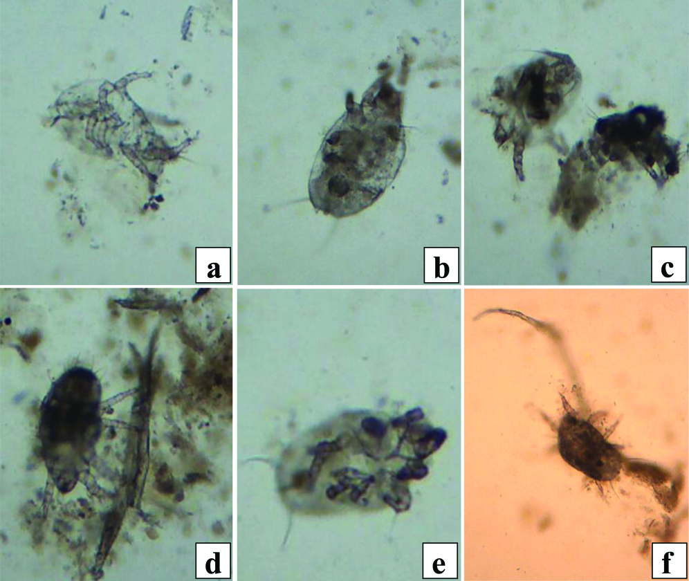
A 25-year-old female patient reported with chief complaint of passage of whitish elongated thread-like material in stool for last 15 days. Small amount of loose yellowish brown stool sample was received in 3 aliquotes; each aliquot measured 1.5 mL approximately. History of ingestion of contaminated food could not be elicited. Stool sample was partly-dried when received in the clinical pathology laboratory for examination. One of the aliquots was scrapped and wet smears were prepared in normal saline. Microscopic stool examination showed fair number of adults, nymphs (larva) and eggs of a tiny arachnid. Adult arthropod was oval turtle-like with hard dorsal cuticle. Adult parasite had 4 pairs of legs/or appendages. Both large and small adult mites were seen. Larger female adult mite measured 450×180 μm approximately. Diminutive mite measured 180×80 μm approximately [Table/Fig-1]. Images of adult mite were suspected to be Dermatophagoides farinae (Dr. YB Chui-personal communication). Videos and images were discussed with Dr. YB Chui for its identification. Dr. Chui is a known immunologist and he has published several articles on mites [1,2]. The patient was advised to take ivermectin 200 μg/Kg body weights once every 10 days×3 doses. The patient could not be followed further.
[a] Ventral aspect of adult mite×400; [b] Shows dorsal aspect of adult female mite; dorsal surface had hard cuticle (×400); [c] Bigger adult female and smaller parasites (×400). Adult mites had 4 pairs of legs; [d] Shows hatching of larva from egg (×400); [e] Side view of an adult mite (×400); [f] Dhows adult parasite with long gnathosoma (×400).
Intestinal acariasis is caused by ingestion of mite-contaminated foods. Mites are small arthropods which are analysed under subclass Acarina. Mites may cause a subclinical infestation [3]. House dust mites, e.g., Dermatophagoides farinae and D. pteronyssinus are commonest among atopic allergens. Rarely, type 1 hypersensitivity reactions leading to bronchial asthma and/or dermatitis may develop against faeces/or secretions of mite [4]. Seventeen allergens have been identified from the extract of D. farinae [5]. In addition, tropomyosin is also an important allergen of D. farinae. The most significant feature of current case was the detection of different developmental stages of mite on stool examination. The patient complained of passage of whitish thread-like structures in stool samples. Somewhat similar case was reported earlier by Zia B et al., [3]. Passage of acaroid mites has also been described in stool samples by Li CP et al., [1,6]. In a previous study, colonoscopy was performed in 16 patients with mites in stool [6]. Examination showed pale mucosa, petechial haemorrhages and exfoliated cells. In addition, live adult mites and eggs were also seen in peripheral zone of ulcer [6]. The mites living in intestinal tract may damage tissues with their gnathosoma, chelicera and feet [1]. They may damage mucosa, submucosa and produce necrosis, inflammation and form ulcer [6]. However, colonoscopy could not be done in present case. Presence of fair number of mites in stool (~10 parasites/smear) of current case suggested intestinal acariasis. Mites living in intestinal tract may also produce diarrhoea, pain in abdomen and burning around anus [2]. Current case also complained of diarrhoea. First case of intestinal acariasis (IA) was reported in the year 1934. It was caused by Tyrophagus longior [7]. Later, three cases of IA were described by Martinez MR; one of these was due to Suidasia mites [8]. Several patients with diarrhoea were caused by Carpoglyphus lactis, presumed to have been transmitted by contaminated sugar. Other studies have reported the cases of abdominal pain, diarrhoea, fatigue, and pyohemofecia due to Dermatophagoides farinae, D.pteronyssinus, Acarus siro, Tyrophagus putrescentiae, Glycyphagus ornatus, Glycyphagus domesticus and Tarsonemus granarius [1]. Recently, a case of IA caused by Tyrophagus putrescentiae has been reported by Khalifa RMA et al., [9]. Allergic IA syndrome (eosinophilic gastroenteropathy) has also been described [10,11]. Moreover, the results of skin prick test on 41 patients with mites in their stools were all positive. It suggested that acaroid mites in intestine may lead to allergy. Rarely, ingestion of foods contaminated with mites may produce anaphylaxis. Common symptoms may be generalised flushing, pruritus, urticaria or angioedema, wheezing and rhinorrhoea beginning after 10 to 20 minutes [2]. Recently, 36 cases of oral mite anaphylaxis have been reported [12]. Thirty four of 36 patients had ingested Okonomiyaki (pancake-mix). It revealed contamination with D. farinae, D. pteronyssinus and Tyrophagus putrescentiae [12]. However, present patient did not give any history of intake of contaminated food. Moreover, urinary acariasis, otoacariasis and vaginal acariasis may also develop [2]. Furthermore, incidence of IA was linked to occupation of patients, being higher in individuals working in traditional Chinese medicine (16%), rice store houses than those in other occupations. Some patients had habit of taking tea immersed in Chinese herbs [6]. The current patient was treated with ivermectin. In addition, family members were advised to get their sera examined for anti-mite IgG antibody by ELISA [13].
Mites belonging to acaroid subclass may produce necrosis of colonic mucosa. In addition, formation of anti-acaroid IgE antibody may sensitize basophils and/or mast cells. Subsequent reaction of allergens (either from cleaved body of acaroid mite or its secretions/or faeces) with IgE antibody may result in degranulation and release of pharmacologically active chemical mediators, resulting in diarrhoea.
[1]. Li CP, Cui YB, Wang J, Yang QG, Tian Y, Diarrhoea and acaroid mites: A clinical studyWorld J of Gastroenterol 2003 9(7):1621-24.10.3748/wjg.v9.i7.162112854179 [Google Scholar] [CrossRef] [PubMed]
[2]. Cui Y, When mites attack: Domestic mites are not just allergensParasit Vectors 2014 7:41110.1186/1756-3305-7-41125175486 [Google Scholar] [CrossRef] [PubMed]
[3]. Zia B, Aftab HB, Zahid MF, Farooqi J, Uddin F, Beg MA, Dust mites in a routine clinical stool sampleAsian Pacific J Trop Biomed 2014 4(suppl 2):5563-64.10.12980/APJTB.4.2014APJTB-2014-0105 [Google Scholar] [CrossRef]
[4]. Terr AI, The atopic diseasesIn ‘Medical Immunology’ 1997 10th edLange Medical books/McGraw Hill Medical:349-369.Parslow et al. [Google Scholar]
[5]. An S, Chen L, Long C, Liu X, Xu X, Lu X, Dermatophagoides farinae allergens diversity identification by proteomicsMol Cell Proteomics 2013 12(7):1818-28.10.1074/mcp.M112.02713623481662 [Google Scholar] [CrossRef] [PubMed]
[6]. Li CP, Cui YB, Wang J, Yang QG, Tian Y, Acaroid mite, intestinal and urinary acariasisWorld J Gastroenterol 2003 9(4):874-77.10.3748/wjg.v9.i4.87412679953 [Google Scholar] [CrossRef] [PubMed]
[7]. Hinman E, Kampmeier RH, Intestinal acariasis due to Tyrophagus longior gervaisAm J Trop Med Hyg 1934 14:355-62.10.4269/ajtmh.1934.s1-14.355 [Google Scholar] [CrossRef]
[8]. Martinez MR, Hoffman A, 3 cases of human intestinal mite infestation in the South of VeracruzRev Invest Salud Publica 1976 36:187-201. [Google Scholar]
[9]. Khalifa RMA, Abdellatif MZM, Ahmad AK, Yones DA, El-Mazary A-AM, First case of intestinal acariasis from EgyptSpringer Plus 2016 5:2810.1186/s40064-015-1584-426788440 [Google Scholar] [CrossRef] [PubMed]
[10]. Scala G, House-dust mite ingestion can induce allergic intestinal syndromeAllergy 1995 50:517-19.10.1111/j.1398-9995.1995.tb01189.x [Google Scholar] [CrossRef]
[11]. Li CP, Wang J, Intestinal acariasis in Anhui provinceWorld J Gatroenterol 2000 6(4):597-600. [Google Scholar]
[12]. Takahashi K, Taniguchi M, Fukutomi Y, Sekiya K, Watai K, Oral mite anaphylaxis caused by mite-contaminated Okonomiyaki/pancake-mix in Japan: 8 case reports and a review of 28 reported casesAllergol Int 2014 63:51-56.10.2332/allergolint.13-OA-057524569151 [Google Scholar] [CrossRef] [PubMed]
[13]. Zhang RB, Huang Y, Li CP, Cui YB, Diagnosis of intestinal acariasis with avidin-biotin system enzyme-linked immunosorbent assayWorld J Gastroenterol 2004 10(9):1369-71.10.3748/wjg.v10.i9.136915112362 [Google Scholar] [CrossRef] [PubMed]