A Study of the Growth of Human Foetal Lung in Relation with the Gestational Age
Rajeev Mukhia1, BP Powar2, Anjali Sabnis3
1 Lecturer, Department of Anatomy, Manipal College of Medical Sciences, Pokhara, Nepal.
2 Retired Professor and Head, Department of Anatomy, Manipal College of Medical Sciences, Pokhara, Nepal.
3 Professor and Head, Department of Anatomy, MGM Medical College, Navi Mumbai, India.
NAME, ADDRESS, E-MAIL ID OF THE CORRESPONDING AUTHOR: Dr. Rajeev Mukhia, Lecturer, Department of Anatomy, Manipal College of Medical Sciences, Kaski, District, Gandaki Zone, Fulbari, Pokhara, Nepal.
E-mail: Rajeev510@yahoo.com
Introduction
Lung is one of the organs of interest for researchers since a long time. Though detailed study about adult lung is there in the literature but study of lungs at different stages in the fetal period is far and few.
Aim
The present study attempted to find out the relationship between the foetal lungs in relation with its development in different gestational weeks.
Materials and Methods
The study was carried out on 40 spontaneously aborted human foetuses of known gestational age ranging from 10 weeks to 40 weeks. The anterior thoracic wall was dissected and the lungs were removed from the thoracic cage. The weight of the foetuses and foetal lung was measured in grams on digital weighing machine. The mean values of all parameters by gestational age were calculated. Data of the study were statistically analysed by using the Microsoft Excel 2007 program.
Results
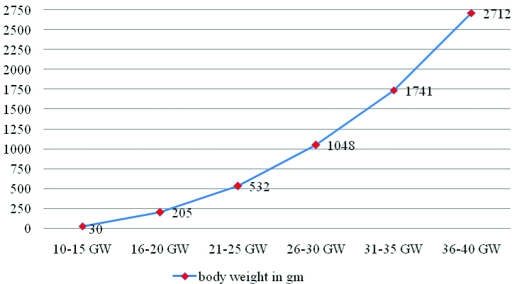
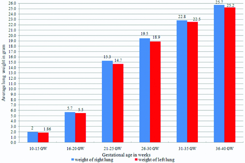
The average weight of right lung at 10th to 15th week and 36th to 40th week was 2.0 and 25.7 gm respectively. The average weight of left lung at 10th to 15th week and 36th to 40th week was 1.8 and 25.2 gm respectively. The body weight of foetuses showed gradual increase from 30 to 2712 gm at 10th week to 40th week of gestation.
Conclusion
Evaluating body and lung weights and measurements against known standards is an important part of perinatal pathology. It also provides new insights to the anatomist and clinician for understanding and developing knowledge in both normal and pathological conditions of pulmonary tissue.
Foetal weight, Gestational week, Lung weight
Introduction
The adult right lung usually weighs 625 g, and the left 565 g, but the range of weights is considerable, because it reflects the amount of blood or serous fluid contained within the lungs when weighed [1,2]. In proportion to body stature, the lungs are heavier in men than in women, and boys have bigger lungs than girls [3]. Lung volumes in children vary significantly according to race [4], and lung volume, air volume and pulmonary tissue volume all increase linearly with body length [5].
In the human embryo, development of lung starts as early as fourth week of embryonic life from a respiratory bud (lung bud) at the caudal end of the laryngotracheal diverticulum and continues into postnatal life up to early adulthood. The lung bud soon divides into two outpouchings, the primary bronchial buds. These primary bronchial buds grow laterally into the pericardio-peritoneal canals, the primordia of the pleural cavities. Secondary and tertiary bronchial buds soon develop. Together with the surrounding splanchnic mesenchyme, the bronchial buds differentiate into bronchi and their ramifications in the lungs. Lung growth and development is a complex process with a host of regulatory factors. The events of maturation of the lungs are divided into four histological stages; the pseudo-glandular, canalicular, terminal sac, and alveolar stages [6,7].
Foetal lung is one of the organs of interest for researchers since a long time. Since the growth of lung in human foetuses with respect to the gestational weeks in the previous studies were found to be incomplete, hence the present study was done to find out the growth of human foetal lung in relation with the gestational week. Though detailed studies of the adult lung is available the studies of the foetal lung have still a large scope for researchers.
Materials and Methods
The present cross-sectional study was carried out on 40 spontaneously aborted human foetuses, aged between 10th to 40th gestational weeks. The normal foetuses were obtained from the Department of Obstetrics and Gynaecology over the period of one and half years (from March 2017 to August 2018). After Ethical committee approval (MEMG/IRC/GA) and permission from the concerned authorities of the Institute, the foetuses were collected in 10% formalin for carrying the study. The foetuses included the spontaneous abortion and still-born foetuses. Cases with any anomaly or pathology were not included in the study. The age of foetuses was calculated from the obstetrical history, Crown Rump Length (CRL) and Crown Heel Length (CHL). The weight of the foetuses was measured in grams on digital weighing machine (SSC-10 Model ISO 9001: 2008).
The dissection was done according to the Cunningham’s manual of practical anatomy 15th edition. The order of dissection was as follows: The sternocleidomastoid and infrahyoid muscles were detached from their sternal and clavicular attachments. The clavicles were cut at their midlength. The costal cartilages and sternum was cut at the level of the xiphisternal joint. The ribs and contents of the intercostal spaces were cut at the midaxillary line. The anterior thoracic wall was then removed. Finger was placed into the pleural cavity between the lung and mediastinum. The lung was pulled laterally from the mediastinum and the structures at the root were cut throughout and the lungs were removed from the thoracic cavity on each side. While retracting the lung, scissors was used to transect the root of the lung halfway between the lung and the mediastinum. Care was taken not to cut into the mediastinum or the lung [8].
The weight of the lungs was measured in grams on digital weighing machine (BS 300+ Model). All the data were represented as mean then analysed with Microsoft Excel 2007 software and represented graphically.
Results
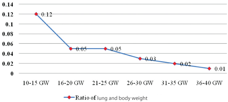
The results of mean body weight, weight of right and left lung and ratio of both lung and foetal weight is shown in [Table/Fig-1]. The average weight of right lung at 10th to 15th week and 36th to 40th week was 2.0 and 25.7 gm respectively. The average weight of left lung at 10th to 15th week and 36th to 40th week was 1.8 and 25.2 gm respectively. The average body weight of foetuses at 10th to 15th week and 36th to 40th week was 30 and 2712 gm respectively. The relative ratio between lung weight and body weight at 10th to 15th week and 36th to 40th week are 0.12 and 0.01 respectively. In the present study the body weight of foetuses showed gradual increase from 10th week to 40th week of gestation. Scatter graph showing weight of foetuses against gestational age is shown in [Table/Fig-2]. Scatter graph showing ratio of both lung and body weight against gestational age is shown in [Table/Fig-3]. Column chart showing mean weight of right and left lung in gram against gestational age is shown in [Table/Fig-4].
Showing mean body weight, weight of right and left lung in gram and ratio of lung and foetal weight.
| Gestational age (GA) in weeks | Number of foetuses | Mean weight in gram | Ratio of lung and body weight |
|---|
| Bodyweight | Weight of right lung | Weight of left lung | Weight of both lung |
|---|
| 10-15 | 7 | 30 | 2 | 1.86 | 3.86 | 0.12 |
| 16-20 | 8 | 205 | 5.7 | 5.5 | 11.2 | 0.05 |
| 21-25 | 7 | 532 | 15.3 | 14.7 | 30 | 0.05 |
| 26-30 | 8 | 1048 | 19.5 | 18.9 | 38.4 | 0.03 |
| 31-35 | 4 | 1741 | 22.8 | 22.5 | 45.3 | 0.02 |
| 36-40 | 6 | 2712 | 25.7 | 25.2 | 50.9 | 0.01 |
Scatter graph showing weight of foetuses in gram against gestational age.
Scatter graph showing ratio of lung and body weight against gestational age.
Column chart showing mean weight of right and left lung in gram against gestational age.
Discussion
Increase in foetal weight as well as organ weight is a good indicator of foetal growth in general. Rate of weight gain during early gestational age differs considerably with late gestational age. In the present study the weight of foetuses showed gradual increase from 10th week to 40th week of gestation. These findings were compared with the findings of others researchers and was in well agreement with that of Moore LK and Persaud TV, while it is less comparable to the findings obtained from the study of Hamilton WJ and Boyd JD and Bocian-Sobkowaka J et al., [9-11]. In the present study, the mean lung weight between 10th to 40th week of gestation was found slightly lower than the studies done by Shepard TH et al., Burdi AR et al., and Golbus MS et al., [12-14].
Tanimura T et al., reported that the lung weight to body weight ratio was found to be increasing and then decreasing and the ratio reached the maximum at 12-14th weeks of gestation [15], which is almost in conformity with the finding of the present study except for some findings in various weeks of gestation. Another study which was done by Mitropoulos G et al., was that all organ weights/body weights was found almost constant after 30 weeks gestation [16], which is found to be in conformity with the finding of the present study. The ratio of lung weight to body weight in this study showed the peak pattern at 10 to 12 weeks of gestation, a finding almost in accordance with Clatworthy HW and Anderson RG, that the ratio reached the maximum at 12-14 weeks of gestation [17]. Shimizu M, also reported the lung growth pattern with a breaking point at 12th week of gestation [18].
To the best of authors’ knowledge, this study has attempted to completely investigate the relationship between the growth of foetal lungs and body from 10th to 40th week gestational age of developing human foetuses.
Limitation
The main limitation of this study was the constricted range of gestational age of foetuses from 10th to 40th week. Hence, it cannot be determined how the early growth of lung must have appeared in the foetuses before 10th week of gestation.
Conclusion
The present study confirms that in the normally developing foetuses the body and lung weight increases with increase in gestational age with more or less difference between the weight of right and left lung. Evaluating body and lung weights and measurements against known standards is an important part of perinatal pathology. It also provides new insights to the anatomist and clinician for understanding and developing knowledge in both normal and pathological conditions of pulmonary tissue.
[1]. Standring S, Borley NR, Gray H, Gray’s anatomy: the anatomical basis of clinical practice 2008 40th EdEdinburghChurchill Livingstone/Elsevier:1428 [Google Scholar]
[2]. Risse C, Gaddum , Rosse P, Hollinshed’s Text book of Anatomy 1997 PhiladelphiaLipincot-Raven:441-61. [Google Scholar]
[3]. Thurlbeck WM, Postnatal human lung growthThorax 1982 37:564-71.10.1136/thx.37.8.5647179184 [Google Scholar] [CrossRef] [PubMed]
[4]. Sylvester KP, Milligan P, Patey RA, Rafferty GF, Greenough A, Lung volumes in healthy Afro-Caribbean children aged 4-17 yearsPediatr Pulmonol 2005 40(2):109-12.10.1002/ppul.2025915965901 [Google Scholar] [CrossRef] [PubMed]
[5]. Rao L, Tiller C, Coates C, Kimmel R, Applegate KE, Granroth-Cook J, Lung growth in infants and toddlers assessed by multi-slice computed tomographyAcad Radiol 2010 17:1128-35.10.1016/j.acra.2010.04.01220542449 [Google Scholar] [CrossRef] [PubMed]
[6]. Moore KL, Persaud TV, The Developing Human Clinically Oriented Embryology 2016 10th editionPhiladelphiaElsevier:196-207. [Google Scholar]
[7]. Krishna Garg, BD Chaurasia Human Embryology 2012 2nd editionCBS Publishers & Distributors:201-207. [Google Scholar]
[8]. Romanes GJ, Cunningham’s manual of practical anatomy 1986 Vol 2 Thorax and Abdomen15th edOxford Medica Publication:34-42. [Google Scholar]
[9]. Moore LK, Persaud TVN, The foetal period Ninth week to birth. Chapter 6: Embryology 2008 8th editionPhiladelphiaElsiver:96 [Google Scholar]
[10]. Hamilton WJ, Boyd JD, Growth of embryo and foetus, development of external form, estimation of embryonic and foetal ages: Human Embryology 1972 4th editionHeffer Cambridge:17510.1007/978-1-349-02796-5_8 [Google Scholar] [CrossRef]
[11]. Bocian-Sobkowaka J, Malendowiez LK, Wozniak T, Weight of organs of foetusesHistopath 1993 [Google Scholar]
[12]. Shepard TH, Shi M, Fellingham GW, Fujinaga M, Fitz Simmons JM, Fantel AG, Organ weight standards for human fetusesPediatr Pathol 1988 8(5):513-24.10.3109/155138188090223073227002 [Google Scholar] [CrossRef] [PubMed]
[13]. Burdi AR, Barr M, Babler WJ, Organ weight patterns in human fetal developmentHum Biol 1981 53:355-66. [Google Scholar]
[14]. Golbus MS, Berry LC, Human fetal development between 90 and 170 days postmensesTeratology 1977 15:103-08.10.1002/tera.1420150114557241 [Google Scholar] [CrossRef] [PubMed]
[15]. Tanimura T, Nelson T, Hollingsworth RR, Shepard TH, Weight standards for organs from early human foetusesAnat Rec 1971 171:227-36.10.1002/ar.10917102035113426 [Google Scholar] [CrossRef] [PubMed]
[16]. Mitropoulos G, Scurry J, Cussen L, Organ weight/body weight ratios: growth rates of fetal organs in the latter half of pregnancy with a simple method for calculating mean organ weightsJ Paediatr Child Health 1992 28(3):236-39.10.1111/j.1440-1754.1992.tb02653.x1605975 [Google Scholar] [CrossRef] [PubMed]
[17]. Clatworthy HW, Anderson RG, Development and growth of the human embryo and fetus. A graphic representation of some aspectsAmer J Dis Child 1944 67:167-75.10.1001/archpedi.1944.02020030002001 [Google Scholar] [CrossRef]
[18]. Shimizu M, Studies on the relative growth of humansHokuryukan, Tokyo, (Japanese) 1946 [Google Scholar]