The extensive use of Global System for Mobile communication (GSM) mobile phones throughout the world raises the possible adverse effects on human health especially on the Central Nervous System (CNS), the brain. In many countries more than half of the population relies/depend on mobiles for wireless communication and internet data [1]. In 2015, more than 7 billion people were using mobiles in the world, estimating to 62.9% of the world’s population. Rapid increase of mobile users in general and specifically upto 80% of youngsters owning a mobile has made communication and technology easier [2].
In this concern, there is a growing interest in scientific community for the potential deleterious effects of Radio Frequency Electro Magnetic Radiation (RF EMR) on the public health, especially much focus on the effects of RF EMR on structural and functional integrity of the brain because the radiation exposure is directly to the head region [3]. In 2006 and 2010, World Health Organisation (WHO) issued a research agenda for high priority research on effects of RF exposure on ageing and neurodegenerative diseases in animals and effects of pre and post-natal RF exposure on development and behaviour in animals [4,5]. The mobile phone releases non-ionising radiation which has low frequency and considered to be safe, but recent studies evidenced that it has an impact on the living tissues especially on the brain which can cause headache, memory loss, heat over the ear, decreased concentration and other cognitive effects [6].
The hippocampus is a part of brain which belongs to the limbic system and is involved in cognitive functions like spatial learning and working memory. It plays a crucial role in the formation of new memories and it is considered as a sensitive region and is affected by mobile phone radiation. The hippocampus is a “S”-shaped folded structure located on the floor of the lateral ventricle on both the cerebral hemispheres. Hippocampal formation consists of hippocampus proper, dentate gyrus and subiculum. Hippocampus proper is also known as Cornu Ammonis (CA), which consists of CA1, CA2, CA3 and CA4 sub-regions [7].
Studies have found that damage to the hippocampal neurons may lead to impairment of memory and learning, behavioural disturbances and impact on Hypothalamo-Pituitary-Adrenal (HPA) axis [3,8,9]. The present study was undertaken to evaluate the long term exposure effect of mobile phone radiofrequency electromagnetic radiation-4G (1800-2100 MHz) on cognitive functions like spatial learning, working memory and hippocampal morphology in adult swiss albino mice.
Materials and Methods
The Experimental study was carried out after the approval of Institutional Animal Ethical Committee (IAEC/PHARMA/SDUMC/2017-18/04). The study was conducted at central animal house Sri Devaraj Urs Medical College, Kolar from November 2017- January 2018, the duration of the study was 3 months.
Animals
Six weeks old healthy male Swiss-Albino Mice were used in this study, the animals were procured from Committee for the Purpose of Control and Supervision of Experiments on Animals (CPCSEA) registered brooders-Invivo Biosciences, Bengaluru.
The Swiss-Albino Mice were kept in polypropylene cages with a temperature of 23±2°C, humidity 55±5% and 10 hours light, 14 hours dark cycle and free access to standard pellet food and water ad libitum. The experimental animal care was taken as per the Committee for the Purpose of Control and Supervision of Experiments on Animals (CPCSEA) guidelines.
Inclusion and Exclusion Criterion
Male healthy active Swiss-Albino mice with average weight of 20 grams when procured were included in this study. Female swiss albino mice and lesser weight mice were excluded from this study.
Experiment Design
A total of 18 Male Swiss-Albino Mice were taken and they were divided into three groups.
Group I: Control group-consists of 6 mice (non-exposed group).
Group II: 30 minutes exposure group-consists of 6 mice which were exposed to Mobile Phones (MP) RF-EMR for 30 minutes/day for 3 months.
Group III: 60 minutes exposure group-consists of 6 mice which were exposed to MP RF-EMR for 60 minutes/day for 3 months.
Mobile phone: 4G android mobile phones (Micromax Bharat-2 with a Specific Absorption Rate (SAR) of 1.6 Watt/Kg) with same specification and with same mobile network were used in this study, keeping a GSM (2100 MHz) mobile phone in silent with auto answer mode. The mobiles were hung down from the roof of the mice cage and the radiation which they emitted during the exposure was quantified by radiation frequency meter (Electrosmog Meter-ED 178 S) which was kept at the periphery, 1950 MHz of RF-EMR was emitting till the periphery of the mice cage during the exposure, so the similar amount of radiation may affect/enters the mice brain.
Exposure technique: Three Mice were kept in each cage during the exposure. Animals of group II and III were exposed to 30 minutes and 60 minutes/day for 3 months respectively. The mobile phones were hung down in the center of the cages during the exposure period for the uniformity of the radiation through out the cage [Table/Fig-1].
Image shows the cage with mice and mobile phone during radiation exposure with Radiation Frequency Meter (Electrosmog Meter-ED 178 S) to quantify the mobile radiation.

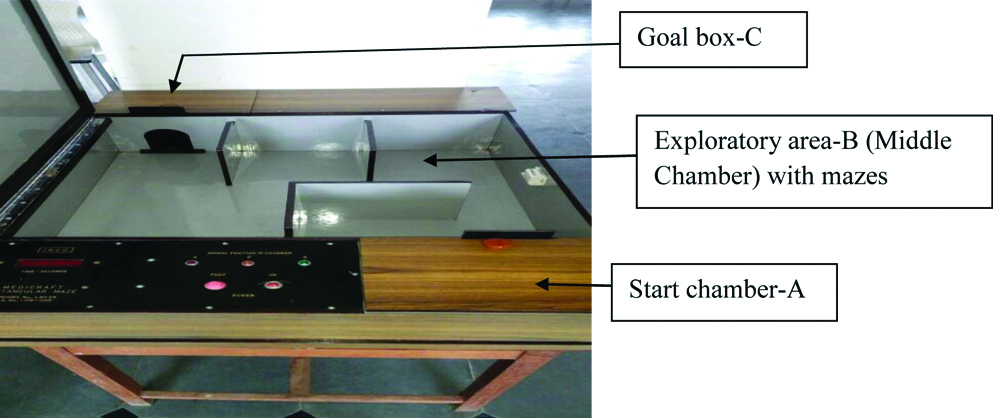
Hebb-Williams Maze: Hebb-Williams Maze is used to test the spatial learning and working memory of the mice. The principle behind the Hebb-Williams Maze test is “The faster the mice navigates the maze, the better its spatial memory”. The Hebb-Williams maze is a square shaped box which measures 60 cm (L)×60 cm (W)×10 cm (H) walls. It consists of start chamber-A (Animal Chamber) which is attached to the exploratory area-B (Middle Chamber) and a goal box-C, located at the opposite end of the start chamber and contains a small food reward. All three chambers were provided with removable doors to allow the animal to move from one chamber to the next.
After 12 hours of fasting, the mice was placed in the start chamber-A and allowed to enter into the exploratory area-B (middle chamber), once the animal enters into middle chamber the door was closed to prevent back entry. The time taken by the animal to reach The Reward Chamber (TRC) from the start chamber was recorded. The animals were trained for 3 days (3 trials/day) and the readings were taken at the 4th day. Low scores indicates better memory, while the high scores indicates poor memory in animals [Table/Fig-2] [10,11].
Hebb-Williams Maze instrument to assess the spatial learning and working memory in Swiss-Albino mice.
Tissue Processing
After the behavioural analysis the mice were euthanized, perfused transcardially with normal saline and the brains were extracted out, fixed in 10% buffered formalin, dehydrated in ascending grades of ethyl based alcohol like 60%, 70%, 80%, 90% and absolute alcohol, cleared in xylene, impregnated in paraffin wax at 60°C, embedded with the help of L-moulds and then 6 μm paraffin sections were taken using rotary microtome at the level of the dorsal hippocampus to assess the hippocampal CA3 cellular architecture with the help of H and E staining. To prevent the bias, every 5th section was taken and the slides were decoded after the histological assessment. Viable neuronal quantification was assessed with the help of ocular micrometer fixed to light microscope (40X).
Statistical Analysis
The results were expressed in Mean±SD and analysed by using one-way ANOVA followed by Least Square Difference (LSD) test for paired wise data. The p<0.05 was considered as statistically significant.
Results
Body Weights of the Mice
The mean body weight of the control group mice was 32.3 grams, 30 min/day radiation exposed mice for 3 months had 31.8 grams and 60 min/day radiation exposed mice for 3 months had 32.7 grams, the mean weight between the three groups didn’t show any significant difference.
Effect of Radiation on Learning Memory in Hebb-Williams Maze
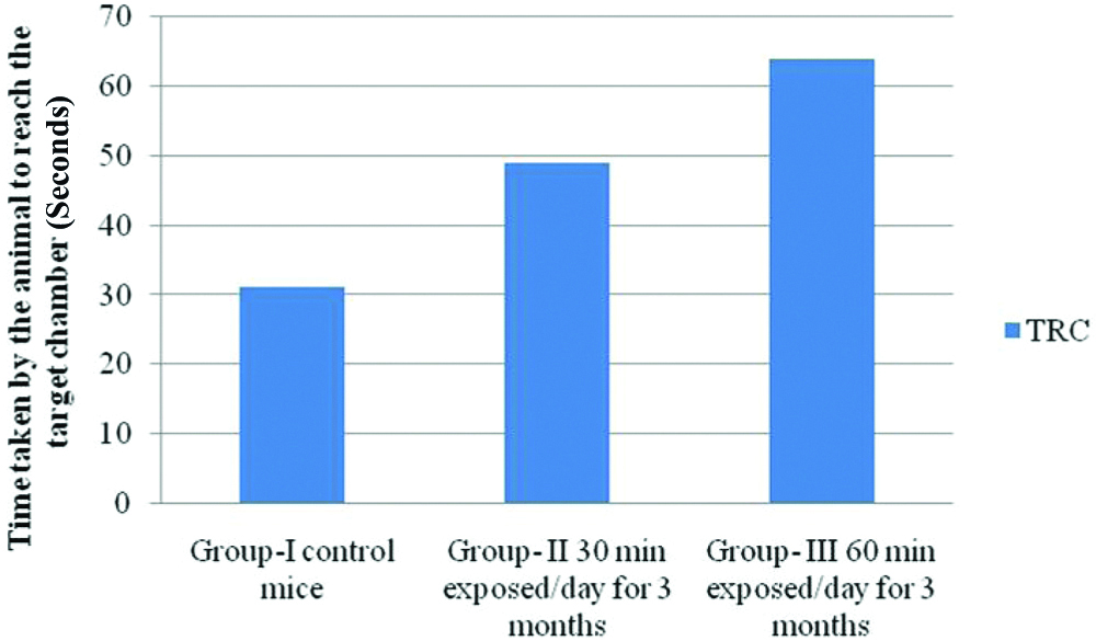
The time taken by the mice to reach the target chamber from the starting chamber was significantly increased in group II (30 min exposed/day) and group III (60 min exposed/day) compared to group I (non-exposed group).
The time taken by the animal to reach The Reward Chamber (TRC) scores in Group I vs Group II (31±15.48 vs 49±17.62 seconds), was not significant (p>0.05); Group I vs Group III (31±15.48 vs 64±22.99 seconds), was statistically significant (p<0.05) [Table/Fig-3].
Effect of Mobile phone radiofrequency-electro-magnetic radiation (MP RF-EMR) on learning and memory by using Hebb-Williams maze- group I, II and III.
Microscopic Anatomy of Hippocampal Cornu Ammonis (CA3) Neurons
Histological sections of haematoxylin and eosin stained hippocampal CA3 pyramidal neurons showed marked difference between control group and RF-EMR exposed groups (group II and III). Sections of control group showed 5-6 layers of compactly arranged pyramidal cells which were healthy with clear nucleus [Table/Fig-4]. Group II (30 min exposure for 3 months) showed less number of pyramidal neurons With darkened nuclei (non-viable neurons) which was scattered when compared to control group [Table/Fig-5]. Group III (60 min exposure for 3 months) showed very less number of pyramidal neurons with more number of darkened nuclei (more non-viable neurons) with vacuolation in cytoplasm and scattered arrangement of pyramidal neurons when compared to group I and group II [Table/Fig-6].
Group I (non-exposed)-Control group H and E stained Hippocampal pyramidal normal neurons (Arrow) in high power (40X)

Group-II (30 min exposed for 3 months) H and E stained Hippocampal pyramidal neurons (Arrow) in high power (40X) showed less in number, Non-Viable and scattered with vacuolation in cytoplasm.

Group- III (60 min exposed for 3 months) H and E stained Hippocampal pyramidal neurons (Arrow) in high power (40X) showed very less in number, Non-Viable and scattered with vacuolation in cytoplasm

Discussion
With advancement of technology like 2G to 3G, 3G to 4G in the telecommunication field, the mobile phones are being used for communication, internet data and as multipurpose device. However over-usage of mobiles with advance multiple features has adverse effects on the brain especially on the hippocampus, which is a sensitive region on the temporal lobe of the brain responsible for spatial learning and working memory, an important cognitive function [7].
In this study, Hebb-Williams maze analysis was used to assess the learning and memory in albino mice exposed to mobile phone radiation frequency and control group [10,11]. In the present study, MP RF-EMR exposed mice took significantly increased time to reach the target chamber in Hebb-Williams maze when compared to the control group, which shows memory retention and memory retrieval is being affected and leads to memory impairment in the mice. Studies have shown that RF EMR exposure will impair the learning and memory, which may be due to neurodegenerative changes and alterations in the morphology of the hippocampus [7,8].
On histological examination, radiation exposed hippocampal CA3 neurons showed less number of pyramidal cells with darkened nuclei (Non-viable), vacuolated cytoplasm and cells were scattered in arrangement. The altered structural integrity in the hippocampus might be the cause for impairment of learning memory. Decrease in pyramidal cell count may be due to inhibition of neurogenesis and this was supported by Odaci E et al., [12]. Bolla SR reported that exposure to 800 MHz mobile radiation for 30 days leads to increased neuronal damage and decreased viable neurons in hippocampal CA3 region [9].
Nittby H et al., reported that exposure to 900 GSM radiation will reduce memory functions in rat, which is similar to our study [1]. MP RF exposure to 900-1800 MHz radiation leads to decrease in nuclear diameter and reduce neuronal density in the hippocampus [13]. Findings on exposure to 50-217 Hz low frequency radiation with television and mobile phone have impact on learning and memory [14]. Fragopoulou AF et al., reported that consolidation and retrieval memory deficits were observed in mice exposed to 9 hr 30 mins for 4 days with 900 MHz non-ionising radiation [15]. Heat shock proteins-HSP 27 and HSP 70 related stress levels are elevated in rat hippocampus exposed to 2450 MHz radiation [16]. A 2.14 GHz Radiation frequency exposure at 4 Watt/kg specific absorption rate increases the body temperature to 1.5°C compared to baseline and upregulates some stress markers like HSP and Heat Shock Transcription Factor (HSF) gene expressions in cerebellum and cerebral cortex [17].
Limitation
The outcome of the present rodent study may not extrapolate with human population due to many reasons like Thickness of the skull bone, Weight/Volume of the brain, Specific Absorption Rate (SAR), Duration of exposure, Frequency of radiation and Lifespan of the human population.
Conclusion
In this present study, we evaluated the chronic exposure effect of MP RF-EMR- 4G (1800-2100 MHz) on cognitive functions like spatial learning, working memory and hippocampal morphology in adult swiss albino mice. We observed that MP RF-EMR exposed mice took significantly increased time to reach the target chamber in Hebb-Williams maze when compared to the control group. Radiation exposed hippocampal CA3 neurons showed less number of pyramidal cells with darkened nuclei (Non-viable), vacuolation in cytoplasm and cells were scattered in arrangement. The altered structural integrity in the hippocampus may alter the spatial learning and memory.