Introduction
Endodontic flare-up is a common complication characterised by pain, swelling or both that occurs within a few hours or days after endodontic procedure and one of the major reasons is the apical extrusion of dental filings, pulp tissue fragments, necrotic tissue, micro-organisms and irrigant during chemo-mechanical preparation of the root canals. Thus, it is important to limit the amount of the debris extruded through the apical foramen into the peri-radicular region for a successful root canal treatment.
Aim
To quantify and compare the amount of debris extruded apically during root canal preparation using conventional hand files, K3 and Kedo-S Rotary files in primary teeth.
Materials and Methods
Thirty six extracted human primary canines were selected and randomly assigned to one of the three groups of twelve teeth each. Myers and Montgomery experimental model was used for instrumentation and debris collection. The teeth were then instrumented with the following file systems: Group 1: Hand files; Group 2: K3 files; Group 3: Kedo-S files. The apical debris which got extruded during instrumentation was collected in Eppendorf tubes which were pre-weighed. The tubes were then placed in an Incubator at 70° Celsius for five days and weighed again to determine the post-instrumentation weight. The weight of the dry debris collected was measured by subtracting the pre-weight of the Eppendorf tubes from the post-instrumentation weight for all the three groups. The data were analysed using one-way ANOVA and Tukey’s post-hoc tests.
Results
A statistically significant difference was observed between hand files and Kedo-S (p<0.05) and between hand files and K3 files (p<0.05). However, there was no statistically significant difference between K3 and Kedo-S files (p=0.069).
Conclusion
Apical debris extrusion was found to be less with Kedo-S files compared to hand files.
Apical debris, Deciduous teeth, Instrumentation
Introduction
Primary teeth play a vital role in proper functioning of masticatory system, growth and development of jaws and muscles, phonation and guides the eruption of permanent teeth. It is thus imperative to retain primary teeth [1]. The primary teeth with irreversible pulpitis or necrosis, or a tooth planned for pulpotomy in which the radicular pulp exhibits clinical signs of irreversible pulpitis are all indicated for pulpectomy, provided the roots exhibit minimal or no resorption [2-4].
The conventional hand instrumentation of primary teeth is time-consuming and causes fatigue to both operator and the child [5]. Barr ES et al., introduced rotary instruments to paediatric endodontics [6]. Rotary instrumentation has made pulpectomy in children faster, consistent, with a predictable root canal shaping [7,8]. The mean time for canal preparation is significantly shorter with rotary files than manual instrumentation in primary molars [9-12].
During root canal preparation, dental shavings, pulpal fragments, necrotic debris, irrigating solution and microorganisms may be inadvertently pushed out of the root canal into the periapical region leading to undesirable consequences like inflammation, postoperative pain, delay of periapical healing, damage to permanent tooth buds and mid treatment flare-ups [1,2]. One of the common issues encountered during root canal treatment is extrusion of intra-canal debris, and there are no instruments or techniques that exist to thoroughly resolve this problem [13]. Irritant that is directed towards the periapical tissues may result in flare-ups and hence, the goal of any instrumentation should be directed towards reducing the risk of extrusion of the debris. Since the last decade, evolution of root canal instruments and irrigation systems have occurred, and it has also been assessed for their potency of extrusion of the apical debris. [1,2,14,15]. In the present study, the three different files were chosen, since hand files serve as the standard instrumentation technique for pulpectomy, Kedo-S files are newly launched exclusive paediatric rotary files and K3 files were proved in a previous study to produce significantly lesser extrusion of debris [16].
The increased flexibility, variable diameter and safe ended tip of the K3 files has shown a drastic decrease in ledging, perforation and zipping [17,18]. Studies have shown that use of K3 files for root canal preparation in primary teeth produces a statistically significant reduction in the instrumentation time [12,19,20].
Kedo-S file system, an exclusive paediatric rotary file for canal instrumentation has been launched and is reported to provide effective instrumentation and uniform quality of obturation in primary teeth [21]. However, no studies have evaluated the apical debris extrusion using Kedo-S file system.
Therefore, the purpose of the present In-Vitro study was to comparatively assess the amount of apically extruded debris following root canal preparation using two different rotary files-K3 files and Kedo files; and hand files in primary canines.
Materials and Methods
The present study is an In-Vitro study conducted in the department of Paediatric and Preventive Dentistry, Saveetha Dental College, Chennai during the month of December 2017 following approval from the Institutional Review Board of Saveetha University, Chennai, India. Sample size was calculated as a total of 36 from a pilot study with 95% power using G power analysis. (α=0.05, 1-β=0.95).
Primary canines extracted due to periapical pathology or trauma in children between 4 and 7 years of age were included. Bucco-lingual and mesio-distal radiographs of each tooth were taken to ensure the presence of single root canal and apical foramen. Teeth with signs of internal or external root resorption, visible root caries, fractures and cracks were excluded from the study.
Preparation of the Sample
The collected teeth were stored in distilled water at room temperature until the experimentation procedure. Access was prepared using a No.6 round bur (Mani Inc; Japan) and complete de-roofing of the pulp chamber was done using high-speed hand-piece under water cooling. After the pulpal tissue was extirpated, a 10 size K file (Dentsply Maillefer, Ballaigues, Switzerland) was used for establishing canal patency. This was followed by working length determination by placing the file 1mm short of the apical foramen [15].
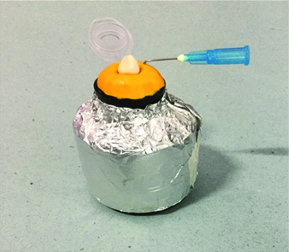
The experimental model described by Myers and Montgomery [22] was used in the present study [Table/Fig-1]. The Eppendorf tube was taken and the tooth was inserted till the cemento-enamel junction. A 27 gauge needle was inserted into the vial as drainage cannula to equalise the pressure inside and outside Eppendorf tubes. The vials were covered by aluminium foils to prevent the operator from viewing the debris extrusion during instrumentation procedure and the Eppendorf tubes were inserted into it. The Eppendorf tubes were preweighed to 10-5 units using a microbalance (Sartorius AG, Gottingen, Germany). Three consecutive measurements were taken for each tube, and mean values were recorded. The teeth were coded and randomly assigned to three groups of 12 each.
Myers and Montgomery experimental model.
Group 1: (n=12) The canal instrumentation was done using 21 mm 0.2 taper stainless steel hand K files (Dentsply Maillefer, Ballaigues, Switzerland) in a quarter pull motion using step back technique. Fresh files from size 15 to 40 were used for each tooth.
Group 2: (n=12) K3 (Syborn Dental, Westcollins, CA, USA) rotary file of 0.30 tip diameter and 0.06% taper was used for canal instrumentation in a crown-down technique according to manufacturer’s instructions [12].
Group 3: (n=12) Kedo-S (Reeganz dental care Pvt., Ltd., India) rotary file-U1 of length 16 mm and 250±rpm, tip diameter of 0.40 mm was used for primary root canal instrumentation according to the manufacturer’s instructions.
The apex was prepared using #30 file in all the three groups for standardisation. The Kedo-S file system consists of three Ni-Ti rotary files-D1, E1, and U1 respectively. The total length of the files was 16 mm with working length 12 mm. All the files have a variably variable taper corresponding to its use in deciduous teeth. U1 with a tip diameter of 0.40 mm indicated for use in primary anterior teeth was used in the present study [21]. The K3 files are rotary files that has a slightly positive rake angle and asymmetrical cross-sectional design [19,20]. The variable pitch in the cutting shank allows for effective cutting of dentinal debris.
The instrumentation of all the teeth was done by a single examiner to eliminate operator bias. The irrigant used was standardised to 10 mL of distilled water for all samples. Following the removal of the Eppendorf tubes from the vials, the external root surface was washed with 1 mL of the distilled water for the collection of the debris adhered to it. For the evaporation of the distilled water and to measure only the weight of the dry debris, the tubes were taken and stored at a temperature of 70° Celsius in an incubator for 5 days. A second examiner who was completely blinded to the study evaluated the amount of apical debris collected. Three consecutive measurements were taken for each tube using the same analytical balance that was used for pre-weighing of the Eppendorf tubes and their mean was calculated. The initial pre-weighed value of the empty Eppendorf tube was subtracted from the final measured gross weight value to arrive at the total net weight of the extruded dry debris.
Statistical Analysis
Statistical analysis was done using SPSS software version 17.0 (SPSS Inc., Chicago, IL, USA). The data was analysed statistically using one-way ANOVA and Tukey’s post-hoc tests.
Results
A statistically significant difference was noted between the three groups with regard to the extrusion of apical debris (p<0.001) [Table/Fig-2].
Comparison of apically extruded debris using hand files, Kedo-S and K3 rotary files.
| Groups | n | Mean±SD (in grams) | p-value |
|---|
| Hand File | 12 | 0.0018893±0.00068844 | 0.001** |
| K3 | 12 | 0.0011500±0.00049162 |
| Kedo-S | 12 | 0.0007267±0.00024159 |
One-way ANOVA (p<0.001)
Post-Hoc analysis showed that the extrusion of apical debris is more with K-files when compared to K3 and Kedo-S Rotary files (p=0.001, p=0.000)
No statistically significant difference was noted between K3 and Kedo-S rotary files (p=0.069) [Table/Fig-3].
Intergroup comparison using Turkey HSD Post-hoc analysis.
| Groups VS. | Mean Difference | Significance |
|---|
| I | II | 0.00073933* | 0.001** |
| III | 0.00116267* | 0.000** |
| II | I | -0.00073933* | 0.001** |
| III | 0.00042333 | 0.069 |
| III | I | -0.00116267* | 0.000** |
| II | -0.00042333 | 0 .069 |
Discussion
Extrusion of dentinal chips, pulpal tissue, micro-organism and irrigation solution are common occurrences during root canal instrumentation of primary teeth due to the use of instruments in apical direction and it plays a major role in determining the success of a root canal treatment, hence it is important to minimise the amount of apical extrusion [23].
Inter-appointment flare-ups and post-operative pain are the most significant complications of apical extrusion of debris and can result in undesirable occurrence for both patient and practitioner [24-26]. Although the virulence of micro-organisms associated with the pathology have been considered as the critical causative factor for occurrence of flare-ups, the apical extrusion of the debris can also initiate an inflammatory reaction that can cause postoperative complications [27]. Even aforementioned studies reported that any form of physical/chemical irritation to peri-radicular tissues caused by the extruded debris can evoke a peri-apical reaction [13]. The factors affecting apical extrusion include instrumentation method, size and the length of canal, instrument type and size, the endpoint of the preparation and the type and amount of irrigant used, of which the type of instrumentation used plays a major role [14]. Hence in the present study, three different instruments (hand K files, K3 and Kedo-S rotary files) with varied designs and meant for usage in primary teeth were selected for canal instrumentation.
Only single-rooted teeth with straight canal were included to overcome morphological variations. Myers and Montgomery method [22] was used in the present study for collection of apically extruded debris. Distilled water was selected as irrigant in the present study to prevent possible alteration in the weight that can occur due to crystallisation of sodium hypochlorite to sodium crystals [5,14]. Floral foam to simulate periapical tissues have been used in few previous studies [28,29], But no attempt was made to simulate periapical tissues in the current study as this approach may alter the results, as the irrigant and debris may get absorbed.
In the present study, hand files produced the largest amount of apical debris extrusion followed by K3 and Kedo-S. The variation in the amount of debris collected may be due to the differences in the cross-sectional designs of the instruments and also due to the dissimilarity in the tapers. However, Post-Hoc Analysis in the present study shows that there was no statistically significant difference between K3 and Kedo-S rotary file. The possible reason for this could be that hand files were used in a sequence, whereas single file system was used in the other two groups. Also the rotary motion of the Ni-Ti files tend to direct debris towards the orifice rather than pushing it apically as it happens with the quarter pull turn motion of K-file. Thus compaction being avoided in the root canal, lesser debris gets extruded apically with rotary files when compared to manual files used in step-back technique [30]. The Kedo-S files have a variably variable taper which prepares the canals less apically and more coronally, leading to lesser apical extrusion of the debris. Ferraz CC et al., reported less extrusion of debris when Profile instruments were used compared to that of the manual technique [31]. Reddy SA et al., reported that hand files produced more debris than rotary Ni-Ti instruments [32]. Thus, the results of our present study is in concordance with the previous studies published depicting that with engine driven instruments, the amount of apical extrusion of debris is lesser when compared with that of the Hand files.
Limitation
A probable limitation of the present study is the smaller sample size used for evaluation of the extruded debris.
On literature search, it was found that there was a lack of studies evaluating the apical debris extrusion in primary teeth using modified rotary file systems. Thus, further research on apical debris extrusion with use of various rotary files in primary teeth is required to determine a safer and a more successful protocol for root canal instrumentation in primary teeth.
Conclusion
Kedo-S rotary file produced the least amount of apical debris extrusion and hence can be considered as close to ideal instrumentation for canal preparation in primary teeth.
One-way ANOVA (p<0.001)
[1]. Kucukyilmaz E, Savas S, Saygili G, Uysal B, Evaluation of Apically Extruded Debris and Irrigant Produced by Different Nickel-Titanium Instrument Systems in Primary TeethJ Contemp Dent Pract 2015 16:864-68.10.5005/jp-journals-10024-177226718292 [Google Scholar] [CrossRef] [PubMed]
[2]. Thakur B, Pawar AM, Kfir A, Neelakantan P, Extrusion of debris from primary molar root canals following instrumentation with traditional and new file systemsJ Contemp Dent Pract 2017 18:1040-44.10.5005/jp-journals-10024-217229109318 [Google Scholar] [CrossRef] [PubMed]
[3]. Fuks AB, Eidelman E, Pulp therapy in the primary dentitionCurr Opin Dent 1991 1:556-63. [Google Scholar]
[4]. Thompson V, Craig RG, Curro FA, Green WS, Ship JA, Treatment of deep carious lesions by complete excavation or partial removal: a critical reviewJ Am Dent Assoc 2008 139:705-12.10.14219/jada.archive.2008.025218519994 [Google Scholar] [CrossRef] [PubMed]
[5]. Topçuoğlu G, Topçuoğlu HS, Akpek F, Evaluation of apically extruded debris during root canal preparation in primary molar teeth using three different rotary systems and hand filesInt J Paediatr Dent 2016 26:357-63.10.1111/ipd.1220826538300 [Google Scholar] [CrossRef] [PubMed]
[6]. Barr ES, Kleier DJ, Barr NV, Use of nickel-titanium rotary files for root canal preparation in primary teethPaediatr Dent 1999 21(7):453-54. [Google Scholar]
[7]. George S, Anandaraj S, Issac JS, John SA, Harris A, Rotary endodontics in primary teeth - A reviewSaudi Dent J 2016 28:12-17.10.1016/j.sdentj.2015.08.00426792964 [Google Scholar] [CrossRef] [PubMed]
[8]. Glickman GN, Koch KA, 21st-century endodonticsJ Am Dent Assoc 2000 131(Suppl):39S-46S.10.14219/jada.archive.2000.040110860344 [Google Scholar] [CrossRef] [PubMed]
[9]. Silva LA, Leonardo MR, Nelson-Filho P, Tanomaru JM, Comparison of rotary and manual instrumentation techniques on cleaning capacity and instrumentation time in deciduous molarsJ Dent Child 2004 71:45-47. [Google Scholar]
[10]. Crespo S, Cortes O, Garcia C, Perez L, Comparison between rotary and manual instrumentation in primary teethJ Clin Paediatr Dent 2008 32:295-98.10.17796/jcpd.32.4.l57l36355u606576 [Google Scholar] [CrossRef]
[11]. Kummer TR, Calvo MC, Cordeiro MM, de Sousa Vieira R, de Carvalho Rocha MJ, Ex vivo study of manual and rotary instrumentation techniques in human primary teethOral Surg Oral Med Oral Pathol Oral Radiol Endod 2008 105:e84-92.10.1016/j.tripleo.2007.12.00818329573 [Google Scholar] [CrossRef] [PubMed]
[12]. Govindaraju L, Jeevanandan G, Subramanian E M, Clinical evaluation of quality of obturation and instrumentation time using two modified rotary file systems with manual instrumentation in primary teethJ Clin Diag Res 2017 11:ZC55-58.10.7860/JCDR/2017/30069.1060229207834 [Google Scholar] [CrossRef] [PubMed]
[13]. Tanalp J, Güngör T, Apical extrusion of debris: a literature review of an inherent occurrence during root canal treatmentInt Endod J 2014 47:211-21.10.1111/iej.1213723711187 [Google Scholar] [CrossRef] [PubMed]
[14]. Verma M, Meena N, Kumari RA, Mallandur S, Vikram R, Gowda V, Comparison of apical debris extrusion during root canal preparation using instrumentation techniques with two operating principles: An in vitro studyJ Conserv Dent 2017 20:96-99.10.4103/0972-0707.21223928855755 [Google Scholar] [CrossRef] [PubMed]
[15]. Madhusudhana K, Mathew VB, Reddy NM, Apical extrusion of debris and irrigants using hand and three rotary instrumentation systems - An in vitro studyContemp Clin Dent 2010 1:234-36. [Google Scholar]
[16]. Ghivari SB, Kubasad GC, Chandak MG, Akarte N, Apical extrusion of debris and irrigant using hand and rotary systems: A comparative studyJ Conserv Dent JCD 2011 14(2):187-90.10.4103/0972-0707.8262221814364 [Google Scholar] [CrossRef] [PubMed]
[17]. Mohammadzade Akhlaghi N, Khalilak Z, Baradaran Mohajeri L, Sheikholeslami M, Saedi S, Comparison of canal preparation pattern of K3 and ProTaper rotary files in curved resin blocksIran Endod J 2008 3:11-16. [Google Scholar]
[18]. Ankrum MT, Hartwell GR, Truitt JE, K3 Endo, ProTaper, and ProFile systems: breakage and distortion in severely curved roots of molarsJ Endod 2004 30:234-37. [Google Scholar]
[19]. Rosa FM, Modesto A, Faraco-Junior IM, Manual and rotary instrumentation techniques for root canal preparation in primary molarDentistry 2014 2:1-5.10.1097/00004770-200404000-0001315085054 [Google Scholar] [CrossRef] [PubMed]
[20]. Romero TO, Gonzalez VM, Reyes HF, Pozos-Guillen AJ, Comparison between rotary and manual instrumentation and obturation times in primary teethJ Clin Paediatr Dent 2011 35:359-64.10.17796/jcpd.35.4.8k013k21t39245n8 [Google Scholar] [CrossRef]
[21]. Jeevanandan G, Kedo-S Paediatric Rotary Files for Root Canal Preparation in Primary Teeth - Case ReportJ Clin Diagn 2017 11:ZR03-ZR05.10.7860/JCDR/2017/25856.950828511532 [Google Scholar] [CrossRef] [PubMed]
[22]. Myers GL, Montgomery S, A comparison of weights of debris extruded apically by conventional filing and Canal Master techniquesJ Endod 1991 17(6):275-79.10.1016/S0099-2399(06)81866-2 [Google Scholar] [CrossRef]
[23]. Hülsmann M, Hahn W, Complications during root canal irrigation--literature review and case reportsInt Endod J 2000 33:186-93.10.1111/j.1365-2591.1968.tb01184.x [Google Scholar] [CrossRef]
[24]. Chapman CE, Collee JG, Beagrie GS, A preliminary report on the correlation between apical infection and instrumentation in endodonticsJ Br Ended Soc 1968 2:7-11. [Google Scholar]
[25]. Seltzer S, Naidorf IJ, Flare-ups in endodontics: etiological factorsJ Endod 1985 11:472-78.10.1016/S0099-2399(85)80220-X [Google Scholar] [CrossRef]
[26]. Naidorf IJ, Endodontic flare-ups: bacteriological and immunological mechanismsJ Endodon 1985 11:462-64.10.1016/S0099-2399(85)80218-1 [Google Scholar] [CrossRef]
[27]. Ingle J, Beveridge E, Endodontics 1985 3rd edPhiladelphiaLea & Febiger:170-80. [Google Scholar]
[28]. Altundasar E, Nagas E, Uyanik O, Serper A, Debris and irrigant extrusion potential of 2 rotary systems and irrigation needlesOral Surg Oral Med Oral Pathol Oral Radiol Endod 2011 112(4):e31-35.10.1016/j.tripleo.2011.03.04421778084 [Google Scholar] [CrossRef] [PubMed]
[29]. Hachmeister DR, Schindler WG, Walker WA III, Thomas DD, The sealing ability and retention characteristics of mineral trioxide aggregate in a model of apexificationJ Endodon 2002 28:386-90.10.1097/00004770-200205000-00010 [Google Scholar] [CrossRef]
[30]. Goerig AC, Michelich RJ, Schultz HH, Instrumentation of root canals in molar using the step-down techniqueJ Endod 1982 8(12):550-54.10.1016/S0099-2399(82)80015-0 [Google Scholar] [CrossRef]
[31]. Ferraz CC, Gomes NV, Gomes BP, Zaia AA, Teixeira FB, Souza-Filho FJ, Apical extrusion of debris and irrigants using two hand and three engine-driven instrumentation techniquesInt Endod J 2001 34(5):354-58.10.1046/j.1365-2591.2001.00394.x11482718 [Google Scholar] [CrossRef] [PubMed]
[32]. Reddy SA, Hicks ML, Apical extrusion of debris using two hand and two rotary instrumentation techniquesJ Endod 1998 24:180e310.1016/S0099-2399(98)80179-9 [Google Scholar] [CrossRef]