Intramuscular Lipoma of the Pectoralis Major Simulating a Breast Lump
Rajendra Benakatti1, Dinesh Bangalore2, Pavan Bhat3
1 Assistant Professor, Department of General Surgery, Kasturba Medical College, Manipal Academy of Higher Education, Manipal, Karnataka, India.
2 Associate Professor, Department of General Surgery, Kasturba Medical College, Manipal Academy of Higher Education, Manipal, Karnataka, India.
3 Junior Resident, Department of General Surgery, Kasturba Medical College, Manipal Academy of Higher Education, Manipal, Karnataka, India.
NAME, ADDRESS, E-MAIL ID OF THE CORRESPONDING AUTHOR: Dr. Rajendra Benakatti, Assistant Professor, Department of General Surgery, Kasturba Medical College, Manipal Academy of Higher Eductaion, Manipal-576104, Karnataka, India.
E-mail: Dr.rajendra.benakatti@gmail.com
Intramuscular lipoma is a deep-seated lipoma that arises in the muscle and is benign and relatively rare. Intramuscular lipomas of the pectoralis major muscle have been reported to mimic malignant breast tumours. Here, we present a 69-year-old female presenting with painless lump in right breast of three months duration progressively increasing in size. Examination revealed a soft, lobulated lump which was freely mobile with no fixity to surrounding structures. Mammogram revealed large radiopaque lesion noted in the retro glandular region with lucent linear striation in the centre. Correlated sonomammography showed a large hyperechoic lesion in retromammary region, anterior to the pectoralis major muscle. On Doppler study there was no vascularity, an ultrasound guided FNAC was done which showed adult adipocytes, suggestive of a lipoma. On exploration, a large encapsulated intramuscular lipoma of the pectoralis major was noted and excised, final histopathological examination also confirmed the diagnosis of lipoma. Lipoma can be considered as a rare differential diagnosis for breast lump.
Excision, Surgery, Swelling
Case Report
A 69-year-old female with no comorbidities presented with a painless lump in right breast of three months duration which was progressively increasing in size. No associated skin abnormality was noted. There was no history of any other lumps in the body. No other significant medical or surgical history was noted.
Examination revealed a single 10×8 cm soft, oval shaped, lobulated lump in the right breast, which was freely mobile with no fixity to surrounding structures, skin over the lump was normal. Patient was admitted for further evaluation of the breast lump.
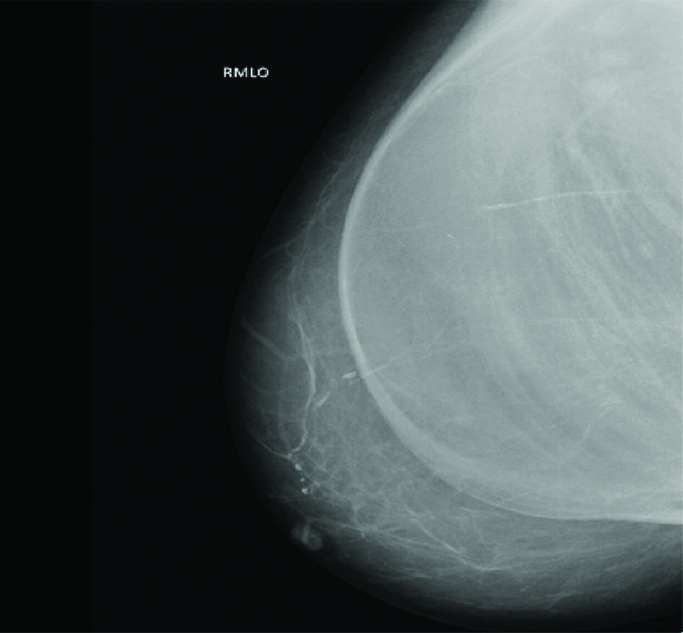
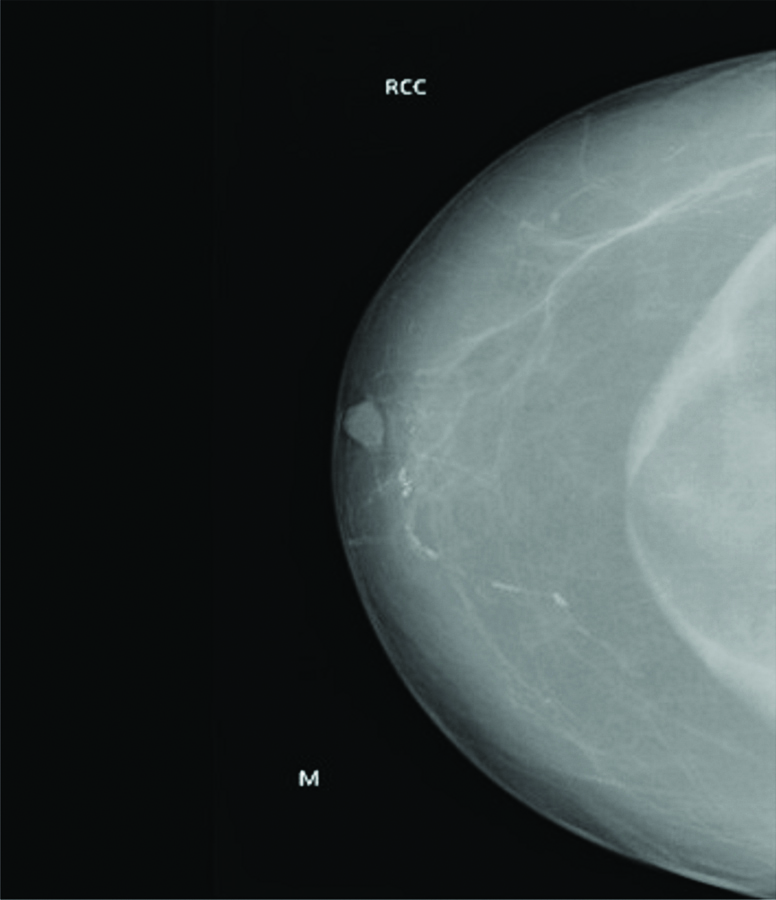
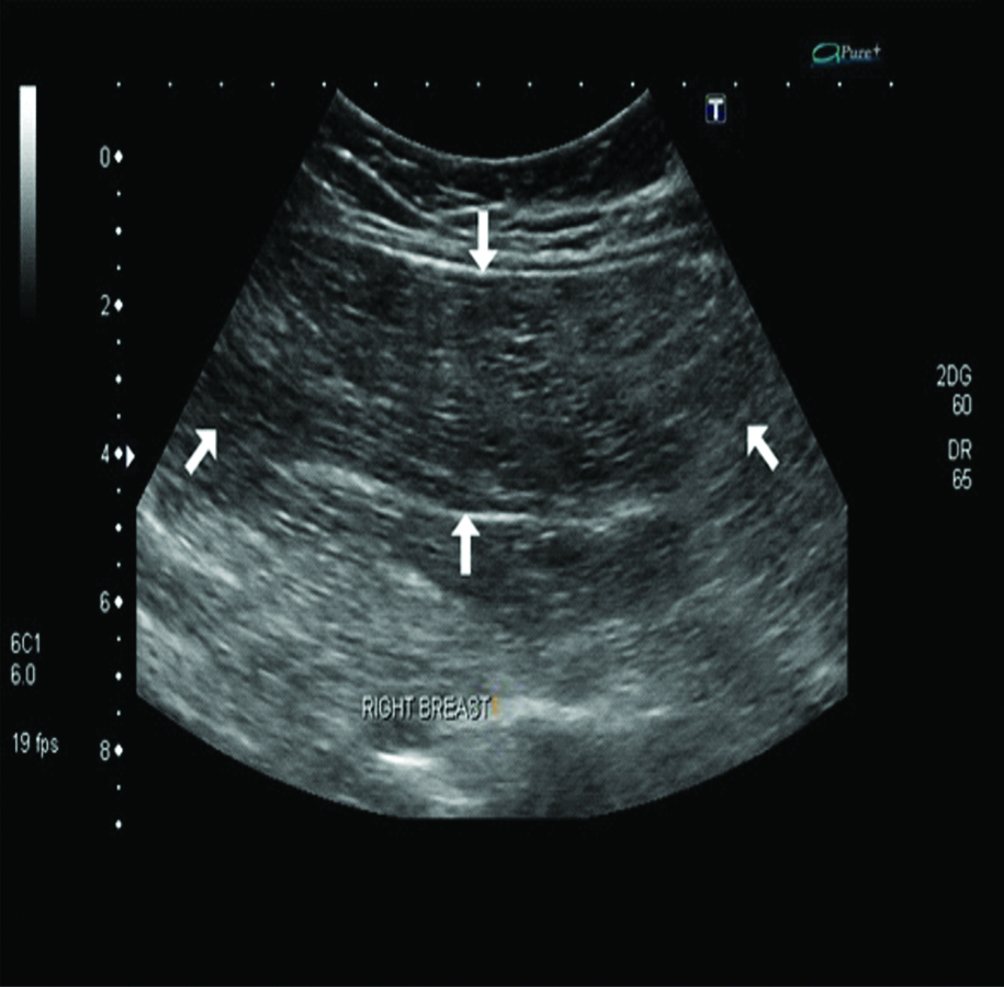
A mammogram was done which revealed large radiopaque lesion noted in the retro glandular region with in the pectoralis major muscles with lucent linear striation in the centre [Table/Fig-1,2], Correlated sonomammography showed a large hyperechoic lesion in retromammary region, within the pectoralis major muscle [Table/Fig-3], On Doppler study, no evidence of vascularity was noted. An ultrasound guided FNAC done, showed adipocyte which was suggestive of a lipoma.
RMLO (Right Mediolateral Oblique) view in mammogram which shows large radiopaque lesion noted in the retro glandular region within the pectoralis major muscle.
RCC (Right Craniocaudal) view of mammogram with radiopaque lesion with linear striation in the centre.
USG showed hyperechoic lesion in retromammary region, within the pectoralis major muscle.
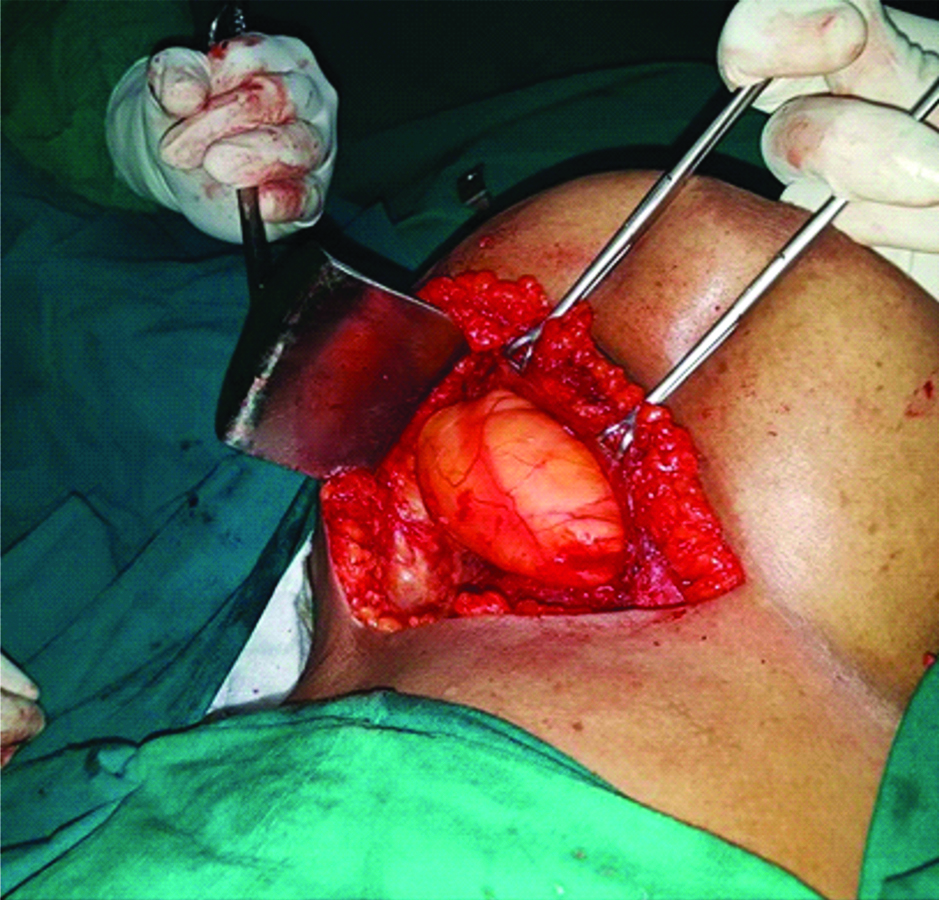
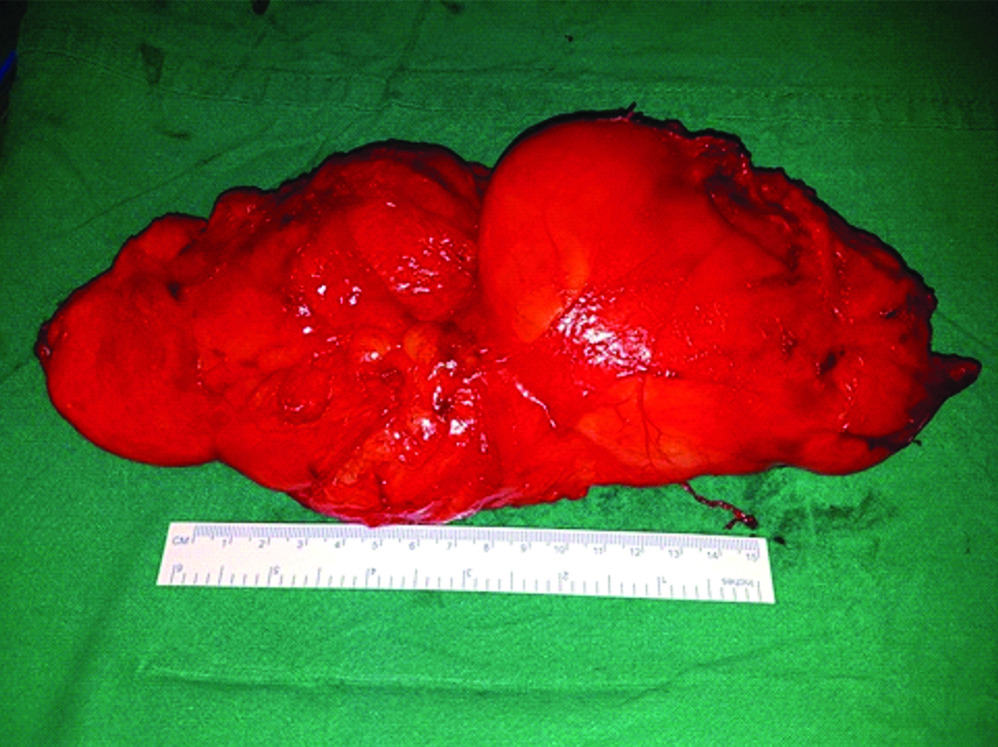
Patient was taken up for surgery, on exploration a large encapsulated intramuscular lipoma noted within the pectoralis major, which was excised [Table/Fig-4,5] and sent for histopathological examination. Final histopathological report also confirmed the diagnosis of lipoma (mature adipocytes were noted).
Intraoperative picture showing a intramuscular lipoma.
Excised specimen showing well-encapsulated lipoma.
Immediate post-operative period was uneventful; suction drain was removed on post-operative day three and patient was discharged on post-operative day five with analgesics. On follow-up after a week wound was healed well and sutures were removed. No recurrence was noted in subsequent six months follow-up.
Discussion
Lipoma is a benign mesenchymal tumour composed of adipocytes. Intramuscular lipomas most likely have neoplastic pathogenesis and represent a true neoplasm directly originating from multipotent mesenchymal cells [1]. They are soft tissue tumours commonly seen in thorax and extremities. They are usually seen in the age group of 50-70 years and most common in obese [2]. Intramuscular lipomas involving the pectoralis major muscle is rare, they may mimic breast malignancies clinically and radiologically [3].
Intramuscular lipomas of the pectoralis muscles are well-encapsulated radiolucent tumours of fat density with displaced pectoralis major on mammogram. Ultrasound shows lipomas of the pectoralis major as well-defined and homogeneously echogenic [4]. CECT and MRI can accurately identify the lipomas.
Intramuscular lipomas are uncommon types of lipoma. Less familiarity with the pathology commonly leads to them misclassified and misdiagnosed with other benign and malignant lesions [3].
In our patient, mammogram and FNAC were used to diagnose lipoma hence MRI was not done. Surgical excision is the treatment of choice as incomplete excision will lead to recurrence [5].
Conclusion
Intramuscular lipoma of pectoralis major muscle are rare tumours that mimic breast tumours both clinically and radiologically. We came across one such case, accurate diagnosis and complete excision is the treatment of choice.
Consent
Prior consent has been obtained from the patient before preparation of the case report.
[1]. McTighe S, Chernev I, Intramuscular lipoma: a review of the literatureOrthopedic Reviews 2014 6(4):5618doi:10.4081/or.2014.561810.4081/or.2014.561825568733 [Google Scholar] [CrossRef] [PubMed]
[2]. Tateishi U, Gladish GW, Kusumoto M, Hasegaw T, Yokoyama R, Chest wall tumours: radiologic findings and pathologic correlation: part. Benign tumoursRadiographics 2003 23:1477-90.10.1148/rg.23601552614615559 [Google Scholar] [CrossRef] [PubMed]
[3]. Britton CA, Subpectoral mass mimicking a malignant breast mass on mammographyAm J Roentgenol 1992 159:22110.2214/ajr.159.1.16097071609707 [Google Scholar] [CrossRef] [PubMed]
[4]. Matsumoto K, Hukuda S, Ishizawa M, Okabe H, MRI findings in intramuscular lipomasSkeletal Radiol 1999 28:145-52.10.1007/s00256005049110231912 [Google Scholar] [CrossRef] [PubMed]
[5]. Gopal U, Patel MH, Wadhwa MK, Intramuscular lipoma of the pectoralis major muscleJ Postgrad Med 2002 48:330-31. [Google Scholar]