Odontomas are benign mixed odontogenic tumours that are detected during routine radiographs since they are asymptomatic and present a very low growth rate. However, when such lesions are not detected early, it can lead to several alterations. Thus, the present case describes a nine-year-old boy presenting an aesthetic complaint related to the prolonged retention of the primary upper central incisors and slight facial oedema. After clinical and radiographic examination, extracting the retained teeth and completely excising the lesion was proposed. After laboratory tests, the diagnosis confirmed a compound odontoma. During the follow-up, a conservative approach was taken, and the spontaneous eruption of the permanent upper central right incisor was observed. According to the patient’s report, there was an increase in self-esteem and satisfaction with his smile. This case highlights the importance of routine exams to detect issues early and to effectively treat dental changes, such as compound odontoma, reducing the risk of sequelae and improving its prognosis. It is important to note that even paediatric patients can complain about dental aesthetics, which can affect their well-being and emotional state.
Case Report
A nine-year-old male patient was referred to the paediatric dental clinic of a public teaching institution presenting with the chief complaint of aesthetic alteration due to the prolonged retention of the upper primary central incisors and an increase in facial volume. Previous medical and dental history was not relevant. The father denied the occurrence of any anterior dental trauma episode and reported that it was the patient’s first visit to the dentist. When questioned, the father and patient reported that the patient was shy and felt quite ashamed about the appearance of his teeth, avoiding smiling.
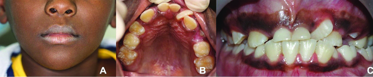
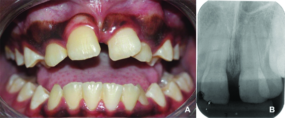
Extraoral clinical examination revealed the presence of oedema in the region between the nose wing and upper lip on the right side of the face, causing slight facial asymmetry [Table/Fig-1a]. The intra-oral examination revealed that the patient had mixed dentition and was caries free [Table/Fig-1b]. He had unsatisfactory oral hygiene, with biofilm on all teeth. A normal overbite and overjet, presence of midline deviation, eruption of tooth 21 in gyroversion, crossbite of teeth 21 and 22 and crowding were verified. The presence of a painless increased volume of hard consistency in the region of the upper central right incisor and the prolonged retention of the primary upper central incisors were observed [Table/Fig-1c].
a) Extraoral view showing slight facial oedema on the right side of the face and asymmetry. b) Intraoral occlusal upper photography showing the prolonged retention of the primary upper central incisors and the tooth 21 in gyroversion. c) Frontal view showing midline deviation and crossbite of teeth 21 and 22.
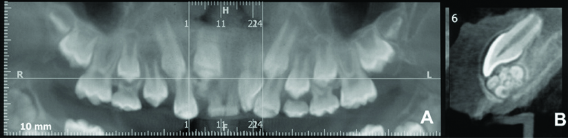
After performing periapical and panoramic radiographs, a cone beam volumetric computed tomography was suggested [Table/Fig-2a]. A circumscribed lesion formed by small hyperdense masses like denticles, surrounded by a hypodense halo [Table/Fig-2b] was found, suggesting the provisional diagnosis of compound odontoma.
a) Panoramic and b) approximated view of the lesion in the cone beam computed tomography.
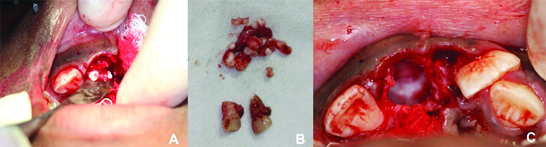
The proposed treatment was the extraction of the retained elements followed by a complete excision of the lesion [Table/Fig-3], to be sent for histopathological examination. After obtaining parents’ consent, the procedure was performed in a clinic ambulatory. Following local anaesthesia, using 2% lidocaine with epinephrine 1:100.000, it was performed by vestibular infiltration with nasopalatine supplementation; the intra-sulcular incision was performed with scalpel and 15c blade. The mucoperiosteal flap of total thickness was bent on the palate to gain access to the lesion. The primary teeth #51 and #61 were extracted with the aid of a 150S forceps. Subsequently, the entire lesion was resected with the aid of a curette. The bone shop was irrigated with saline solution (0.9%) and inspected. Finally, the margins were sutured with absorbable ac. Polygalactinvicryl 4-0 (Ethicon®).
(a) Surgical procedure b) showing the removal of the retained primary central incisors and the lesion and c) its complete excision.
The patient presented cooperative behaviour throughout the procedure. At the end, the postoperative hygiene and dietary guidelines were explained to the patients. A 0.12% chlorhexidine solution was prescribed to be used twice a day for seven days for cleaning the affected area and an analgesic (dipyrone) to be used for three days.
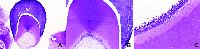
The macroscopical exam revealed 12 pieces of tooth-like structures with hard consistency and different sizes, measuring 15×11×7 mm in total. In addition, another part of the lesion was formed by soft tissue with fibroelastic consistency and brownish colour, measuring 10×7×6 mm. Microscopically, the lesion presented a constitution similar to normal dental tissues, with enamel, dentin, and dental pulp cells [Table/Fig-4]. The analysis confirmed the diagnosis of compound odontoma.
a,b) Histopathological aspects of compound odontoma, showing a malformed tooth germ composed of enamel, dentin, and pulp. c) Presence of odontoblasts forming dentin (stained with H&E; a: 40X, b: 100X, c: 400X).
One week after surgery, patient visited for suture removal and evaluation of the local tissue healing, which was satisfactory. Further clinical and radiographic follow-up evaluations were carried out every six months, and there was spontaneous eruption of the permanent upper central right incisor. The patient was happy and has shown to be more confident and more smiling during consultations. The parents were instructed to seek orthodontic treatment at the end of the exfoliation of the primary teeth. Meanwhile, he continued to receive preventive health care and clinical and radiographic evaluations, that completed 18 months of follow-up [Table/Fig-5].
a) Intraoral frontal view and b) periapical radiography after 18 months of follow-up.
Discussion
Odontomas are frequently detected by routine radiographs, usually between 10 to 19 years of age. Its incidence is of 0.4%, being the complex type the most frequent, occurring in 62% of the cases [1]. They are asymptomatic and may be associated with clinical signs, such as unerupted or impacted teeth, prolonged retention of primary teeth, tooth displacement, and/or malocclusion [1-3].
The presence of delayed eruption or dental impaction should always be considered as a warning sign [4]. It could be caused by a physical obstruction, due to supernumerary tooth, ectopic eruption of other teeth, cysts or tumours, scars from previous surgeries or dental trauma, ankylosis, gingival hyperplasia or premature loss of primary tooth, or by other causes, as cleidocranial dysplasia, arch-length deficiency, sclerosteosis, Gardner syndrome, genetic predisposition, nutritional deficiency or traumatic displacement of tooth germ [5]. In the present case, the prolonged retention of the deciduous tooth and the impaction of the permanent successor was associated with the presence of an odontoma, which was verified through the radiographs, as done in most of cases [1,3,4,6-9].
The early diagnosis and multi-discipline approach are essential to ensure a favourable prognosis in the treatment of eruptive alterations [4]. Numerous dental disorders can be detected by clinical and radiological routine examinations [10]. However, in the present report, despite the patient being nine-year-old, he had never been to the dentist, which delayed the detection of his problem. The American Academy of Paediatric Dentistry recommends that the first examination should be done at the time of the eruption of the first tooth and no later than 12 months of age. The occlusion and dentofacial development must be monitored throughout regular clinical evaluations and radiographs [10].
It is important to focus on early oral disease prevention, especially due to its excellent cost-effectiveness [11]. Even so, in the present case, it was possible to obtain a satisfactory result with conservative management after the removal of the lesion. One of the factors to be considered to assess the difficulty of spontaneous eruption of a permanent tooth is the position of the contralateral element. In the present case, the authors waited for the spontaneous eruption, which occurred satisfactorily, rather than planning an orthodontic intervention from the first moment, as was observed in similar cases [4,9].
The spontaneous eruption of the impacted permanent tooth was different from other reports with patients at ages close to the present case, in which the orthodontic traction of the impacted tooth had to be done [4,7,9]. In addition, as in the present case, the patients presented delayed eruption of incisor teeth while the contralateral teeth had already erupted [4,9], showing a difficulty for the spontaneous eruption of these elements. However, it occurred in the case reported.
Although the panoramic and intraoral radiographs could be used to establish a diagnostic hypothesis, cone beam computed tomography has played an important role in the identification of lesions [12], being a more appropriate tool for planning the excision of the lesion [13]. This justified the use of this advanced diagnostic imaging technique for the present case.
Aesthetics is an important factor to consider in current dentistry. The self-perception of facial aesthetics is the main motivational factor that leads patients to seek orthodontic treatment [14]. This report shows a paediatric patient with a compound odontoma that produced dental and facial changes, causing discontentment with aesthetics, which prompted him to seek dental care. In similar reports, despite the absence of permanent anterior dental elements, the authors did not report complaints regarding aesthetics by the patients or their relatives [4,9].
Moreover, it is known that dental problems can affect psychological and social aspects of the patients, especially the adolescents, leading to consequences that interfere in their quality of life [15]. In the present case, the patient only sought dental care due to an aesthetic complaint. It highlights the importance of carrying out an integrated treatment plan that covers the patient’s and/or family’s complaints and wishes.
Conclusion
The present report shows satisfactory management of a compound odontoma in a nine-year-old boy with prolonged retention of the primary upper central incisors, through a minimally invasive approach after a complete surgical excision of the lesion. The spontaneous eruption of the permanent impacted tooth and an improvement in self-esteem and satisfaction of the patient were observed. This emphasises the importance of routine exams to conduct an early diagnosis and solve aesthetic problems related to dental treatment among children and adolescents.
[1]. Tekkesin MS, Pehlivan S, Olgac V, Aksakalli N, Alatli C, Clinical and histopathological investigation of odontomas: review of the literature and presentation of 160 casesJ Oral Maxillofac Surg 2012 70:1358-61.10.1016/j.joms.2011.05.02421840103 [Google Scholar] [CrossRef] [PubMed]
[2]. Satish V, Prabhadevi MC, Sharma R, Odontome: A brief overviewInt J Clin Pediatr Dent 2011 4(3):177-85.10.5005/jp-journals-10005-110627678223 [Google Scholar] [CrossRef] [PubMed]
[3]. Kämmerer PW, Schneider D, Schiegnitz E, Schneider S, Walter C, Frerich B, Clinical parameter of odontoma with special emphasis on treatment of impacted teeth-a retrospective multicentre study and literature reviewClin Oral Invest 2016 20:1827-35.10.1007/s00784-015-1673-326612404 [Google Scholar] [CrossRef] [PubMed]
[4]. Da Silva Bastos VDA, Freitas-Fernandes LB, Soares DN, Neto OC, Abrahão AC, Farinhas JA, Management of over retention of permanent incisor impacted by compound odontoma: Clinical, radiological, and microscopic evaluationPediatr Dent J 2018 28:68-72.10.1016/j.pdj.2018.03.001 [Google Scholar] [CrossRef]
[5]. Suri L, Gagari E, Vastardis H, Delayed tooth eruption: Pathogenesis, diagnosis, and treatment. A literature reviewAm J Orthod Dentofacial Orthop 2004 126(4):432-45.10.1016/j.ajodo.2003.10.03115470346 [Google Scholar] [CrossRef] [PubMed]
[6]. Shetty L, Gangwani K, Kulkarni D, Londhe U, Odontome, cyst, impacted tooth, and space infection in a single patient: All-in-one diagnostic dilemmaAnn Maxillofac Surg 2018 8(1):127-30.10.4103/ams.ams_211_1729963439 [Google Scholar] [CrossRef] [PubMed]
[7]. Lacarbonara M, Lacarbonara V, Cazzolla AP, Spinelli V, Crincoli V, Lacaita MG, Odontomas in developmental age: confocal laser scanning microscopy analysis of a caseEur J Paediatr Dent 2017 18(1):77-79. [Google Scholar]
[8]. Jain A, Karuna YM, Baliga M, Suprabha BS, Natarajan S, Surgical management of complex odontoma associated with agenesis of a molarContemporary clinical dentistry 2018 9(Suppl 2):S388-90.10.4103/ccd.ccd_789_1730294179 [Google Scholar] [CrossRef] [PubMed]
[9]. Baldawa RS, Khante KC, Kalburge JV, Kasat VO, Orthodontic management of an impacted maxillary incisor due to odontomaContemp Clin Dent 2011 2(1):37-40.10.4103/0976-237X.7931222114453 [Google Scholar] [CrossRef] [PubMed]
[10]. American Academy of Pediatric DentistryGuideline on periodicity of examination, preventive dental services, anticipatory guidance/counseling, and oral treatment for infants, children, and adolescentsPediatr Dent 2016 38(6):133-41. [Google Scholar]
[11]. Bhaskar V, McGraw KA, Divaris K, The importance of preventive dental visits from a young age: systematic review and current perspectivesClin Cosmet Investig Dent 2014 6:21-27.10.2147/CCIDE.S4149924672258 [Google Scholar] [CrossRef] [PubMed]
[12]. Anuraag B, Choudhary M, Utility of cone beam CT in maxillo-facial radiologySF Dent Oral Res J 2017 1:110.23959/sfdorj-1000006 [Google Scholar] [CrossRef]
[13]. Santos LAN, Lopez LJ, Roque-Torees GD, Oliveira VF, Freitas DQ, Complex odontoma: a case report with micro-computed tomography findingsCase Rep Dent 2016 2016:3584751DOI:http://dx.doi.org/10.1155/2016/358475110.1155/2016/358475127293913 [Google Scholar] [CrossRef] [PubMed]
[14]. Samsonyanova L, Broukal Z, A systematic review of individual motivational factors in orthodontic treatment: facial attractiveness as the main motivational factor in orthodontic treatmentInt J Dent 2014 2014:938274DOI: http://dx.doi.org/10.1155/2014/93827410.1155/2014/93827424963296 [Google Scholar] [CrossRef] [PubMed]
[15]. Boeira GF, Salas MMS, Araújo DC, Masotti AS, Demarco FF, Factors influencing dental appearance satisfaction in adolescents: a cross-sectional study conducted in Southern BrazilBraz J Oral Sci 2016 15(1):8-15.10.20396/bjos.v15i1.8647091 [Google Scholar] [CrossRef]