Normal Sonographic Optic Nerve Sheath Diameter among Children in South Western Nigeria
Ayodeji Anike Olatunji1, Olubunmi O Bodunde2
1 Senior Lecturer/Consultant, Department of Radiology, Olabisi Onabanjo Univeristy Teaching Hospital, Sagamu, Ogun State, Nigeria.
2 Senior Lecturer/Consultant, Department of Ophthalmology, Olabisi Onabanjo Univeristy Teaching Hospital, Sagamu, Ogun State, Nigeria.
NAME, ADDRESS, E-MAIL ID OF THE CORRESPONDING AUTHOR: Dr. Ayodeji Anike Olatunji, Senior Lecturer/Consultant, Department of Radiology, Olabisi Onabanjo University Teaching Hospital, Sagamu, Ogun State, Nigeria.
E-mail: Ayodeji.olatunji@yahoo.com
Introduction
Optic Nerve Sheath Diameter (ONSD) correlates with intracranial pressure and can be utilised for monitoring in emergency situations.
Aim
To determine the reference values of optic nerve sheath diameter in healthy school going children in South western Nigeria by using B-mode sonography.
Materials and Methods
A cross-sectional study was conducted in children from three selected schools after consent and approval from the parents and Institutional Health Research Ethics committee, respectively. B-mode sonography using a 4-12 MHz probe was carried out on children with normal visual acuity and fundoscopy. Demographics of the children and the optic nerve sheath diameter were recorded in a proforma. Data was analysed with SPSS version 21 and were represented as mean and standard deviation. Paired t-test was used to compare ONSD between the right and left eye. Independent t-test was used to compare ONSD between males and females. Significance was set at p-value <0.05.
Results
Optic nerve sheath diameter was measured in both eyes of 468 children. There were 235 (50.2%) boys and 233 (49.8%) girls (M:F Ratio=1.01: 1) and age range was 2 to 16 years. Mean ONSD in the right eyes was 0.56±0.08 cm while in the left eye it was 0.58±0.09 cm for boys, while in girls in the right eyes it was 0.55±0.08 cm and in left eye it was 0.57±0.07 cm. The mean ONSD of the left eyes was longer than the right eyes. (p=<0.001). There was no significant difference in ONSD measurement between males and females (p=0.275).
Conclusion
Mean ONSD in children was longer in the left eye than the right, and increases with age.
Emmetropes, Eyes, School children, Ultrasonography
Introduction
Anatomically, the optic nerve is a continuation of the brain and hence, carries along with it the membranes of the brain as well as the cerebrospinal fluid. It is surrounded by Cerebrospinal Fluid (CSF) and dura mater, which forms the Optic Nerve Sheath (ONS) [1]. This makes it vulnerable to the changes within the brain, surrounding coverings and the fluid [2,3].
Neurological disorders or diseases often result in raised intracranial pressure which impacts on the optic nerve resulting in its swelling and resultant papilloedema. Raised intracranial pressure is usually diagnosed with invasive methods like Computerised Tomography (CT), Magnetic Resonance Imaging (MRI) or Lumber puncture. CT and MRI are quite expensive for the majority of the populace in a developing economy like ours, apart from their unavailability in most hospitals except tertiary care centres which are mostly located in cities. Plain film findings involving erosion of the clinoid processes and thumb printing appearances on the skull are part of the radiological ways of assessing raised intracranial pressure in most health care facilities in the country, which is grossly inadequate especially in emergency situations. However, optic nerve sheath dilatation has been shown to be a much earlier manifestation of increased intracranial pressure [4]. Several studies [1-13] have shown a correlation between ONSD measurement and raised intra cranial pressure. A mean ONSD of >5/5.2 mm is considered abnormal in some of the cases and thus increased intracranial pressure should be suspected [3,8]. This knowledge can be useful in intracranial hypertension which is a common life threatening syndrome caused by a variety of neurological and non-neurological diseases [5]. The rapid diagnosis of intracranial hypertension is usually needed for therapeutic reasons which in various clinical settings can rarely be achieved without invasive procedures [1-3].
However, Zaidi SJH et al., has suggested that there is need for establishment of a normal range that is not age or size dependent, if the ONSD must be used as a screening measure [14]. This is because ONSD can easily be done using ultrasonography. B scan ultrasonography was used in the ONSD assessment of 1998 study by Beatty S et al., and was found to be accurate [15]. This accuracy has been verified by other researchers like Ballantyne SA et al., and Lochner P et al., [16,17]. The distension of Optic Nerve Sheath is maximal 3 mm behind the eye. This became the landmark used in the measurement of the ONSD globally [18].
Ultrasound is readily available, affordable, portable, non-invasive, and comfortable and does not usually require sedation in children. It is also said that ultrasound provides a quick, accurate well-tolerated, non-invasive tool for evaluating potentially vision threatening conditions at the bedside [12]. This study therefore aims at producing baseline data of normal range value of ONSD for children in the country (Nigeria) and contributing to the body of knowledge worldwide.
Materials and Methods
The cross-sectional observational study was done in a South Western Nigerian tertiary hospital, from August 2016-March 2017. Children from three schools within the Local Government Area of the Hospital, including both public and private institutions were recruited for the study after a certificate was obtained from the Institutions Health Research Ethics Committee. The Local Government Education board was informed and the Head teachers of the schools involved were duly informed and educated about the study. Informed consents were duly obtained from the parents of all participants. No child was coerced into the study. All the children had the normality of their eyes ascertained by the Ophthalmologist in the study who checked their visual acuity, did ocular examination, fundoscopy and refraction for them. A total of 495 children were screened for the study, however, 468 children were scanned (involving 936 eyes), aged 2 years to 16 years. Twenty-five children who had refractive errors after examination by the Ophthalmologist were excluded. Two children were dropped off the study because of lack of cooperation.
The radiologist performed the sonography from the most upper part of the upper eyelid and the patient was asked to gently close the eyes. A linear probe with 4-12 MHz power was used on a Philips 550 clear Vue model, manufactured in 2013. Ultrasound scans were performed through the closed eye lid from the temporal side each time performing an axial cut through the eyes and including a longitudinal section of the optic nerve. The optic disc, and as long a section of optic nerve as possible, was included in the freeze-rame. The option to toggle through the last 10 seconds of images was often used to obtain the best image. Measurements were made with electronic calipers 3 mm behind the posterior sclera surface of the globe. Single Radiologist took all the dimensions to avoid inter observer error. Both eyes were measured despite the fact that previous studies have shown the presence of intraocular symmetry between the OND/ONSD of fellow eyes [16,19,20].
A protocol sheet approved for the study was filled on behalf of each child including the age, last birthday, gender, sonographic dimensions of the optic nerves and any other interesting findings seen within the eyes during the scan.
Statistical Analysis
Analysis of data was done with SPSS version 21.0 for windows software. (SPSS, Inc. Chicago, Illinois). Continuous variables were expressed as mean±standard deviation (SD) with student t-test analysis for comparison. Categorical variables were expressed as percentages and comparison was done by chi-square analysis. The p-value <0.05 was considered significant.
Results
A total number of 468 children involving 936 eyes were studied. The youngest been 2 years while the oldest was 16-year-old. The mean age of the participants was 8.81±3.82 years.
[Table/Fig-1] shows the mean ONSD for both the eyes in boys and girls. There was no statistically significant difference in the mean ONSD between the two genders. ANOVA; F=1.194; p=0.275 for right eyes and F=0.654; p=0.419 for the left eyes.
Optic nerve sheath diameter and gender.
| Gender | n | Mean optic nerve sheath diameter±SD (in cm) |
|---|
| Right eye | Left eye |
|---|
| Male | 235 | 0.5575±0.07538 | 0.5756±0.08547 |
| Female | 233 | 0.5529±0.07791 | 0.5738±0.07021 |
| Total | 468 | 0.5552±0.07660 | 0.5747±0.07817 |
The mean ONSD of the right eyes in the total population studied was 0.56±0.08 cm and 0.57±0.08 cm on the left eyes. The difference was statistically significant (p<0.001).
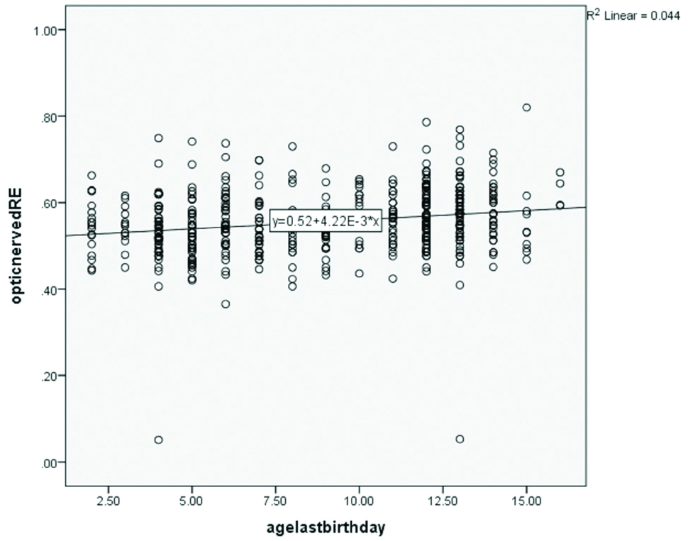
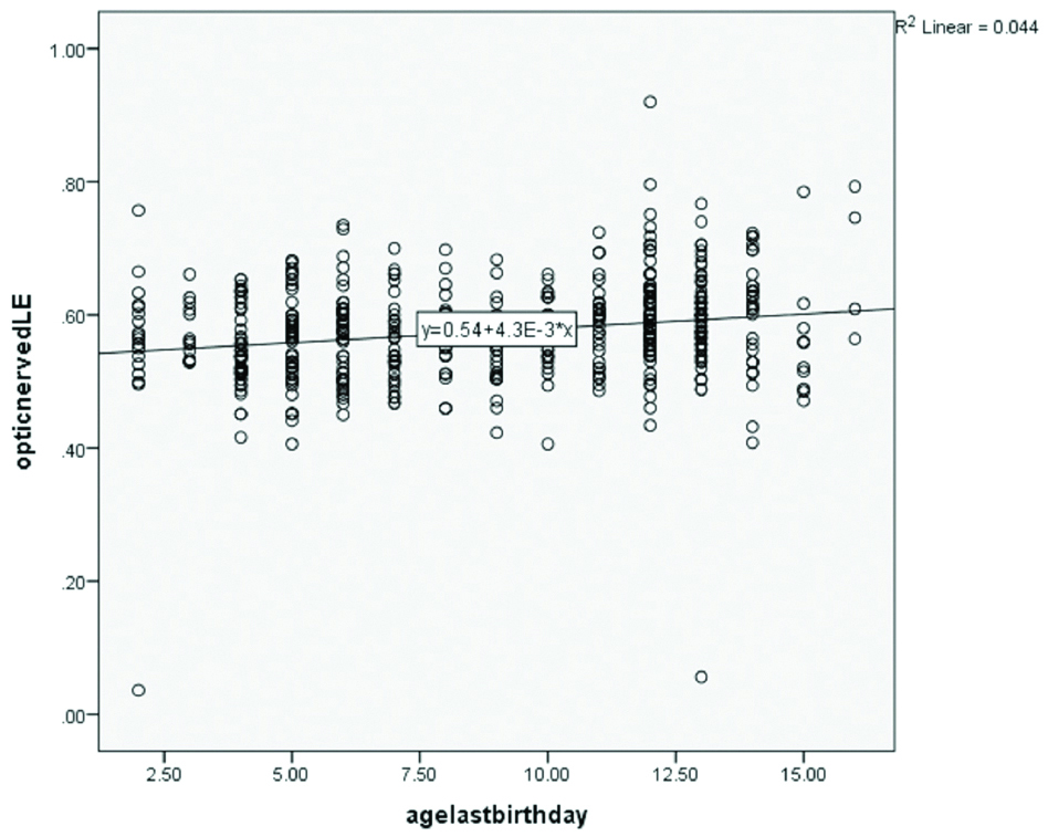
[Table/Fig-2] shows the mean ONSD in different age groups for the right eyes. The ANOVA right eyes; F value=5.4, p=<0.001, and eta value=0.212. The mean ONSD in the left eyes shows ANOVA; F=5.46, p=<0.0001 and the eta value=0.212. Optic nerve diameter increases with age [Table/Fig-3,4].
Optic nerve sheath diameter in different age groups.
| Age group (years) | n | Optic nerve sheath diameter±SD (cm) |
|---|
| Right eye | Left eye |
|---|
| 2-4 | 78 | 0.5373±0.08185 | 0.5546±0.08292 |
| 5-7 | 123 | 0.5409±0.06846 | 0.5614±0.06633 |
| 8-10 | 66 | 0.5474±0.06811 | 0.5620±0.06211 |
| 11-13 | 157 | 0.5716±0.07935 | 0.5937±0.08162 |
| 14-16 | 44 | 0.5800±0.07554 | 0.5983±0.09055 |
| Total | 498 | 0.5552±0.07660 | 0.5747±0.07817 |
Scatter plot of right eye optic nerve sheath diameter versus age-last-birthday.
Scatter plot of left eye optic nerve diameter versus age-last-birthday.
When comparing the means of the right eyes with that of the left eyes, the t-value is 3.93 while the p-value=0.0001 which means the left eye mean ONSD was significantly more than the right eye mean diameter.
Discussion
Generally, there was a dearth of literature concerning the normal measurement of optic nerve diameter in the black children except of cause, where they are been compared with the caucasian population or the children born by adults of African descent in the Americas or other countries of the world. In Nigeria, where this study was being done, the adults were more in focus than the children and only one study was done in the northern part [20] of the country. This study found that the mean optic nerve diameter of the right eyes was 0.55 cm±0.08 cm in girls and 0.56 cm±0.08 cm in boys compared well with the value that Steinborn M et al., got in their study i.e., 5.86±0.71 mm, 5.75±0.52 mm, and 5.77±0.48 mm [21-23]. There was a similarity too in the age group considered in that study as in the present study. This is relatively higher than the value that Ballantyne SA et al., quoted in their study as 2.1-4.3 mm with a mean of 3.08 mm in British children [16], Malayeri AA et al., in Iranian children 2.0-4.3 mm (mean of 3.3 mm) [24] and Beare NAV et al., in Malawian children of 2.5-4.1 mm (mean of 3.5 mm) most probably due to equipment especially the model in use [13]. Zaidi SJH et al., has documented that the intrinsic value of using the ultrasound as a means of measuring optic nerve sheath diameter can be affected by model of machine, portion of the globe where the probe is placed as well as the experience of the operator [14]. The other reason may be the dissimilarity in the ages studied which in their study involved infants, while the influence of race, environment, and nutritional status of participants may also be responsible.
The Steinborn M study [21-23], also agreed with our finding of no significant difference between the values for boys and girls (p=0.517 right eyes, 0.805 left eyes). The current study showed an absence of statistically significant values in the sexes as documented in both children and adult population of studies available to the authors by Beatty S et al., and Anas I, Maude RR et al., [15,20,25]. However, Ballantyne SA et al., did not find a statistically significant difference in values between the left and the right eyes, and this may not be unrelated with the possibility of the dominant use of the left hemisphere of the brain among these children [16]. Maude RR et al., also concluded that there was no relationship between ONSD and age (>4 years), gender, head circumference and no difference in left versus right eye or horizontal beam [25].
The ONSD measured in both eyes in this study showed a linearity of diameter with age, although no statistical difference was observed in the different age groups. This is in agreement with Newman WD et al., (2002) who also found that the mean ONSD increases with age [26].
Limitation
The study is from one local government area of Nigeria and majority of them are from one ethnic group of the country; the study does not include ammetropes.
Conclusion
The optic nerve sheath diameter can be done in the Nigerian children; it has a value of 5.6 mm in the normal children. It increases gradually with age. It is slightly higher in the left eyes when compared with the right. No significant sex difference.
[1]. Moretti R, Pizzi B, Ultrasonography of the optic nerve in neurocritically ill patientsActa Anaesthesiolscand 2011 55(6):644-52.10.1111/j.1399-6576.2011.02432.x21463263 [Google Scholar] [CrossRef] [PubMed]
[2]. Helmke K, Hansen HC, Fundamentals of transorbital sonographic evaluation of optic nerve sheath expansion under intracranial hypertension II. Experimental studyPediatric Radiology 1996 26:706-10.10.1007/BF013833848805600 [Google Scholar] [CrossRef] [PubMed]
[3]. Helmke K, Hansen HC, Fundamentals of transorbital sonographic evaluation of optic nervesheath expansion under intracranial hypertension I. Experimental studyPediatric radiology 1996 26:701-05.10.1007/BF013833838805599 [Google Scholar] [CrossRef] [PubMed]
[4]. Chacko J, Optic nerve sheath diameter: An Ultrasonographic window to view raised intracranial pressure?Indian J Crit Care Med 2014 18(11):707-08.10.4103/0972-5229.14400725425835 [Google Scholar] [CrossRef] [PubMed]
[5]. Luberda M, Stachura K, Moskala M, Optic nerve Sonography-the non-invasive evaluation of intracranial pressurePrzegl Lek 2013 70(11):983-85. [Google Scholar]
[6]. Maissan IM, Dirven PJ, Haitsma IK, Hoeks SE, Gommers D, Stolker RJ, Ultrasonographic measured optic nerve sheath diameter as an accurate and quick monitor for changes in intracranial pressureJ Neurosurg 2015 123(3):743-47.10.3171/2014.10.JNS14119725955869 [Google Scholar] [CrossRef] [PubMed]
[7]. Ohle R, McIsaac SM, Woo MY, Perry JJ, Sonography of the optic nerve sheath diameter for detection of raised intracranial pressure compared to computed tomography: a systematic review and meta-analysisJ Ultrasound Med 2015 34(7):1285-94.10.7863/ultra.34.7.128526112632 [Google Scholar] [CrossRef] [PubMed]
[8]. Frumin E, Schlang J, Wiechmann W, Hata S, Rosen S, Anderson C, Prospective analysis of single operator sonographic optic nerve sheath diameter measurement for diagnosis of elevated intracranial pressureWest J Emerg Med 2014 15(2):217-20.10.5811/westjem.2013.9.1619124672615 [Google Scholar] [CrossRef] [PubMed]
[9]. Sangani SV, Parikh S, Can sonographic measurement of optic nerve sheath diameter be used to detect raised intracranial pressure in patients with tuberculous meningitis? A prospective observational studyIndian J Radiol Imaging 2015 25(2):173-76.10.4103/0971-3026.15586925969641 [Google Scholar] [CrossRef] [PubMed]
[10]. Raffiz M, Abdullah JM, Optic nerve sheath diameter measurement: a means of detecting raised ICP in adult traumatic and non-traumatic neurosurgical patientsAm J Emerg Med 2017 35(1):150-53.10.1016/j.ajem.2016.09.04427852525 [Google Scholar] [CrossRef] [PubMed]
[11]. Haratz K, Viñals F, Lev D, Feit H, Ben-Sira L, Lerman-Sagie T, Fetal optic nerve sheath measurement as a non-invasive tool for assessment of increased intracranial pressureUltrasound Obstet Gynecol 2011 38(6):646-51.10.1002/uog.905021584889 [Google Scholar] [CrossRef] [PubMed]
[12]. Le A, Hoehn ME, Smith ME, Spentzas T, Schlappy D, Pershad J, Bedside sonographic measurement of optic nerve sheath diameter as a predictor of increased intracranial pressure in childrenAnn Emerg Med 2009 53(6):785-91.10.1016/j.annemergmed.2008.11.02519167786 [Google Scholar] [CrossRef] [PubMed]
[13]. Beare NAV, Kampondeni S, Glover SJ, Molyneux E, Taylor TE, Harding SP, Detection of raised intracranial pressure by ultrasound measurement of optic nerve sheath diameter in African childrenTrop Med Int Health 2008 13(11):1400-04.10.1111/j.1365-3156.2008.02153.x18983275 [Google Scholar] [CrossRef] [PubMed]
[14]. Zaidi SJH, Yamamoto LG, Optic nerve sheath diameter measurements by CT scan in ventriculoperitoneal shunt obstructionHawaii J Med Public Health 2014 73(8):251-55. [Google Scholar]
[15]. Beatty S, Good PA, McLaughlin J, O’Neill EC, Echographic measurements of the retrobulbar optic nerve in normal and glaucomatous eyesBr J Ophthalmol 1998 82(1):43-47.10.1136/bjo.82.1.439536879 [Google Scholar] [CrossRef] [PubMed]
[16]. Ballantyne SA, O’Neill G, Hamilton R, Hollman AS, Observer variation in the sonographic measurement of optic nerve sheath diameter in normal adultsEur J Ultrasound 2002 15(3):145-49.10.1016/S0929-8266(02)00036-8 [Google Scholar] [CrossRef]
[17]. Lochner P, Coppo L, Cantello R, Nardone R, Naldi A, Leone MA, Intra- and inter observer reliability of transorbital sonographic assessment of the optic nerve sheath diameter and optic nerve diameter in healthy adultsJ Ultrasound 2014 19(1):41-45.10.1007/s40477-014-0144-z26941872 [Google Scholar] [CrossRef] [PubMed]
[18]. Hassen GW, Bruck I, Donahue J, Mason B, Sweeney B, Saab W, Accuracy of optic nerve sheath diameter measurement by emergency physicians using bedside ultrasoundThe Journal of Emergency Medicine 2015 48(4):450-57.10.1016/j.jemermed.2014.09.06025497897 [Google Scholar] [CrossRef] [PubMed]
[19]. Karim S, Clark RA, Poukens V, Demer JL, Demonstration of systemic variation in human intraorbital optic nerve size by quantitative magnetic resonance imaging and histologyInvest Ophthalmol Vis Sci 2004 45(4):1047-51.10.1167/iovs.03-124615037567 [Google Scholar] [CrossRef] [PubMed]
[20]. Anas I, Transorbital sonographic measurement of normal optic sheath nerve diameter in Nigerian adult populationMalays J Med Sci 2014 21(5):24-29. [Google Scholar]
[21]. Steinborn M, Fiegler J, Ruedisser K, Hapfelmeier A, Denne C, Macdonald E, Measurement of the optic nerve sheath diameter in children: comparison between transbulbar sonography and magnetic resonance imagingUltraschall Med 2011 [Epub ahead of print]10.1055/s-0031-1273491 [Google Scholar] [CrossRef]
[22]. Steinborn M, Friedmann M, Hahn H, Hapfelmeier A, Macdonald E, Warncke K, Normal values for transbulbar Sonography and Magnetic resonance imaging of the optic nerve sheath diameter (ONSD) in children and adolescentsUltraschall Med 2015 36(1):54-58.10.1055/s-0034-138501225140497 [Google Scholar] [CrossRef] [PubMed]
[23]. Steinborn M, Friedmann M, Makowski C, Hahn H, Hapfelmeier A, Juenger H, High resolution transbulbar Sonography in children with suspicion of increased intracranial pressureChilds Nerv Syst 2016 32(4):655-60.10.1007/s00381-015-3001-226759020 [Google Scholar] [CrossRef] [PubMed]
[24]. Malayeri AA, Bavarian S, Mehdizadeh M, Sonographic evaluation of optic nerve diameter in children with raised intracranial pressureJ Ultrasound Med 2005 24(2):143-47.10.7863/jum.2005.24.2.14315661943 [Google Scholar] [CrossRef] [PubMed]
[25]. Maude RR, Hossain MA, Hassan MU, Osbourne S, Sayeed KL, Karim MR, Transorbital sonographic evaluation of normal optic nerve sheath diameter in healthy volunteers in BangladeshPLos One 2013 8(12):e8101310.1371/journal.pone.008101324312515 [Google Scholar] [CrossRef] [PubMed]
[26]. Newman WD, Hollman AS, Dutton GN, Carachi R, Measurement of optic nerve sheath diameter by ultrasound: a means of detecting acute raised intracranial pressure in hydrocephalusBr J Ophthalmol 2002 86:1109-13.10.1136/bjo.86.10.110912234888 [Google Scholar] [CrossRef] [PubMed]